
Beyond Feelings: Quantifying Therapy's Impact with Brain Imaging

Welcome to Mind Care Guide.
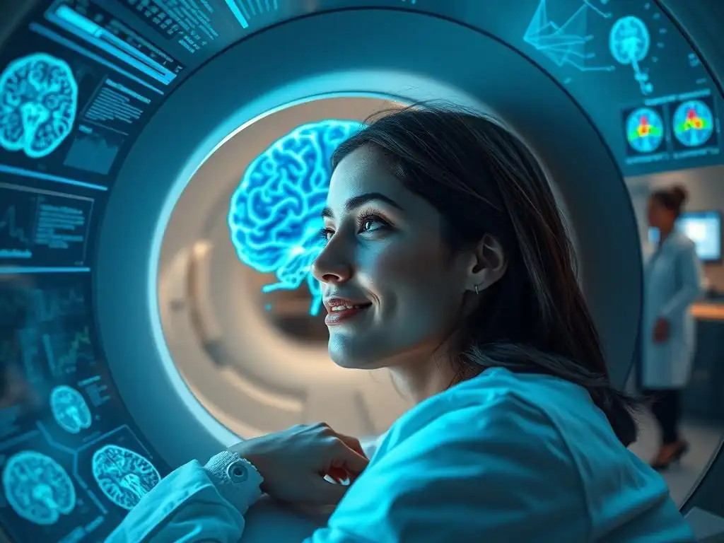
Feeling lost in the maze of mental health treatments? We understand. In a world of subjective experiences, it's natural to crave tangible evidence of progress. That's why we're diving deep into the fascinating world of neuroscience to explore a groundbreaking question: Just how effective is therapy? Our latest article, "Beyond Feelings: Quantifying Therapy's Impact with Brain Imaging," sheds light on the brain imaging therapy effectiveness, offering a glimpse into how science is helping us understand and validate the journey to mental well-being. Curious to see what the scans reveal? Let's explore together.
Beyond Feelings: Quantifying Therapy's Impact with Brain Imaging
- Introduction: The Quest to Objectively Measure Therapy Outcomes
-
Brain Imaging Techniques Used to Evaluate Therapy
- Functional Magnetic Resonance Imaging (fMRI): Detecting Brain Activity Changes
- Electroencephalography (EEG): Measuring Brainwave Patterns and Therapy's Influence
- Positron Emission Tomography (PET): Examining Neurochemical Changes After Therapy
- Magnetic Resonance Spectroscopy (MRS): Assessing Neurotransmitter Levels
-
How Brain Imaging Reveals Therapy's Impact on Specific Mental Health Conditions
- Brain Imaging and Depression Treatment: Mapping Changes in the Prefrontal Cortex and Amygdala
- Anxiety Disorders and Brain Activity: Visualizing the Effects of Therapy on Fear Circuits
- PTSD Therapy and the Brain: Examining Amygdala and Hippocampus Modulation
- Schizophrenia: Assessing Cognitive Function Changes with Brain Imaging
- Addiction Therapy: Tracking Reward System Changes with Brain Imaging
-
Case Studies: Real-World Examples of Brain Imaging Validating Therapy Success
- Case Study 1: Obsessive-Compulsive Disorder - fMRI Evidence of Improved Executive Function After Exposure and Response Prevention (ERP)
- Case Study 2: Social Anxiety Disorder - EEG Confirmation of Reduced Anxiety Symptoms with Cognitive Behavioral Therapy (CBT)
- Case Study 3: Major Depressive Disorder - PET Scan Showing Increased Serotonin Receptor Binding After Selective Serotonin Reuptake Inhibitor (SSRI) Therapy and Psychotherapy
- The Future of Brain Imaging in Mental Healthcare
- Ethical Considerations in Using Brain Imaging for Therapy Evaluation
- Conclusion: Brain Imaging - A Powerful Tool for Understanding and Optimizing Therapy's Effectiveness
-
Frequently Asked Questions
- What is brain imaging therapy, and how does it differ from traditional talk therapy?
- How can brain imaging help determine the effectiveness of therapy?
- What are the benefits of using brain imaging in mental health treatment?
- Are there any risks or limitations associated with using brain imaging in therapy?
- What types of mental health conditions can benefit from brain imaging therapy?
- Final Reflection: The Evolving Landscape of Mental Healthcare
- Unlock Your Brain's Potential: Take Charge of Your Mental Wellbeing
Introduction: The Quest to Objectively Measure Therapy Outcomes
The Limitations of Subjective Reports in Assessing Therapy Effectiveness
Traditionally, the success of psychotherapy has been largely measured through subjective reports from patients and therapists. Questionnaires asking about symptom severity, mood levels, and overall well-being provide invaluable insights into a person's lived experience before, during, and after therapy. Similarly, therapists rely on their clinical observations and the patient's self-reporting to gauge progress.
While essential, this reliance on subjective data presents inherent limitations. Individual biases, memory recall issues, the desire to please the therapist, or difficulty articulating internal states can all influence the accuracy of these reports. Furthermore, these methods may not fully capture subtle, underlying changes in emotional regulation or cognitive processing that occur as a result of effective treatment.
The challenge in relying solely on how someone feels is that feelings are fluid and can be influenced by many factors outside of therapy. A patient might report feeling better one week due to external circumstances, even if the core neurological patterns associated with their condition haven't significantly shifted. This makes it difficult to definitively attribute improvements solely to the therapeutic intervention. Consequently, researchers and clinicians have long sought more objective methods to complement subjective assessments, aiming to provide a clearer, more verifiable picture of therapy's true impact and better understand the effectiveness of brain imaging in therapy evaluation.
Introducing Brain Imaging: A Window into the Brain's Response to Therapy
Brain imaging techniques offer a powerful, non-invasive way to look inside the living brain and observe its structure and function. By capturing images of brain activity, connectivity, or chemical composition, these technologies provide an objective lens through which to examine the neural correlates of mental health conditions and how they change in response to therapeutic interventions. Unlike subjective reports, brain scans provide data that is less susceptible to conscious bias or reporting inaccuracies, offering a complementary perspective on treatment outcomes. This is where the field of brain imaging therapy effectiveness research truly shines, providing tangible evidence of neurological shifts.
By comparing brain scans taken before and after a course of therapy, researchers can identify specific changes in brain regions, networks, or neurochemical levels that correlate with clinical improvements. For example, if therapy for depression leads to a reduction in activity in brain areas associated with negative emotions, this provides objective evidence that the treatment is impacting the underlying neural circuitry.
This ability to visualize and quantify these changes moves the assessment of therapy's impact beyond just "feeling better" to understanding the biological mechanisms of recovery, bolstering the evidence base for various psychotherapies and improving the understanding of their brain imaging therapy effectiveness.
Brain Imaging Techniques Used to Evaluate Therapy
Functional Magnetic Resonance Imaging (fMRI): Detecting Brain Activity Changes
Functional Magnetic Resonance Imaging (fMRI) is a widely used neuroimaging technique that measures brain activity by detecting changes in blood flow. When a brain region becomes more active, it requires more oxygenated blood, and fMRI can detect the subtle magnetic differences between oxygenated and deoxygenated blood. By observing these changes while an individual performs a task (like recalling memories or processing emotions) or even while resting, researchers can create maps of brain activity and connectivity patterns. This makes fMRI particularly valuable for studying the neural circuits involved in thoughts, feelings, and behaviors associated with mental health conditions.
In the context of evaluating therapy effectiveness, fMRI allows researchers to see how the brain is working differently after treatment. Studies might examine changes in the activity levels of specific brain regions (e.g., the amygdala in anxiety disorders or the prefrontal cortex in depression) or alterations in the connectivity between different brain networks (e.g., the default mode network or executive control network). Observing a normalization of activity or connectivity patterns that were previously atypical in a patient population provides objective evidence for the effectiveness of brain imaging in therapy assessment, showing tangible neural changes alongside reported symptom reduction.
Electroencephalography (EEG): Measuring Brainwave Patterns and Therapy's Influence
Electroencephalography (EEG) measures electrical activity in the brain using electrodes placed on the scalp. This technique is excellent at capturing rapid changes in brain activity, measured in milliseconds, reflecting the synchronized firing of large groups of neurons. Different patterns of electrical oscillations, known as brainwaves (e.g., alpha, beta, theta, delta), are associated with different states of consciousness or cognitive processes. EEG is relatively inexpensive and portable compared to fMRI, making it a valuable tool for certain types of research and potentially for future clinical applications in assessing brain imaging therapy effectiveness.
EEG studies can assess the impact of therapy by looking at changes in specific brainwave frequencies or event-related potentials (ERPs), which are characteristic brainwave responses to specific stimuli (like viewing emotional images). For example, research might examine how therapy alters the amplitude or latency of ERPs associated with processing emotional information in patients with anxiety or depression. Changes in overall brainwave power or connectivity between different brain regions, as measured by EEG, can also provide insights into how therapy is normalizing neural activity patterns, offering a different temporal perspective compared to fMRI on the brain's response to treatment.
Positron Emission Tomography (PET): Examining Neurochemical Changes After Therapy
Positron Emission Tomography (PET) is a neuroimaging technique that uses radioactive tracers to visualize and measure metabolic processes or the distribution of specific molecules in the brain, such as neurotransmitter receptors or transporters. A radioactive substance, often linked to a molecule of interest (like a glucose analog to measure metabolic activity or a ligand that binds to serotonin receptors), is injected into the bloodstream. As the substance decays, it emits positrons, which are detected by the PET scanner, allowing researchers to map the concentration of the tracer throughout the brain.
PET scans are particularly useful for investigating the neurochemical underpinnings of mental health disorders and how pharmacotherapy and psychotherapy might alter these systems. For instance, studies using PET have examined changes in serotonin transporter availability in depression or dopamine receptor density in addiction following treatment. While less common for evaluating therapy alone compared to fMRI or EEG due to the use of radiation and higher cost, PET can provide unique insights into the molecular targets affected by therapeutic interventions, contributing valuable objective data to the understanding of brain imaging therapy effectiveness at a chemical level.
Magnetic Resonance Spectroscopy (MRS): Assessing Neurotransmitter Levels
Magnetic Resonance Spectroscopy (MRS), a technique related to MRI, allows researchers to measure the concentration of specific chemical compounds within the brain, including certain neurotransmitters and metabolites. Unlike standard MRI which images water protons to visualize structure, MRS detects signals from other molecules, such as N-acetylaspartate (NAA), choline, creatine, glutamate, GABA, and lactate. By analyzing the spectrum of signals emitted by these molecules, researchers can estimate their relative or absolute concentrations in a particular brain region.
MRS can provide direct evidence of how therapy influences the levels of key neurotransmitters involved in mood, anxiety, and cognitive function. For example, studies might use MRS to investigate changes in glutamate or GABA levels in the prefrontal cortex or other regions in patients with depression or anxiety disorders before and after receiving psychotherapy. Observing a normalization in the concentrations of these excitatory and inhibitory neurotransmitters provides objective data about the biochemical impact of therapy, complementing functional imaging findings and enhancing the understanding of the neurobiological basis of brain imaging therapy effectiveness.
How Brain Imaging Reveals Therapy's Impact on Specific Mental Health Conditions

Brain Imaging and Depression Treatment: Mapping Changes in the Prefrontal Cortex and Amygdala
Major Depressive Disorder (MDD) is often associated with disrupted activity and connectivity in brain networks involved in emotion regulation, reward processing, and cognitive control. Neuroimaging studies have consistently pointed to areas like the prefrontal cortex (PFC), particularly the ventromedial PFC (vmPFC) and dorsolateral PFC (dlPFC), and the amygdala as key players. The amygdala is often hyperactive in depression, processing negative stimuli excessively, while certain areas of the PFC, crucial for regulating emotions and executive functions, may show reduced activity or altered connectivity with the amygdala.
Studies using fMRI have shown that successful psychotherapy for depression, such as Cognitive Behavioral Therapy (CBT) or Interpersonal Therapy (IPT), is often associated with a normalization of activity in these regions. For instance, research has demonstrated decreased activity in the amygdala and increased activity in the vmPFC following successful CBT, suggesting improved top-down control over emotional responses.
These changes in neural circuitry provide objective evidence for the effectiveness of brain imaging in therapy for depression, correlating with clinical improvements reported by patients and therapists. PET studies have also shown increases in serotonin receptor binding in certain brain areas after successful treatment, pointing to neurochemical restoration.
Anxiety Disorders and Brain Activity: Visualizing the Effects of Therapy on Fear Circuits
Anxiety disorders, including Generalized Anxiety Disorder (GAD), Social Anxiety Disorder (SAD), and panic disorder, are characterized by excessive fear and worry. Neuroimaging research has implicated a "fear circuit" involving the amygdala, insula, and anterior cingulate cortex (ACC), often showing heightened reactivity to potential threats and altered connectivity with prefrontal regulatory regions. The insula is involved in processing bodily states associated with anxiety, while the ACC plays a role in conflict monitoring and emotional processing.
Psychotherapies like CBT, exposure therapy, and mindfulness-based therapies aim to help individuals confront feared situations, challenge anxious thoughts, and regulate physiological arousal. fMRI studies evaluating the brain imaging therapy effectiveness for anxiety disorders often look for reductions in amygdala and insula reactivity during the processing of anxiety-provoking stimuli after therapy. They may also observe increased activity or connectivity in prefrontal regions, suggesting enhanced cognitive control over fear responses. For example, studies on SAD have shown reduced amygdala responses to social cues after CBT, providing neural validation for the therapy's ability to dampen excessive fear reactions.
PTSD Therapy and the Brain: Examining Amygdala and Hippocampus Modulation
Post-Traumatic Stress Disorder (PTSD) involves persistent re-experiencing of a traumatic event, avoidance behaviors, negative alterations in cognition or mood, and hyperarousal. Neuroimaging findings in PTSD frequently include increased amygdala reactivity to trauma-related cues, reduced volume and activity in the hippocampus (a brain region critical for memory and contextual processing), and altered activity in the medial prefrontal cortex (mPFC), which is involved in fear extinction and emotional regulation.
Trauma-focused therapies, such as Prolonged Exposure (PE) or Eye Movement Desensitization and Reprocessing (EMDR), aim to help individuals process traumatic memories in a safe environment. Brain imaging studies investigating the effectiveness of brain imaging in therapy for PTSD have shown promising results. For example, fMRI studies have reported decreased amygdala reactivity and increased mPFC activity or connectivity after successful trauma-focused therapy, suggesting improved emotional regulation and fear extinction.
While hippocampal volume changes are less frequently observed post-therapy, some studies indicate functional normalization or increased connectivity with other brain regions. These findings underscore how therapy can help rewire the brain's response to trauma.
Schizophrenia: Assessing Cognitive Function Changes with Brain Imaging
Schizophrenia is a complex disorder characterized by disruptions in thought processes, perceptions, emotional responses, and social interactions. While primarily treated with antipsychotic medication, psychosocial therapies like CBT, social skills training, and cognitive remediation are crucial for improving functional outcomes. Neuroimaging in schizophrenia has revealed structural abnormalities (like reduced gray matter volume in certain areas), functional alterations (disrupted connectivity in various networks), and neurochemical imbalances (particularly involving dopamine).
Evaluating the brain imaging therapy effectiveness in schizophrenia often focuses on cognitive and social cognitive improvements targeted by specific therapies. For instance, cognitive remediation therapy aims to improve attention, memory, and executive functions, which are often impaired in schizophrenia. fMRI studies can assess changes in brain activity patterns during cognitive tasks (e.g., working memory or problem-solving) after this type of therapy.
Similarly, social skills training might be evaluated by examining changes in brain responses to social stimuli using fMRI. While structural changes are less likely to be observed due to therapy alone, functional and connectivity changes related to improved cognitive processing or social interaction can provide objective markers of therapy's impact on specific symptom domains.
Addiction Therapy: Tracking Reward System Changes with Brain Imaging
Substance Use Disorders (Addiction) are chronic conditions characterized by compulsive drug seeking and use despite harmful consequences. Addiction is understood as a disease of the brain's reward, motivation, memory, and related circuitry. Key brain regions involved include the ventral striatum (including the nucleus accumbens), prefrontal cortex, amygdala, and hippocampus. PET studies have famously shown reduced dopamine D2 receptor availability in the striatum of individuals with chronic addiction, correlating with reduced sensitivity to natural rewards and increased susceptibility to craving.
Behavioral therapies like CBT, Contingency Management, and Motivational Interviewing, as well as pharmacotherapies, aim to reduce drug-seeking behavior, manage cravings, and restore healthy decision-making. Brain imaging, particularly PET and fMRI, is used to assess the impact of these treatments on the reward system and prefrontal control regions.
Studies evaluating brain imaging therapy effectiveness in addiction might look for changes in dopamine receptor binding using PET, or measure changes in the fMRI response to drug cues or natural rewards after therapy. Observing a normalization of activity in reward pathways or increased activity in prefrontal control regions associated with inhibitory control provides objective evidence for the therapy's ability to help individuals regain control over compulsive behaviors and restore brain function.
Case Studies: Real-World Examples of Brain Imaging Validating Therapy Success
Case Study 1: Obsessive-Compulsive Disorder - fMRI Evidence of Improved Executive Function After Exposure and Response Prevention (ERP)
Obsessive-Compulsive Disorder (OCD) is characterized by intrusive thoughts (obsessions) and repetitive behaviors or mental acts (compulsions). Neuroimaging studies have implicated circuits involving the orbitofrontal cortex (OFC), anterior cingulate cortex (ACC), and striatum (caudate nucleus) in the pathophysiology of OCD, often showing heightened activity or abnormal connectivity. Exposure and Response Prevention (ERP) therapy is a highly effective behavioral treatment for OCD, involving confronting feared situations or thoughts without engaging in compulsions.
A seminal study using fMRI examined patients with OCD before and after successful ERP therapy. Patients were scanned while performing tasks related to executive function or symptom provocation. Before therapy, patients showed abnormal activity patterns in the OFC, ACC, and caudate nucleus.
Following approximately 10-20 sessions of ERP and significant symptom reduction (e.g., a ~40-50% reduction on the Yale-Brown Obsessive Compulsive Scale), post-treatment fMRI scans revealed a normalization of activity in these key brain regions. Specifically, studies have shown decreased activity in the OFC and caudate, and altered activity in the ACC, resembling patterns seen in healthy controls. This provides compelling fMRI evidence for the effectiveness of brain imaging in therapy for OCD, demonstrating that behavioral therapy can directly alter the neural circuits underlying the disorder.
Social Anxiety Disorder (SAD) involves intense fear of social situations, driven by concerns about being judged or scrutinized. While fMRI is often used, EEG offers insights into the rapid processing of social cues. Individuals with SAD often show heightened neural responses to social threat cues, such as critical faces, which can be measured using EEG through event-related potentials (ERPs). One such ERP, the Error-Related Negativity (ERN), is sometimes heightened in anxiety, reflecting increased sensitivity to potential mistakes or negative feedback.
A study investigating the neural effects of CBT for SAD used EEG to measure participants' brain responses to social stimuli before and after a course of therapy. Participants showed typical SAD symptoms and neural profiles pre-treatment. After approximately 12-16 sessions of group or individual CBT, participants reported significant reductions in social anxiety symptoms (e.g., scores on social anxiety scales decreased by ~30-50%).
Post-treatment EEG recordings showed a decrease in the amplitude of ERP components associated with processing negative social feedback or monitoring errors in social contexts, compared to pre-treatment levels. While specific ERP findings can vary across studies, observing a reduction in neural reactivity to social threats, as captured by EEG, provides objective confirmation of the brain imaging therapy effectiveness in reducing the brain's hypervigilance towards perceived social dangers.
Case Study 3: Major Depressive Disorder - PET Scan Showing Increased Serotonin Receptor Binding After Selective Serotonin Reuptake Inhibitor (SSRI) Therapy and Psychotherapy
Major Depressive Disorder is associated with dysregulation of neurotransmitter systems, including serotonin. Selective Serotonin Reuptake Inhibitors (SSRIs) are common pharmacological treatments that increase serotonin availability in the synapse. While often used in conjunction with medication, psychotherapy also impacts neurotransmitter systems indirectly. PET scans can measure the density and binding of neurotransmitter receptors in the brain, providing a direct window into these systems.
A study examining the combined impact of SSRI therapy and psychotherapy in patients with MDD used PET imaging with a tracer that binds to serotonin receptors (specifically, 5-HT1A receptors, which are involved in regulating serotonin release). Patients received a combination of antidepressant medication and regular psychotherapy sessions over several months. Before treatment, many patients with MDD show altered binding potential in certain brain regions, such as the raphe nuclei (where serotonin neurons originate) or limbic areas. Following successful treatment, indicated by significant clinical improvement (e.g., remission in a substantial percentage of patients), post-treatment PET scans revealed an increase in 5-HT1A receptor binding potential in key brain regions compared to baseline. This objective neurochemical change, visualized by PET, provides strong evidence that successful treatment, whether pharmacological or combined with therapy, can restore aspects of the brain's serotonin system, contributing to the understanding of brain imaging therapy effectiveness at a molecular level.
The Future of Brain Imaging in Mental Healthcare
Personalized Therapy Approaches Guided by Brain Imaging Data
The current approach to selecting therapy for a patient often relies on diagnosis, symptom presentation, and therapist expertise, sometimes involving trial and error. Brain imaging holds significant promise for moving towards more personalized mental healthcare.
By identifying specific neural signatures associated with different subtypes of mental disorders or different response patterns to treatment, clinicians may one day be able to use pre-treatment brain scans to guide therapy selection. For instance, research might reveal that patients with a particular pattern of connectivity in the prefrontal cortex respond better to CBT, while those with a different pattern might benefit more from IPT or a specific medication.
Integrating brain imaging data into clinical decision-making could allow therapists to tailor interventions to an individual's unique brain profile, potentially increasing the likelihood of successful outcomes and reducing the time spent on ineffective treatments. This could involve selecting the most appropriate type of therapy, adjusting the intensity or duration of treatment, or combining different modalities based on objective neurobiological markers. While still largely a research area, the potential for brain imaging to inform personalized therapy is a major driving force behind continued investigation into brain imaging therapy effectiveness.
Predicting Treatment Response Using Pre-Treatment Brain Scans
Another exciting area of research involves using brain imaging performed before therapy begins to predict how well a patient will respond to a specific treatment. Different patterns of brain activity, connectivity, or structure observed at baseline might indicate whether a person is likely to be a good responder to CBT, an SSRI, or another intervention. For example, studies have explored whether the level of activity in the anterior cingulate cortex predicts response to antidepressant medication or whether connectivity patterns in the amygdala predict response to exposure therapy for anxiety.
Identifying reliable neural predictors of treatment response could revolutionize clinical practice by allowing clinicians to proactively choose the most effective treatment for a patient, rather than waiting weeks or months to see if a chosen therapy is working. This could save valuable time, reduce patient suffering, and optimize resource allocation. While finding robust and generalizable biomarkers remains challenging and requires large-scale studies, the ability to predict brain imaging therapy effectiveness based on pre-treatment scans is a significant goal in neuroscience and psychiatry, offering the potential for more efficient and targeted care.
Integrating Brain Imaging into Routine Clinical Practice: Challenges and Opportunities
Despite its potential, integrating brain imaging into routine mental healthcare faces significant challenges. Cost is a major barrier; fMRI and PET scans are expensive and not readily available in many clinical settings. The interpretation of brain imaging data requires specialized expertise, and translating complex research findings into clinically actionable information is an ongoing process. Furthermore, variability in scanning protocols, data analysis methods, and individual brain differences needs to be addressed to ensure reliability and validity in a clinical context.
Nevertheless, the opportunities presented by brain imaging are substantial. As technology becomes more accessible and affordable, and as research identifies clearer, more reliable biomarkers, brain imaging could become a standard tool for diagnosing subtypes of disorders, monitoring treatment progress objectively, and guiding personalized care. Simpler, more portable technologies like advanced EEG or near-infrared spectroscopy (NIRS) might offer more practical clinical applications in the near future. The ongoing research into brain imaging therapy effectiveness is paving the way for a future where objective neurobiological information complements clinical assessment, leading to more precise and effective mental health treatment for everyone.
Ethical Considerations in Using Brain Imaging for Therapy Evaluation
The use of brain imaging in mental healthcare raises important ethical considerations, particularly regarding privacy and data security. Brain scans contain highly sensitive and unique information about an individual's brain structure and function, potentially revealing not only clinical conditions but also predispositions or other neurological traits. As brain imaging data is increasingly collected and stored, ensuring its protection from unauthorized access, use, or disclosure is paramount. Robust data security measures, including anonymization or de-identification protocols, secure storage systems, and strict access controls, are essential to protect patient privacy.
Moreover, the potential for misuse of this data is a concern. Information about an individual's brain could potentially be used in discriminatory ways, such as in employment, insurance, or legal contexts. Establishing clear ethical guidelines and legal frameworks around the collection, storage, sharing, and use of brain imaging data is critical to building trust and ensuring that this powerful technology is used responsibly and solely for the benefit of the patient, while upholding the principles behind evaluating brain imaging therapy effectiveness ethically.
Informed Consent and Patient Understanding of Brain Imaging Procedures
Obtaining truly informed consent is crucial when using brain imaging in either a research or clinical setting. Patients must fully understand the nature of the procedure, its purpose (whether for diagnosis, research, or evaluating brain imaging therapy effectiveness), potential risks (though minimal for fMRI/EEG, PET involves radiation), and the implications of the data collected. Explaining complex neuroimaging concepts and their relevance to their mental health condition in an understandable way can be challenging but is essential for respecting patient autonomy.
Patients should be clearly informed about how their brain data will be used, who will have access to it, and whether it will be stored for future research or clinical purposes. They should also understand that while brain imaging provides objective data, it is only one piece of information and does not define their entire identity or prognosis. Ensuring patients feel comfortable asking questions and have their concerns addressed is vital for ethical practice in the burgeoning field of using brain imaging to evaluate therapy outcomes.
Avoiding Misinterpretation and Over-Interpretation of Brain Imaging Results
Brain imaging data is complex and requires sophisticated analysis and interpretation. There is a significant risk of misinterpreting or over-interpreting findings, both by clinicians and the general public. A single brain scan provides a snapshot in time and place; variations can occur due to many factors (e.g., mood, fatigue, scanner noise). Attributing specific behaviors or complex mental states solely to localized brain activity patterns is often an oversimplification. Furthermore, group-level findings from research studies do not always directly translate to individual patients.
Clinicians utilizing brain imaging data for evaluating brain imaging therapy effectiveness must have adequate training to interpret the results within the broader clinical context, integrated with subjective reports and behavioral observations. Educating patients about the limitations of brain imaging and avoiding deterministic language ("your brain scan shows you are depressed") is also crucial. Responsible communication about brain imaging results is necessary to prevent undue anxiety, stigma, or a reductionist view of mental health, ensuring that this powerful tool enhances, rather than overshadows, the holistic understanding of the patient.
Conclusion: Brain Imaging - A Powerful Tool for Understanding and Optimizing Therapy's Effectiveness
The Potential of Brain Imaging to Revolutionize Mental Health Treatment
Brain imaging has emerged as a powerful tool that is fundamentally changing how we understand the brain basis of mental health conditions and, increasingly, how we evaluate the impact of therapeutic interventions. By moving beyond subjective reports, techniques like fMRI, EEG, PET, and MRS provide objective evidence of the neural changes that occur during successful therapy.
This ability to visualize and quantify therapy's effects on brain activity, connectivity, and neurochemistry is not only validating the effectiveness of brain imaging in therapy but also deepening our understanding of the mechanisms of recovery. This objective insight holds immense potential to revolutionize mental healthcare.
Looking ahead, brain imaging could transform diagnosis by helping to identify distinct subtypes of disorders based on neural profiles, leading to more precise interventions. It could guide personalized treatment selection by predicting which therapies are most likely to be effective for a given individual. It could also provide objective markers for monitoring progress and adjusting treatment plans in real-time. While challenges related to cost, accessibility, and data interpretation remain, the ongoing advancements in neuroimaging technology and research are steadily bringing this potential closer to reality, promising a future of more data-driven, personalized, and effective mental health treatment.
Moving Beyond Feelings: The Importance of Objective Measurement in Therapy Research
While subjective experience and self-report will always be cornerstones of mental healthcare, the integration of objective measures like brain imaging is crucial for advancing the field. Understanding brain imaging therapy effectiveness requires looking at both how a person feels and how their brain is functioning. Objective data helps to validate subjective reports, identify the underlying biological changes associated with clinical improvement, and distinguish between placebo effects and the specific impacts of therapeutic interventions.
The continued integration of brain imaging into therapy research and, eventually, clinical practice represents a significant step forward in our ability to understand, treat, and optimize mental health outcomes. By providing a window into the brain's response to therapy, brain imaging is helping us move beyond feelings alone to a more comprehensive, objective, and ultimately more effective approach to mental well-being. This interdisciplinary effort, combining neuroscience with clinical psychology and psychiatry, is paving the way for a future where the impact of therapy is not only felt but also seen.
Frequently Asked Questions
What is brain imaging therapy, and how does it differ from traditional talk therapy?
Brain imaging therapy uses techniques like fMRI or PET scans to visualize brain activity during therapy. This provides objective data about how the brain responds to treatment, unlike the subjective reports in traditional talk therapy. It helps tailor treatment approaches based on individual brain responses.
How can brain imaging help determine the effectiveness of therapy?
Brain imaging can reveal changes in brain activity patterns that correlate with therapeutic progress. By comparing scans before and after therapy, clinicians can identify areas where the brain has reorganized or improved function, providing tangible evidence of effectiveness. This helps ensure personalized and effective treatment plans.
What are the benefits of using brain imaging in mental health treatment?
Brain imaging offers a more objective and quantifiable way to assess therapy effectiveness. It can lead to more targeted interventions, personalized treatment plans, and a better understanding of the underlying neurological mechanisms of mental health disorders.
Are there any risks or limitations associated with using brain imaging in therapy?
Brain imaging can be expensive and not always accessible. The interpretation of brain scans requires specialized expertise. Furthermore, correlation does not equal causation, so brain changes may not always directly indicate therapy effectiveness.
What types of mental health conditions can benefit from brain imaging therapy?
Conditions like depression, anxiety, PTSD, and OCD, which involve specific brain circuitry, can potentially benefit from brain imaging. It can help in understanding the neurological basis of these disorders and monitoring the impact of various treatments on brain function, leading to more tailored and effective approaches.
Final Reflection: The Evolving Landscape of Mental Healthcare
The integration of brain imaging into therapeutic assessment is no longer a futuristic concept; it's a vital tool reshaping our understanding of mental health and paving the way for truly personalized treatment. This shift promises a future where treatment efficacy is not merely inferred from subjective reports, but objectively measured and continuously refined, leading to significantly improved patient outcomes.
The journey from relying solely on self-reported symptoms to visualizing the neurological underpinnings of mental illness has been transformative. It's allowed us to move beyond simplistic categorizations and delve into the intricate neural mechanisms driving conditions like anxiety and depression. This deeper understanding has profound implications for treatment development and refinement. The greatest advances in medicine have come from understanding the mechanisms of disease, not just the symptoms.
This quote underscores the power of objective measurement in advancing mental healthcare.
Consider the implications of this for your own well-being. Are you ready to advocate for more precise, data-driven approaches to your mental health journey? The future of therapy lies in this collaborative approach, where patients and clinicians work together, guided by objective data, to achieve lasting mental wellness. Embrace this evolving landscape, and demand the most effective, personalized care possible. Your brain, your health, your future – it's all interconnected.
Unlock Your Brain's Potential: Take Charge of Your Mental Wellbeing
Thanks for exploring the fascinating world of brain imaging and therapy with us! We hope you gained a clearer understanding of how we can measure progress and personalize treatment. Now, let's put that knowledge into action.
- Start a conversation: Discuss this article's insights with your therapist. Ask about the possibilities of brain imaging to track your progress and refine your treatment plan. This is a powerful step towards a more personalized approach to mental health.
- Share your thoughts: Let's build a community around this topic. Share your reflections, questions, or experiences in the comments section below! Your insights could help others on their journey.
- Explore further: Research resources on brain imaging and mental health in your area. You might be surprised by the advancements and opportunities available.
What new questions does this understanding of brain imaging in therapy bring to mind? Let’s continue the conversation!