
Mapping Emotional Regulation: Brain Scan Insights from Therapy

Welcome to Mind Care Guide, a place where understanding meets support on your journey to mental well-being.
Are you grappling with overwhelming emotions? What if we told you there's a new frontier in understanding and managing them? Mapping Emotional Regulation: Brain Scan Insights from Therapy delves into the groundbreaking potential of emotional regulation brain scan therapy. Join us as we explore how this innovative approach is illuminating the neural pathways of emotion and offering hope for a more balanced, fulfilling life. Intrigued? Let's dive in and uncover the science behind emotional regulation.
Mapping Emotional Regulation: Brain Scan Insights from Therapy
- Introduction: Understanding Emotional Regulation Through Brain Scans
- Emotional Regulation: A Deep Dive
- Brain Scan Techniques for Emotional Regulation Research
- Emotional Regulation Brain Scan Therapy: The Process
- Brain Scan Findings: What We're Learning About Therapy's Impact
- Case Studies: Emotional Regulation Therapy and Brain Scan Results
- Benefits of Using Brain Scans in Emotional Regulation Therapy
- Limitations and Future Directions of Emotional Regulation Brain Scan Therapy
- Conclusion: Brain Scans and the Future of Emotional Regulation Treatment
-
Frequently Asked Questions
- What is Emotional Regulation Brain Scan Therapy and what does it involve?
- How can brain scans help in improving emotional regulation?
- What are the potential benefits of using brain scan therapy for emotional regulation compared to traditional methods?
- Are there any risks or limitations associated with Emotional Regulation Brain Scan Therapy?
- Who is a good candidate for Emotional Regulation Brain Scan Therapy?
- Final Reflection: Charting the Course to Emotional Wellbeing
- Unlock Your Emotional Intelligence: Take Control Now!
Introduction: Understanding Emotional Regulation Through Brain Scans
The Importance of Emotional Regulation in Mental Well-being
Emotional regulation is a fundamental aspect of mental well-being, referring to our ability to influence which emotions we have, when we have them, and how we experience and express them. Effective emotional regulation allows individuals to navigate challenging situations, maintain healthy relationships, and pursue goals without being overwhelmed by intense feelings. When this ability is impaired, it can contribute significantly to the onset and maintenance of various mental health conditions, including anxiety disorders, depression, and personality disorders. Difficulties in regulating emotions can manifest as impulsivity, mood swings, chronic worry, or an inability to recover from emotional distress.
The capacity for emotional regulation is not fixed; it is a skill that can be learned and improved through various therapeutic interventions. Understanding the underlying neural mechanisms involved in emotional processing and regulation is crucial for developing more effective treatments. Research consistently highlights the intricate interplay between different brain regions that support our emotional lives and our capacity to manage them. Insights derived from studying these neural circuits provide a deeper understanding of why some individuals struggle with emotional control and how therapeutic approaches can foster positive change.
How Brain Scans are Revolutionizing Therapy for Emotional Dysregulation
Traditional therapy methods have long relied on self-report and behavioral observations to assess progress in emotional regulation. While valuable, these methods offer a limited view of the internal processes occurring within the brain. The advent of advanced neuroimaging techniques, commonly referred to as brain scans, is transforming our understanding of emotional dysregulation and how therapy impacts the brain. Techniques like fMRI and EEG provide non-invasive ways to observe brain activity and structure, offering objective insights into the neural correlates of emotional states and regulatory processes.
Integrating brain scan data into the therapeutic process, often termed emotional regulation brain scan therapy in a broader sense, allows clinicians and researchers to gain a more nuanced picture of a patient's challenges. By mapping brain activity patterns associated with emotional responses before and after therapy, it's possible to see tangible evidence of how interventions are working at a neural level.
This objective feedback can inform treatment strategies, personalize interventions, and potentially increase patient motivation by providing visible proof of positive brain changes accompanying improved emotional control. While still an evolving field, the use of brain scans holds immense promise for enhancing the precision and efficacy of treatments for emotional dysregulation.
Understanding Neuroscience for Better Mental Health
A basic understanding of the neuroscience behind emotions and their regulation can empower individuals to take a more active role in their mental health journey. Knowing that emotional responses involve specific brain areas and pathways helps demystify feelings that can sometimes feel overwhelming or uncontrollable. It shifts the perspective from viewing emotional difficulties as personal failings to understanding them as challenges involving complex biological systems that can be influenced and modified.
Neuroscience research emphasizes the brain's remarkable plasticity – its ability to change and adapt throughout life in response to experiences, including therapeutic interventions. This understanding provides a biological basis for hope and change, reinforcing the idea that skills like emotional regulation can be learned and strengthened, leading to lasting improvements in mental well-being.
By shedding light on how therapeutic techniques like cognitive restructuring or mindfulness impact brain networks, neuroscience validates these approaches and encourages their adoption. This knowledge can be particularly valuable for individuals undergoing emotional regulation brain scan therapy, as it helps them interpret their scan results and understand the physiological basis of their progress.
Emotional Regulation: A Deep Dive
Defining Emotional Regulation: Skills and Processes
Emotional regulation encompasses a wide range of processes by which individuals influence their emotional experiences and expressions. These processes can be automatic or controlled, conscious or unconscious. Psychologists often categorize regulatory strategies into several types, including:
- Situation Selection: Choosing to avoid or approach certain situations based on their likely emotional impact.
- Situation Modification: Actively changing aspects of a situation to alter its emotional effect.
- Attentional Deployment: Directing attention away from or towards emotional aspects of a situation (e.g., distraction, rumination).
- Cognitive Change: Altering how one appraises a situation or one's capacity to cope with it (e.g., reappraisal, acceptance).
- Response Modulation: Directly influencing physiological, behavioral, or experiential aspects of emotional responses (e.g., suppressing expression, relaxation techniques).
Effective emotional regulation involves flexibility in applying these strategies depending on the context and desired outcome. Difficulties often arise from a limited repertoire of strategies, rigid application of ineffective strategies (like suppression in situations where it's unhelpful), or an inability to initiate or maintain regulatory efforts when distressed. Therapies targeting emotional dysregulation aim to expand an individual's toolkit of strategies and improve their ability to select and implement them effectively.
The Neuroscience of Emotion: Key Brain Regions Involved
Emotional experiences and their regulation involve a complex network of interconnected brain regions. At the core of this network is the limbic system, often considered the brain's emotional center. Key players include:
- Amygdala: This almond-shaped structure is critical for processing salient emotional information, particularly fear and threat. An overactive amygdala is often associated with anxiety and heightened emotional reactivity.
- Hippocampus: Involved in memory formation, the hippocampus provides context to emotional experiences, linking feelings to specific events and places.
- Insula: This region processes bodily states associated with emotions, contributing to our subjective feeling of emotions (e.g., a racing heart with fear).
- Anterior Cingulate Cortex (ACC): Plays a role in detecting conflicts and monitoring emotional states, contributing to conscious emotional processing.
Beyond the limbic system, the prefrontal cortex (PFC), particularly the ventromedial PFC (vmPFC) and dorsolateral PFC (dlPFC), is crucial for executive functions, including the conscious control and regulation of emotions. The PFC can modulate activity in the amygdala and other limbic areas, allowing us to reappraise situations, inhibit impulsive reactions, and make goal-directed decisions even when experiencing strong emotions. Understanding the communication pathways between these regions is central to mapping how emotional regulation brain scan therapy might induce positive changes.
Emotional Dysregulation: Impact on Mental Health Conditions
Emotional dysregulation is not a standalone diagnosis but a transdiagnostic feature present in a wide range of mental health conditions. Its severity and manifestation can vary greatly.
- In Anxiety Disorders, dysregulation often involves an inability to decrease or tolerate feelings of fear and worry, leading to avoidance behaviors and chronic distress. Brain scans in individuals with anxiety often show heightened amygdala reactivity and altered connectivity patterns involving the PFC.
- Depression can involve difficulty regulating sadness or anhedonia (inability to experience pleasure), sometimes manifesting as emotional numbness or irritability. Neuroimaging studies in depression frequently report reduced activity in areas associated with reward processing and altered communication within emotional regulation networks.
- Borderline Personality Disorder (BPD) is characterized by severe emotional dysregulation, leading to intense, unstable moods, impulsive behavior, and difficulties in relationships. Brain scan findings in BPD often indicate heightened amygdala activity, reduced volume in areas like the hippocampus, and altered connectivity between emotional and regulatory brain regions.
Addressing emotional dysregulation through targeted therapeutic interventions is therefore critical for improving outcomes across these and other conditions like PTSD, eating disorders, and substance use disorders. Emotional regulation brain scan therapy approaches seek to identify and track the neural signatures of these difficulties and the impact of treatment on these patterns.
Brain Scan Techniques for Emotional Regulation Research
Functional Magnetic Resonance Imaging (fMRI): Capturing Brain Activity During Emotion
Functional Magnetic Resonance Imaging (fMRI) is one of the most widely used neuroimaging techniques for studying emotional regulation. It measures brain activity by detecting changes in blood flow. When a brain region is more active, it requires more oxygen, leading to increased blood flow to that area. fMRI detects the ratio of oxygenated to deoxygenated hemoglobin (the BOLD signal - Blood-Oxygenation-Level Dependent).
In emotional regulation research, participants are often asked to perform tasks while in the fMRI scanner, such as viewing emotional images, recalling emotional memories, or attempting to regulate their emotional response using specific strategies (e.g., reappraisal). By comparing brain activity during emotional states versus neutral states, or during successful versus unsuccessful regulation attempts, researchers can pinpoint the brain regions and networks involved.
For instance, fMRI studies have consistently shown that reappraisal strategies increase activity in the prefrontal cortex and decrease activity in the amygdala, providing neural evidence for the effectiveness of this cognitive regulation technique. This capability makes fMRI a powerful tool for mapping the effects of emotional regulation brain scan therapy.
Electroencephalography (EEG): Measuring Brainwaves and Emotional Responses
Electroencephalography (EEG) measures the electrical activity of the brain using electrodes placed on the scalp. Unlike fMRI, which measures blood flow changes over seconds, EEG provides excellent temporal resolution, capturing rapid changes in brain activity occurring in milliseconds. This makes EEG particularly useful for studying the timing of emotional responses and regulatory processes.
EEG data is analyzed in terms of brainwaves (e.g., alpha, beta, theta, delta, gamma) which represent different states of brain activity. Specific patterns of brainwave activity, such as frontal alpha asymmetry or event-related potentials (ERPs) elicited by emotional stimuli, have been linked to emotional processing and regulation. For example, studies using EEG have identified early brain responses (within 100-300 ms) to emotional stimuli in areas related to attention and initial emotional evaluation.
EEG can provide insights into how quickly and efficiently the brain responds to emotional cues and how these responses might change with therapy. While it offers less spatial precision than fMRI, its ability to track rapid neural dynamics makes it a valuable complementary technique in emotional regulation brain scan therapy research.
Other Neuroimaging Methods: PET Scans and SPECT Scans
Beyond fMRI and EEG, other neuroimaging techniques contribute to our understanding of emotional regulation, although they are used less frequently in typical emotional regulation brain scan therapy settings due to cost or invasiveness.
- Positron Emission Tomography (PET): PET measures metabolic activity or the distribution of specific molecules in the brain by tracking radioactive tracers injected into the bloodstream. PET can be used to study glucose metabolism (an indicator of brain activity) or to image the distribution of neurotransmitter receptors (like serotonin or dopamine receptors) which are heavily involved in mood and emotional processing. PET studies have revealed altered neurotransmitter system function in conditions characterized by emotional dysregulation, such as depression and anxiety.
- Single-Photon Emission Computed Tomography (SPECT): Similar to PET, SPECT uses radioactive tracers but measures gamma rays. It is often used to assess blood flow to brain regions. SPECT has been employed to study activity patterns in conditions like PTSD, where altered blood flow in areas like the amygdala and prefrontal cortex is sometimes observed.
While not as commonly used for tracking real-time regulation processes as fMRI or EEG, PET and SPECT provide valuable insights into the neurochemical and metabolic underpinnings of emotional states and dysregulation, complementing the functional and structural information obtained from other brain scan techniques in the broader context of understanding emotional regulation and the impact of therapy.
Emotional Regulation Brain Scan Therapy: The Process
Pre-Therapy Brain Scan: Establishing a Baseline
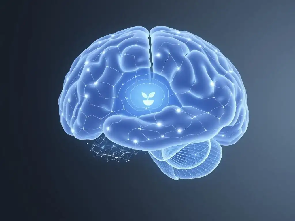
The process of utilizing brain scan insights in emotional regulation therapy often begins with a pre-therapy brain scan. This initial scan serves as a baseline, providing a snapshot of the individual's brain structure and/or function before therapeutic intervention begins. For example, an fMRI scan might measure the reactivity of the amygdala to emotional stimuli or the connectivity between the prefrontal cortex and limbic regions while the patient attempts a simple regulation task.
Establishing this baseline is critical. It allows clinicians and researchers to quantify the specific neural patterns associated with the individual's emotional dysregulation. For someone with severe anxiety, the baseline fMRI might show exaggerated amygdala responses to threat cues compared to a control group. For an individual with BPD, the baseline might reveal weaker functional connectivity between the vmPFC and the amygdala, suggesting impaired top-down control. This initial data provides objective targets for the therapy and a point of comparison against which future changes can be measured, forming the foundation for personalized emotional regulation brain scan therapy.
Types of Therapy Used in Conjunction with Brain Scans: CBT and Neurofeedback
Brain scan insights can be integrated with various therapeutic modalities known to be effective for emotional regulation. Two prominent examples include Cognitive Behavioral Therapy (CBT) and Neurofeedback.
- Cognitive Behavioral Therapy (CBT): CBT is a widely used therapy that helps individuals identify and change negative thought patterns and behaviors. Techniques like cognitive restructuring (changing maladaptive appraisals) and behavioral experiments directly target processes believed to involve the prefrontal cortex and its modulation of limbic activity. Brain scans, particularly fMRI, can track how CBT-induced changes in cognitive strategies correlate with changes in brain activity patterns, such as increased PFC activation during regulation tasks and decreased amygdala reactivity. Studies have shown, for instance, that successful CBT for anxiety is associated with normalization of activity in threat processing circuits.
- Neurofeedback: Neurofeedback is a specific type of emotional regulation brain scan therapy that uses real-time brain activity data to train individuals to self-regulate their brain function. Using fMRI or EEG, the patient receives feedback (e.g., a visual display) representing the activity level of a specific brain region (like the amygdala) or the strength of connectivity between regions. The patient is then instructed to try to change this activity (e.g., decrease amygdala activation) using mental strategies, and the feedback signal indicates their success. This direct brain training approach is specifically designed to modify the neural patterns identified as problematic.
Real-Time Feedback and Neurofeedback Techniques
Real-time neurofeedback, particularly using fMRI (rt-fMRI neurofeedback), is a cutting-edge approach within emotional regulation brain scan therapy. In a typical rt-fMRI session, the patient lies in the scanner while the fMRI system processes brain activity data in near real-time. This data is then presented back to the patient, often as a simple visual display like a thermometer or a moving ball, representing the activity level of a target brain region (e.g., the right amygdala).
The therapist guides the patient to use mental strategies (e.g., recalling calm memories, reappraising a situation, mindfulness) to try to change the feedback signal in a desired direction (e.g., lower the thermometer to decrease amygdala activity). Through repeated practice and feedback, the patient learns to voluntarily modulate the activity of the target brain region.
EEG neurofeedback works on similar principles but targets specific brainwave frequencies or patterns. While still largely experimental for many applications, studies are exploring its potential to directly train individuals to regulate key brain areas involved in emotional reactivity, offering a potentially powerful tool for emotional regulation brain scan therapy.
Post-Therapy Brain Scan: Assessing Changes in Brain Activity
Following a course of emotional regulation therapy, whether it's traditional therapy informed by initial scans or a direct neurofeedback protocol, a post-therapy brain scan is often conducted. This scan uses the same technique and protocol as the baseline scan to allow for direct comparison.
The purpose of the post-therapy scan is to objectively assess whether the therapy has resulted in measurable changes in brain structure, connectivity, or activity patterns that are hypothesized to underlie improved emotional regulation. For example, a post-therapy fMRI might show reduced amygdala reactivity to emotional stimuli or increased functional connectivity between the prefrontal cortex and the amygdala during regulation tasks compared to the baseline scan. Structural MRI might reveal increases in gray matter density in areas like the hippocampus or prefrontal cortex.
These objective measures provide empirical evidence of therapy's impact on the brain, complementing the patient's self-report of reduced symptoms and improved emotional control. This comparison is a core component of evaluating the effectiveness of emotional regulation brain scan therapy approaches.
Brain Scan Findings: What We're Learning About Therapy's Impact
Structural Brain Changes: Increased Gray Matter in Key Areas
Neuroimaging studies, particularly using structural MRI, are revealing that successful emotional regulation therapy can lead to observable changes in the physical structure of the brain. Gray matter consists of neuronal cell bodies, dendrites, and synapses. An increase in gray matter density or volume in specific areas is often associated with learning, skill acquisition, and adaptation.
Research indicates that therapies like mindfulness-based cognitive therapy (MBCT) and dialectical behavior therapy (DBT), which target emotional regulation skills, can lead to increases in gray matter volume in regions critical for emotional processing and regulation. For instance, studies have reported increased gray matter in the insula, which is involved in body awareness and emotional feeling, and in parts of the prefrontal cortex, associated with executive control.
Some findings also suggest increases in hippocampal volume, a region often reduced in conditions like chronic stress and depression. While the exact mechanisms are complex, these structural changes provide compelling evidence that emotional regulation brain scan therapy can literally reshape the brain, enhancing its capacity for emotional control.
Functional Connectivity: Enhanced Communication Between Brain Regions
Beyond structural changes, brain scans using fMRI and EEG are showing that therapy for emotional dysregulation alters the way different brain regions communicate with each other – their functional connectivity. Effective emotional regulation relies on coordinated activity across distributed brain networks, particularly the interaction between prefrontal control regions and subcortical emotional centers like the amygdala.
Studies have consistently found that various forms of emotional regulation therapy can enhance functional connectivity in key circuits. For example, CBT for anxiety has been associated with increased resting-state connectivity between the prefrontal cortex and the amygdala, suggesting improved top-down regulatory control even when not actively regulating emotions.
Similarly, mindfulness training has been shown to increase connectivity within networks related to attention and self-awareness (like the default mode network and salience network) in ways that support emotional detachment and non-reactivity. These findings from emotional regulation brain scan therapy research highlight that therapy doesn't just change individual regions but optimizes the communication between them, leading to more integrated and effective emotional processing.
Changes in Amygdala Activity: Reduced Emotional Reactivity
One of the most consistent findings in emotional regulation brain scan therapy research is the impact of therapy on amygdala activity. The amygdala, central to processing threat and generating fear responses, is often hyperactive in conditions characterized by excessive fear, anxiety, and emotional reactivity, such as PTSD and anxiety disorders.
Therapeutic interventions aimed at reducing emotional reactivity and improving regulation frequently lead to a decrease in amygdala activity, particularly when individuals are exposed to emotional stimuli. For instance, studies using fMRI have demonstrated that successful CBT for social anxiety disorder results in significantly reduced amygdala responses to social threat cues. Similarly, mindfulness practices have been shown to attenuate amygdala activation in response to negative images.
This reduction in the "alarm signal" from the amygdala is a key neural correlate of improved emotional regulation, indicating that the brain is becoming less reactive to perceived threats after therapy. Measuring this change via brain scans provides objective support for the effectiveness of emotional regulation brain scan therapy.
Prefrontal Cortex Activation: Improved Cognitive Control of Emotions
The prefrontal cortex (PFC), particularly the lateral and medial regions, is vital for cognitive control over emotions. It's involved in evaluating emotional stimuli, selecting appropriate regulatory strategies, and inhibiting unwanted emotional responses. In emotional dysregulation, activity in these PFC regions may be diminished or their connectivity with subcortical areas may be impaired.
Emotional regulation therapies often aim to strengthen these cognitive control mechanisms. Brain scan studies using fMRI show that effective therapy increases activation in prefrontal areas during tasks requiring emotional regulation, such as reappraising negative images. For example, studies on depression treatment have found that clinical improvement is associated with increased activity in the dorsolateral PFC during emotional tasks.
This heightened PFC engagement suggests that the brain is better able to recruit executive resources to manage emotional responses. Increased activity and improved connectivity within the prefrontal cortex represent key brain changes observed in emotional regulation brain scan therapy research, reflecting enhanced top-down control over the emotional brain.
Case Studies: Emotional Regulation Therapy and Brain Scan Results
Case Study 1: Anxiety Disorder and fMRI Results
Consider a hypothetical case study of a 35-year-old individual, "Anna," diagnosed with generalized anxiety disorder (GAD), characterized by chronic worry and difficulty controlling anxious thoughts. Before starting therapy, Anna undergoes an fMRI scan. During the scan, she is shown a series of images, including neutral and anxiety-provoking scenes. The pre-therapy fMRI reveals heightened activity in Anna's amygdala when viewing anxiety-provoking images compared to neutral ones, and relatively low activity in her ventromedial prefrontal cortex (vmPFC) during attempts to reappraise the images. This pattern is consistent with impaired top-down control over threat processing.
Anna then completes a 12-week course of CBT focused on cognitive restructuring and worry management. After therapy, she reports a significant reduction in worry and anxiety symptoms. A follow-up fMRI scan is performed using the same protocol.
The post-therapy scan shows a noticeable decrease in amygdala activity when viewing anxiety-provoking images. Furthermore, there is increased activation in her vmPFC during the reappraisal task, and enhanced functional connectivity between the vmPFC and the amygdala. These fMRI results provide objective neural evidence supporting Anna's subjective report of reduced anxiety and illustrate how emotional regulation brain scan therapy, specifically using fMRI to track CBT outcomes, can map improvements in brain function.
Case Study 2: Borderline Personality Disorder and EEG Findings
Let's look at a conceptual case involving a 24-year-old individual, "Ben," diagnosed with Borderline Personality Disorder (BPD), experiencing intense emotional lability and reactivity. Ben participates in a study using EEG to assess his rapid emotional responses. The baseline EEG analysis reveals atypical patterns, such as exaggerated early event-related potentials (ERPs) linked to emotional salience (like the P1 or EPN components) when presented with emotionally charged words, suggesting hyper-vigilance to emotional cues. There might also be reduced frontal alpha asymmetry patterns typically associated with approach motivation or positive affect regulation.
Ben engages in Dialectical Behavior Therapy (DBT), which includes modules on mindfulness, distress tolerance, and emotion regulation skills. After several months of consistent DBT practice, Ben reports improved emotional stability and reduced impulsive behaviors. A follow-up EEG is conducted. The post-therapy EEG shows a reduction in the amplitude of the early emotional ERPs, indicating less initial hyper-vigilance to emotional stimuli.
Changes in frontal alpha asymmetry might also be observed, suggesting shifts in emotional processing styles. While EEG provides less spatial detail than fMRI, its temporal resolution offers unique insights into the rapid neural processing that contributes to emotional reactivity. This example demonstrates how emotional regulation brain scan therapy utilizing EEG can track changes in the timing and intensity of neural responses following skills-based interventions like DBT.
Analyzing the Data: Common Themes and Individual Differences
Analyzing brain scan data from multiple individuals undergoing emotional regulation therapy reveals both common themes and important individual differences. Across various conditions and therapies, a recurring pattern is the shift towards a more balanced interplay between subcortical emotional regions (like the amygdala) and prefrontal regulatory areas. This often manifests as decreased reactivity in the amygdala and increased engagement or connectivity of the PFC during emotional tasks or even at rest. These common findings reinforce the idea that strengthening top-down control is a core mechanism of effective emotional regulation therapy.
However, brain scan data also highlights significant individual variability. Not everyone shows the exact same pattern of change, and the degree of change can vary. Factors such as the specific diagnosis, the severity of symptoms, individual differences in brain structure and function before therapy, and adherence to the therapy protocol likely contribute to these variations.
Analyzing these differences is crucial for advancing emotional regulation brain scan therapy. It can help identify which individuals are most likely to benefit from specific interventions, predict treatment response, and potentially tailor therapeutic approaches based on an individual's unique neural profile. Future research aims to refine these analyses to make personalized emotional regulation brain scan therapy a more widespread reality.
Benefits of Using Brain Scans in Emotional Regulation Therapy

Personalized Treatment Plans Based on Brain Activity
One of the most significant potential benefits of incorporating brain scans into emotional regulation therapy is the ability to personalize treatment plans. Brain scan data can provide objective information about the specific neural deficits or patterns contributing to an individual's emotional dysregulation. For example, an fMRI might reveal that a patient struggles particularly with engaging the vmPFC during reappraisal, suggesting that therapy should heavily focus on cognitive change strategies. Another patient might show excessive amygdala reactivity but relatively intact PFC function, indicating that strategies focused on modulating the emotional response itself (like mindfulness or relaxation) might be more beneficial.
By identifying these specific neural targets, clinicians can potentially tailor the therapeutic approach to address the individual's unique brain profile. This moves beyond a one-size-fits-all approach and aims to match the right intervention to the right person, potentially increasing the efficiency and effectiveness of emotional regulation brain scan therapy. While this is still an area of active research, the promise of using neuroimaging to guide personalized mental healthcare is substantial.
Objective Measurement of Therapy Progress
Traditional therapy relies heavily on subjective reports from patients and observations by therapists to gauge progress. While essential, these measures can be influenced by various factors and may not always capture the full extent of change. Brain scans offer a more objective measure of how therapy is impacting the neural circuits involved in emotional regulation.
Comparing pre- and post-therapy brain scans provides empirical evidence of changes in brain structure, function, or connectivity that correlate with symptom improvement. For instance, if a patient reports feeling less anxious, a post-therapy fMRI showing reduced amygdala activity provides objective validation for that report. This objective data can complement subjective reports, offering a more comprehensive picture of treatment outcomes. For the field of emotional regulation brain scan therapy, this objective measurement capability is vital for research validation and potential clinical application.
Increased Patient Engagement and Motivation
Receiving objective feedback about their brain activity can be a powerful motivator for patients undergoing emotional regulation therapy. Seeing a visual representation (e.g., in neurofeedback) or receiving a report showing positive changes in brain patterns after therapy can make the often abstract process of emotional regulation feel more tangible and achievable.
Patients might feel more engaged in the therapeutic process when they understand the biological basis of their difficulties and can see evidence that their efforts are leading to real changes in their brain. For example, being shown that their amygdala activity has decreased after practicing mindfulness can reinforce the value of the technique and encourage continued effort. This increased understanding and visible progress can enhance hope and self-efficacy, crucial factors in successful therapy outcomes. Incorporating brain scan insights can transform emotional regulation brain scan therapy into a more collaborative and empowering experience.
Early Identification of Treatment Response
Brain scans may hold the potential to identify early indicators of whether a patient is likely to respond to a particular type of emotional regulation therapy. Changes in brain activity or connectivity can sometimes be observed relatively early in the course of treatment, potentially before significant symptomatic improvement is reported.
If early brain scan data can predict later clinical outcomes, it could allow clinicians to adjust treatment plans sooner for non-responders, saving time and resources and preventing prolonged engagement in ineffective therapies. For example, if a patient's brain scan after a few weeks of CBT shows no change in the targeted neural circuit, it might signal the need to switch to a different therapeutic approach or modality. While research in this area is ongoing, the possibility of using brain scans for early prediction of treatment response is a promising avenue for optimizing emotional regulation brain scan therapy.
Limitations and Future Directions of Emotional Regulation Brain Scan Therapy
Cost and Accessibility of Brain Scan Technology
Despite the promising insights offered by brain scans, a major limitation for their widespread use in clinical emotional regulation therapy is the high cost and limited accessibility of the technology. fMRI scanners, in particular, are expensive to purchase and operate, and they are typically located in large medical centers or research institutions. This makes routine scanning impractical for most therapy practices and cost-prohibitive for many patients.
EEG is generally more accessible and less expensive than fMRI, but still requires specialized equipment and expertise. For emotional regulation brain scan therapy to become a standard clinical tool, the technology needs to become more affordable, portable, and easier to use in diverse clinical settings. Developing more cost-effective or mobile neuroimaging solutions is a critical step for expanding the reach of brain scan-informed therapy beyond research contexts.
Ethical Considerations in Neuroimaging
The use of brain scans in mental health therapy raises several important ethical considerations. One concern relates to the interpretation and communication of scan results to patients. Brain imaging data is complex and can be easily misinterpreted or oversimplified, potentially leading to anxiety or deterministic thinking about one's mental state ("My brain looks like this, so I will always struggle"). Ensuring that scan results are communicated accurately, empathetically, and within the context of the brain's plasticity is crucial.
Privacy and data security are also paramount. Brain scan data is highly personal and potentially identifiable. Robust measures are needed to protect patient confidentiality and ensure that sensitive neuroimaging data is stored and used responsibly. Furthermore, there are broader questions about the potential societal implications of neurotechnology and the need for clear guidelines on its appropriate use in clinical settings. Addressing these ethical challenges is essential for the responsible development and implementation of emotional regulation brain scan therapy.
The Need for Larger, Controlled Studies
While promising findings exist, the field of emotional regulation brain scan therapy is still relatively young. Many studies to date have involved small sample sizes or have been exploratory in nature. To firmly establish the efficacy of brain scan-informed or brain scan-based therapies (like neurofeedback) and to understand who benefits most, there is a critical need for larger, well-designed randomized controlled trials.
These studies should compare emotional regulation brain scan therapy approaches to established, evidence-based therapies without the neuroimaging component. They need to rigorously assess clinical outcomes, long-term effects, and cost-effectiveness. Larger studies across diverse populations are also needed to ensure that findings are generalizable and applicable to the wide range of individuals experiencing emotional dysregulation. Building a robust evidence base through rigorous research is key to integrating brain scan technology effectively and safely into clinical practice.
Future Research: Exploring the Potential of Neurostimulation
Beyond using brain scans to monitor or train brain activity, future directions in emotional regulation brain scan therapy research include exploring the potential of direct neurostimulation techniques. Techniques like Transcranial Magnetic Stimulation (TMS) or Transcranial Direct Current Stimulation (tDCS) use magnetic fields or weak electrical currents, respectively, applied non-invasively to the scalp to modulate activity in specific brain regions.
Research is investigating whether targeting areas involved in emotional regulation, such as the prefrontal cortex, with neurostimulation can enhance the effects of therapy or directly improve regulatory capacity. For example, applying TMS to the dorsolateral PFC is being explored as an adjunctive treatment for depression and anxiety, conditions marked by emotional dysregulation. Combining brain stimulation with therapy or neurofeedback, guided by initial brain scan assessments, represents a potential future frontier for more targeted and powerful interventions in emotional regulation brain scan therapy.
Conclusion: Brain Scans and the Future of Emotional Regulation Treatment
Summarizing the Key Findings of Emotional Regulation Brain Scan Studies
Research utilizing brain scan techniques like fMRI, EEG, and structural MRI has profoundly advanced our understanding of emotional regulation and how therapy impacts the brain. Key findings consistently show that emotional dysregulation is associated with altered activity and connectivity in a network of brain regions, including the amygdala, insula, and prefrontal cortex. Crucially, emotional regulation therapies are not just changing behavior or thoughts; they are associated with measurable changes at the neural level.
These changes include increases in gray matter volume in relevant areas, enhanced functional connectivity between regulatory and emotional brain regions, reduced reactivity in areas like the amygdala, and increased activation in prefrontal control areas during regulation tasks. These brain scan insights provide objective evidence that therapy fosters greater neural capacity for managing emotions, validating the effectiveness of various therapeutic approaches aimed at improving emotional control. The body of evidence from emotional regulation brain scan studies is growing, painting a clearer picture of the brain's remarkable ability to change in response to targeted interventions.
Promoting Innovative Mental Health Treatments
The insights gained from brain scan research are paving the way for more innovative and potentially more effective mental health treatments for emotional dysregulation. By providing a deeper, objective understanding of the neural underpinnings of emotional difficulties, neuroimaging informs the development of new therapeutic strategies and refines existing ones. Approaches like neurofeedback, which directly use brain scan data to train self-regulation, represent a direct application of this knowledge.
Furthermore, brain scan findings can help destigmatize mental health challenges by highlighting their basis in brain function, shifting the narrative from blame to biological understanding and the potential for recovery through targeted intervention. Promoting emotional regulation brain scan therapy, in its various forms, contributes to a future where mental health treatment is increasingly personalized, data-driven, and grounded in a sophisticated understanding of the brain.
Empowering Individuals with Knowledge About Emotional Regulation and Brain Health
Ultimately, understanding the connection between emotional regulation, brain function, and therapy outcomes empowers individuals. Learning that emotional struggles have correlates in the brain and that these neural patterns can be positively influenced through therapeutic effort can reduce feelings of helplessness and instill hope.
Providing patients with information about how emotional regulation brain scan therapy works, what brain scans can reveal, and how therapy is shown to change the brain can enhance their engagement and motivation. It encourages a perspective where mental health is viewed through the lens of brain health and plasticity – the idea that the brain is capable of change and growth. By demystifying the neuroscience and highlighting the tangible impact of therapy on the brain, we can empower individuals to actively participate in their recovery and build lasting skills for emotional well-being.
Frequently Asked Questions
What is Emotional Regulation Brain Scan Therapy and what does it involve?
This refers to a therapeutic approach that combines traditional therapy with brain imaging technology (like fMRI) to understand and improve emotional regulation. The scan provides visual insights into brain activity related to emotions, which can then be used to guide therapy.
How can brain scans help in improving emotional regulation?
Brain scans offer a visual representation of which brain regions are involved in emotional responses. This allows therapists to tailor interventions to specifically target and strengthen areas crucial for effective emotional regulation.
What are the potential benefits of using brain scan therapy for emotional regulation compared to traditional methods?
It offers a more personalized and targeted approach to therapy. By visualizing brain activity, therapists can gain a deeper understanding of individual emotional processes, potentially leading to faster and more effective outcomes.
Are there any risks or limitations associated with Emotional Regulation Brain Scan Therapy?
It can be expensive and not always accessible. Additionally, the interpretation of brain scans requires specialized expertise, and the results should be considered alongside other clinical information.
Who is a good candidate for Emotional Regulation Brain Scan Therapy?
Individuals struggling with significant emotional dysregulation, such as those with anxiety disorders, depression, or PTSD, who haven't responded well to traditional therapy might be considered. It's important to consult with a qualified mental health professional to determine suitability.
Final Reflection: Charting the Course to Emotional Wellbeing
The intricate dance between our brains and our emotions, once a mysterious realm, is now being illuminated by the powerful lens of brain scan technology. Understanding how therapy impacts emotional regulation, as detailed in this article, is not merely an academic pursuit; it's a crucial step towards developing more effective and personalized mental healthcare for everyone. The insights gleaned from brain scans are transforming our understanding of therapeutic interventions, paving the way for a future where mental health treatment is more precise and effective than ever before.
The journey of emotional regulation is deeply personal, a lifelong process of learning and growth. Witnessing the tangible changes in brain activity during therapy, as revealed through these scans, underscores the profound impact of therapeutic interventions on our inner landscape. The mind is not a vessel to be filled, but a fire to be kindled.
– Plutarch. This quote resonates deeply with the transformative power of therapy, as it ignites the potential for positive change within the brain itself, a change that is demonstrably visible through neuroimaging techniques.
Consider the implications of this research. You, the reader, are now equipped with a deeper understanding of how your brain responds to therapeutic interventions aimed at improving emotional regulation. This knowledge empowers you to actively participate in your own mental health journey. Seek out therapists who utilize evidence-based practices, and don't hesitate to ask questions about how your progress is being monitored and measured. Embrace the potential for change, and remember that the path to emotional wellbeing is a journey worth undertaking, one illuminated by the ever-advancing science of the brain.
Unlock Your Emotional Intelligence: Take Control Now!
Thanks for diving deep into the fascinating world of emotional regulation with us! We hope you gained valuable insights into how your brain processes emotions and how therapy can help. Ready to put this knowledge into action?
- Start practicing a new mindfulness technique: Try the body scan meditation we mentioned – even 5 minutes a day can make a difference!
- Share this article with a friend struggling with emotional regulation. Helping others can be a powerful way to reinforce your own learning.
- Explore additional resources on our website: We have a wealth of articles and guides on emotional well-being that you might find helpful.
What specific emotional regulation strategy are you most excited to try, and why? Let's discuss your experiences in the comments below!