
The Brain After OCD Therapy: What Scans Reveal About Change

Welcome to Mind Care Guide, a sanctuary for exploring the intricacies of mental well-being. Ever wondered what truly happens in the brain during OCD therapy? Join us as we delve into the fascinating world of neuroscience with a deep dive into OCD therapy brain scans, uncovering the remarkable changes that can occur and offering a glimpse of hope for those navigating this often-challenging condition. Intrigued? Let's explore the science behind healing.
The Brain After OCD Therapy: What Scans Reveal About Change
- Introduction: Understanding OCD and the Potential of Therapy
- OCD Therapy Brain Scans: Unveiling Neurological Changes
- Brain Regions Affected by OCD: Before and After Treatment
- How OCD Therapy Reshapes the Brain: Specific Changes Observed
- Different OCD Therapy Approaches and Their Effects on the Brain
- The Long-Term Impact of OCD Therapy on the Brain: Sustainability of Changes
- Benefits of Using Brain Scans in OCD Therapy Research
- Limitations of OCD Therapy Brain Scans
- Future Directions in OCD Therapy and Brain Scan Research
- Conclusion: The Future of OCD Treatment and Brain-Based Understanding
-
Frequently Asked Questions
- What are brain scans showing us about the effectiveness of OCD therapy?
- Which types of brain scans are most commonly used to study the effects of OCD therapy?
- How do brain scans help personalize OCD therapy approaches for individuals?
- Do brain scans predict the long-term success of OCD therapy?
- What are the limitations of using brain scans to evaluate the success of OCD therapy?
- Final Reflection: Reimagining the OCD Brain: A Journey of Recovery and Resilience
- Unlock Your Brain's Potential: Discover the Next Steps
Introduction: Understanding OCD and the Potential of Therapy
The Impact of OCD on Brain Function
Obsessive-Compulsive Disorder (OCD) is a complex mental health condition characterized by persistent, intrusive thoughts (obsessions) and repetitive behaviors or mental acts (compulsions) that individuals feel driven to perform. These symptoms are not merely worries; they are often distressing and significantly interfere with daily life. Research, including studies using advanced neuroimaging techniques, has consistently shown that OCD is associated with specific patterns of activity and connectivity in certain brain circuits.
Key areas implicated include the cortico-striatal-thalamo-cortical (CSTC) loops, particularly involving the orbitofrontal cortex (OFC), anterior cingulate cortex (ACC), and striatum. These regions are crucial for decision-making, error detection, habit formation, and reward processing.
In individuals with untreated OCD, these brain circuits often exhibit heightened activity or abnormal connectivity. For example, the OFC, involved in assessing risk and reward, can show excessive activation, contributing to the feeling that something is "not right" or incomplete.
Similarly, the ACC, which plays a role in detecting errors and resolving conflict, may be overactive, potentially driving the urge to perform compulsions to alleviate distress. This persistent dysregulation in these critical brain areas forms the neurological basis for the intrusive thoughts and compulsive behaviors that define OCD, highlighting the biological underpinnings of the disorder and the potential for interventions targeting these circuits.
The Promise of OCD Therapy: A Path to Recovery and Brain Rewiring
Despite the significant challenges posed by OCD, effective therapies offer a powerful path toward recovery and, importantly, can lead to observable changes in brain function. The primary goal of OCD therapy is not just symptom management but also to help individuals gain control over their thoughts and behaviors, ultimately reducing the distress and interference caused by the disorder. This therapeutic process, particularly modalities like Cognitive Behavioral Therapy (CBT) and Exposure and Response Prevention (ERP), actively engages the brain's capacity for neuroplasticity – its ability to reorganize itself by forming new neural connections throughout life.
The promise of OCD therapy lies in its potential to "rewire" the dysfunctional brain circuits associated with the disorder. By learning new ways to respond to obsessive thoughts and resist compulsive urges, individuals are, in essence, training their brains to adopt healthier patterns of activity.
This process is not instantaneous but unfolds over time with consistent effort and practice. Modern research utilizing OCD therapy brain scans has provided compelling visual evidence that these psychological interventions can indeed lead to measurable neurological changes, offering hope and validation for those undergoing treatment and reinforcing the understanding that recovery involves both mind and brain.
OCD Therapy Brain Scans: Unveiling Neurological Changes
What are OCD Therapy Brain Scans? Exploring Neuroimaging Techniques
OCD therapy brain scans refer to the use of neuroimaging technologies before, during, and after treatment to observe and measure changes in brain structure, function, and connectivity in individuals with Obsessive-Compulsive Disorder. These techniques provide a non-invasive window into the living brain, allowing researchers and clinicians to gain insights into how therapeutic interventions influence neural activity. By comparing scans taken at different time points, scientists can identify specific brain regions or circuits whose activity levels or communication patterns are altered following successful therapy. This approach moves beyond simply tracking symptom reduction to understanding the underlying biological mechanisms of recovery.
The application of OCD therapy brain scans is crucial for advancing our understanding of both the disorder and the efficacy of its treatments. These scans can help to objectively validate the impact of psychological therapies, demonstrating that they lead to tangible changes in brain function, similar to how medication can affect brain chemistry.
Furthermore, this research helps to refine theoretical models of OCD and its treatment, providing a neurobiological basis for the observed clinical improvements. While the technology is complex and requires specialized interpretation, the information gleaned from these scans is invaluable for illustrating the profound impact that effective therapy can have on the brain.
Types of Brain Scans Used in OCD Research: fMRI, PET, and SPECT
Several neuroimaging techniques are commonly employed in OCD therapy brain scans research, each offering unique insights into different aspects of brain activity and structure. Functional Magnetic Resonance Imaging (fMRI) is perhaps the most widely used technique. It measures brain activity by detecting changes in blood flow. When a brain region is more active, it requires more oxygenated blood, which fMRI can detect. Studies using fMRI in OCD research often examine activity levels in specific brain areas during tasks related to obsessions or compulsions, or at rest, before and after therapy.
Other important techniques include Positron Emission Tomography (PET) and Single-Photon Emission Computed Tomography (SPECT). PET scans use a radioactive tracer, often a form of glucose, to measure metabolic activity in the brain. Areas that are more active consume more glucose.
SPECT scans also use radioactive tracers but measure blood flow. Both PET and SPECT have been used to identify areas of altered metabolism or blood flow in individuals with OCD compared to healthy controls, and to observe how these patterns change after successful treatment. While fMRI provides better spatial resolution for mapping activity, PET and SPECT can be useful for studying neurotransmitter systems, offering complementary information in the study of OCD and the effects of OCD therapy brain scans.
Brain Regions Affected by OCD: Before and After Treatment
The Orbitofrontal Cortex (OFC) and OCD: Activity Levels and Therapy's Influence
The orbitofrontal cortex (OFC), located just above the orbits of the eyes, plays a critical role in evaluating the emotional significance of stimuli, decision-making, and processing reward and punishment. In individuals with OCD, the OFC is often found to be hyperactive, particularly in the context of triggers related to their obsessions. This excessive activity is thought to contribute to the persistent feeling of unease, doubt, and the need for certainty that characterizes many OCD symptoms. For example, someone with contamination fears might experience heightened OFC activity when encountering perceived contaminants, signaling a strong "danger" signal.
Following successful OCD therapy, particularly ERP, studies using OCD therapy brain scans have frequently observed a significant reduction in this hyperactivity within the OFC. Research, such as early work utilizing PET scans, indicated that metabolic activity in the OFC decreased after effective behavioral therapy.
More recent fMRI studies corroborate this, showing normalized activity patterns in response to disorder-relevant stimuli. This reduction in OFC overactivity is a key neural signature of therapeutic success, suggesting that therapy helps to recalibrate the brain's threat detection system, allowing individuals to process information more adaptively and reducing the intensity of distress associated with obsessive thoughts.
The Anterior Cingulate Cortex (ACC) in OCD: Modulation Through Therapy
The anterior cingulate cortex (ACC) is another central player in the brain circuitry associated with OCD. It is involved in error detection, conflict monitoring, and modulating emotional responses. In OCD, the ACC is often hyperactive, particularly when individuals are struggling with intrusive thoughts or resisting compulsions.
This heightened activity might reflect the brain's exaggerated response to perceived errors or conflicts, such as the feeling that something isn't "just right" or the internal struggle between an obsession and the desire to resist a compulsion. The ACC's role in driving attention and action may contribute to the difficulty individuals with OCD have in disengaging from obsessive thoughts and urges.
Evidence from OCD therapy brain scans indicates that effective treatment can modulate activity in the ACC. Studies have shown that after successful therapy, ACC activity tends to decrease or normalize, especially during tasks that previously elicited an exaggerated response. This suggests that therapy helps individuals with OCD to better regulate their responses to internal conflict and distress.
The reduced ACC activity may reflect a diminished "alarm signal" in response to obsessive thoughts and a decreased sense of urgency to perform compulsions. This modulation is crucial for breaking the cycle of obsessions and compulsions, allowing individuals to experience less internal conflict and greater control over their actions.
The Striatum and OCD: How Therapy Normalizes Activity
The striatum, a part of the basal ganglia, is deeply involved in habit formation, motor control, and reward-related learning. In OCD, the striatum, particularly its dorsal regions, is implicated in the formation and maintenance of rigid, habitual compulsive behaviors. Abnormal activity or connectivity within the striatum can contribute to the difficulty individuals with OCD have in stopping repetitive actions, even when they recognize them as irrational. This region is thought to be part of the "habit loop" that becomes dysregulated in OCD, making compulsions feel automatic and difficult to resist.
OCD therapy brain scans have revealed that successful treatment can help to normalize activity within the striatum. Studies using fMRI have shown changes in striatal activity and connectivity after therapy, often indicating a shift away from the rigid, habitual patterns associated with compulsions. Therapy, particularly ERP, which involves intentionally breaking the link between obsessions and compulsions, appears to help individuals develop new, more flexible behavioral responses. This normalization in the striatum supports the behavioral changes observed in therapy, suggesting that treatment helps to restore more adaptive habit learning and executive control over actions.
How OCD Therapy Reshapes the Brain: Specific Changes Observed
Reduced Hyperactivity in Key Brain Regions Following OCD Treatment
A consistent finding across numerous studies using OCD therapy brain scans is the reduction of hyperactivity in specific brain regions previously identified as overactive in individuals with OCD. As discussed, the orbitofrontal cortex (OFC) and anterior cingulate cortex (ACC) are prime examples. Before therapy, these areas often show excessive activation in response to triggers.
For instance, one study using fMRI showed a significant reduction in OFC and ACC activity when patients were exposed to stimuli related to their fears after completing a course of ERP therapy. This reduction is not just statistical; it correlates with a decrease in the severity of OCD symptoms reported by patients.
This observed decrease in neural activity suggests that effective therapy helps to dampen the brain's exaggerated threat response and conflict signals. It's as if the brain learns to stop sounding the alarm so intensely every time an intrusive thought occurs or a situation triggers anxiety. This normalization of activity in key emotional and cognitive processing centers is a direct neural correlate of the clinical improvements seen in patients undergoing successful treatment. It provides tangible evidence that psychological interventions can lead to profound, measurable changes in how the brain functions at a fundamental level.
Increased Connectivity and Communication Between Brain Areas
Beyond changes in the activity levels of individual regions, OCD therapy brain scans also reveal alterations in the connectivity and communication between different brain areas. In untreated OCD, there can be abnormal connectivity within the cortico-striatal-thalamo-cortical circuits, sometimes showing hyperconnectivity within these loops and hypoconnectivity between these loops and other brain networks, such as those involved in cognitive control and emotional regulation. This imbalanced connectivity might contribute to the feeling of being "stuck" in obsessive-compulsive cycles.
Following successful therapy, studies using advanced fMRI techniques (like resting-state fMRI or diffusion tensor imaging) have shown evidence of altered connectivity patterns. Some research suggests a normalization of connectivity within the CSTC loops or increased connectivity between these loops and frontal regions involved in executive control and cognitive flexibility. This could indicate that therapy strengthens the brain's ability to regulate and control the activity in the areas driving obsessions and compulsions. Improved communication between brain networks is essential for flexible thinking and adaptive behavior, suggesting that therapy helps to restore a more balanced and efficient neural network architecture.
Neuroplasticity and OCD: The Brain's Ability to Adapt
The changes observed in OCD therapy brain scans are powerful demonstrations of neuroplasticity – the brain's remarkable ability to change and adapt throughout life in response to experience. Therapy provides a structured and consistent experience that challenges the brain's ingrained patterns of thinking and behaving.
By repeatedly facing fears without engaging in compulsions (as in ERP) or by learning to reframe negative thoughts (as in CBT), individuals are actively shaping their neural pathways. Each time a person resists a compulsion and tolerates the resulting anxiety, they are, in a sense, strengthening new neural connections that support adaptive coping and weakening the old connections that support the compulsive habit loop.
This process of neuroplasticity is the fundamental mechanism by which OCD therapy leads to lasting change. It is not just about "talking about problems"; it is about actively retraining the brain. The evidence from OCD therapy brain scans provides concrete support for this concept, showing that the brain is not a static entity but is capable of significant reorganization. Understanding neuroplasticity offers a hopeful perspective: even deeply ingrained patterns associated with conditions like OCD can be modified through focused therapeutic effort, leading to structural and functional changes that support recovery and improved well-being.
Different OCD Therapy Approaches and Their Effects on the Brain
Cognitive Behavioral Therapy (CBT) and Brain Changes: A Closer Look
Cognitive Behavioral Therapy (CBT) is a widely effective psychological treatment for OCD. It focuses on identifying and challenging distorted thoughts (cognitions) and changing maladaptive behaviors. In the context of OCD, CBT helps individuals recognize obsessive thoughts for what they are – intrusive thoughts, not necessarily reflective of reality or danger – and develop more helpful responses than engaging in compulsions. While ERP is often considered a specific form of CBT for OCD, general CBT principles about challenging thought patterns also play a role.
Studies using OCD therapy brain scans have investigated the neural effects of CBT. While ERP has been more extensively studied with neuroimaging, research on broader CBT approaches for OCD also indicates observable brain changes. These changes often overlap with those seen in ERP studies, including reductions in activity in the OFC and ACC.
This suggests that the cognitive restructuring component of CBT, which helps individuals re-evaluate the threat level associated with their obsessions, also impacts the brain's fear and error detection circuits. The specific neural pathways affected by CBT might be related to the prefrontal cortex's role in cognitive control and reappraisal, helping to exert top-down regulation over limbic and striatal areas involved in emotional and habitual responses.
Exposure and Response Prevention (ERP) Therapy: Remapping Neural Pathways
Exposure and Response Prevention (ERP) is considered the gold standard behavioral therapy for OCD and has been the most studied therapeutic approach using neuroimaging. ERP involves gradually exposing individuals to their feared thoughts, images, objects, or situations (exposure) while preventing them from engaging in their usual compulsive rituals (response prevention). The goal is to learn that the feared outcome does not occur or that the anxiety naturally decreases over time without performing the compulsion.
The neural changes observed with ERP therapy using OCD therapy brain scans are particularly compelling. As mentioned earlier, ERP is strongly associated with reductions in OFC and ACC hyperactivity and normalization of activity in the striatum. This is thought to reflect the remapping of neural pathways that previously linked obsessional triggers directly to compulsive actions.
By breaking this link through repeated exposure and response prevention, ERP helps the brain create new pathways that allow for anxiety tolerance and non-engagement in compulsions. The process essentially retrains the brain's fear response and habit system, providing a clear neurobiological basis for why ERP is so effective in breaking the OCD cycle.
Medication and Brain Activity: The Role of Serotonin Reuptake Inhibitors (SSRIs)
Pharmacological treatment, primarily with selective serotonin reuptake inhibitors (SSRIs), is another common approach for managing OCD symptoms, often used in conjunction with therapy or as a standalone treatment. SSRIs are believed to work by increasing the levels of serotonin in the brain, a neurotransmitter that plays a role in mood, anxiety, and obsessive-compulsive symptoms. While psychological therapies like ERP target dysfunctional circuits through behavioral and cognitive means, SSRIs target neurotransmitter systems, which in turn can influence the activity within those same circuits.
OCD therapy brain scans have also been used to investigate the effects of SSRI medication on brain function in individuals with OCD. Studies have shown that effective SSRI treatment can lead to similar patterns of neural change as therapy, including decreased activity in the OFC, ACC, and striatum.
This suggests that both effective psychological therapy and effective medication can normalize activity in the core brain regions implicated in OCD, albeit potentially through different initial mechanisms (behavioral/cognitive retraining vs. neurochemical modulation). Understanding the overlapping and distinct neural effects of therapy and medication is an ongoing area of research, informing decisions about combined treatment approaches.
The Long-Term Impact of OCD Therapy on the Brain: Sustainability of Changes
Maintaining Brain Health After Therapy: Strategies for Long-Term Well-being
A crucial question regarding the impact of OCD therapy on the brain is the sustainability of the observed changes. Research suggests that the positive neural adaptations achieved through effective treatment, particularly ERP, can be long-lasting, provided that individuals continue to apply the skills learned in therapy. Maintaining brain health after successful OCD treatment involves actively reinforcing the new, adaptive neural pathways that were strengthened during therapy. This isn't just about avoiding relapse; it's about continuing to practice healthy coping mechanisms and challenging the ingrained patterns that characterized the disorder.
Strategies for maintaining these brain changes and promoting long-term well-being often mirror the principles of therapy itself. This includes:
- Continued use of ERP principles: Facing minor fears or triggers without engaging in compulsions.
- Mindfulness practices: Cultivating awareness of thoughts without judgment, reducing their power.
- Stress management: High stress levels can potentially reactivate vulnerable circuits.
- Healthy lifestyle: Regular exercise, adequate sleep, and a balanced diet all support overall brain health and resilience.
- Utilizing learned cognitive skills: Challenging unhelpful thought patterns when they arise.
By consistently applying these strategies, individuals reinforce the neural pathways associated with emotional regulation, cognitive flexibility, and adaptive responses, helping to maintain the positive changes observed in OCD therapy brain scans over time.
Relapse Prevention and Brain Function: Recognizing and Addressing Potential Setbacks
While OCD therapy can lead to significant and lasting brain changes, the possibility of setbacks or relapse exists. Understanding the neurological basis of relapse can be empowering. Relapse doesn't necessarily mean the brain has reverted entirely to its pre-treatment state, but it might indicate a reactivation of some of the previously hyperactive or abnormally connected circuits, perhaps triggered by stress, major life changes, or discontinuation of practice of learned skills. Recognizing early signs of potential relapse and having strategies to address them is vital for long-term recovery.
From a brain perspective, recognizing a setback might involve noticing the re-emergence of intense intrusive thoughts, a stronger urge to perform compulsions, or heightened anxiety in triggering situations – symptoms that correlate with increased activity in areas like the OFC or ACC. Addressing these potential setbacks involves re-engaging with the skills learned in therapy. This might mean intentionally initiating mini-exposures, actively resisting compulsions, or revisiting cognitive strategies.
Early intervention, essentially "exercising" the adaptive neural pathways again, can help prevent a full return of severe symptoms and reinforce the positive brain changes achieved through initial OCD therapy brain scans guided treatment. Having a "relapse prevention plan" that includes recognizing triggers and specific steps to take is a practical application of maintaining brain health.
Benefits of Using Brain Scans in OCD Therapy Research
Objective Measurement of Treatment Effectiveness
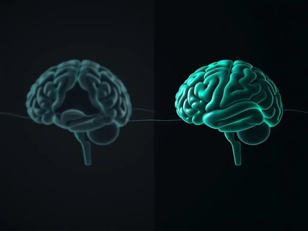
One of the significant benefits of using OCD therapy brain scans in research is that they provide an objective measure of treatment effectiveness that goes beyond self-report symptom scales. While patient reports are crucial, brain scans offer an independent, biological measure of how the brain is responding to therapy. Observing a decrease in activity in the OFC and ACC or changes in connectivity patterns after treatment provides tangible evidence that the therapy is having a real impact on the underlying neural dysfunction associated with OCD.
This objective data strengthens the scientific understanding of why certain therapies work and provides compelling evidence for their efficacy. It helps to validate psychological treatments as interventions that produce measurable biological changes, just like pharmacological treatments. This can be particularly validating for patients, offering concrete proof that their hard work in therapy is leading to physical changes in their brain, fostering hope and motivation for continued recovery.
Identifying Potential Biomarkers for OCD
OCD therapy brain scans research also holds promise for identifying potential biomarkers for the disorder. Biomarkers are measurable indicators of a biological state or condition. In the context of OCD, researchers are looking for specific brain activity patterns, structural features, or connectivity profiles that are characteristic of the disorder or predict treatment response. For example, certain patterns of activity in the striatum might be associated with a greater propensity for developing compulsive habits, or the degree of OFC hyperactivity might correlate with symptom severity.
Identifying such biomarkers could revolutionize the diagnosis and treatment of OCD. If specific brain signatures could predict who is most likely to respond to a particular therapy (e.g., ERP vs. SSRIs), treatment could be personalized from the outset, leading to more efficient and effective care. While this research is still evolving, the ability to use OCD therapy brain scans to potentially predict treatment outcomes or identify subtypes of OCD based on brain profiles is a major step towards precision mental healthcare.
Improving and Personalizing OCD Treatment Approaches
Ultimately, the insights gained from OCD therapy brain scans are aimed at improving and personalizing treatment approaches. By understanding exactly how different therapies affect the brain, researchers can refine existing treatments and develop new, more targeted interventions. For instance, if scans reveal that a specific type of therapy is particularly effective at normalizing connectivity in a certain circuit, this information can be used to enhance that therapy or develop novel techniques that specifically target that connection.
Furthermore, as mentioned regarding biomarkers, brain scan data might eventually help clinicians tailor treatment plans to individual patients based on their unique brain profiles. Imagine a future where a patient's initial brain scan could help determine whether ERP, CBT, medication, or a combination is most likely to be effective for them. This personalized approach, guided by neurobiological data from OCD therapy brain scans, has the potential to significantly increase treatment success rates and reduce the time and frustration associated with trial-and-error treatment selection, making recovery more accessible and efficient for individuals living with OCD.
Limitations of OCD Therapy Brain Scans
Cost and Accessibility of Neuroimaging Technology
Despite their immense value, the use of OCD therapy brain scans in both research and potential clinical application faces significant limitations, primarily related to cost and accessibility. Neuroimaging equipment like fMRI, PET, and SPECT scanners are extremely expensive to purchase and maintain. Running a scan requires highly trained personnel, including technicians and radiologists or neuroscientists to interpret the data. This makes routine brain scanning cost-prohibitive for most clinical settings and limits its availability primarily to well-funded research institutions.
For the average individual seeking OCD therapy, getting a brain scan before, during, and after treatment is simply not a practical or affordable option. The high cost also restricts the scale of research studies that can be conducted, limiting the sample sizes and diversity of populations studied. Until these technologies become more affordable and widely available, their primary role will remain in research settings to advance understanding, rather than as a routine tool in clinical practice for guiding individual patient care or assessing treatment outcomes directly for every patient.
Interpreting Brain Scan Data: Complexity and Challenges
Interpreting brain scan data is a complex process that requires specialized expertise and is not always straightforward. Brain activity is dynamic and influenced by many factors, including the individual's state of mind, what they were thinking about during the scan, and even small movements. Distinguishing meaningful patterns related to OCD and treatment effects from noise or individual variability can be challenging. Furthermore, different studies may use slightly different scanning parameters, tasks, or analysis techniques, which can make comparing results across studies difficult and sometimes lead to seemingly contradictory findings.
The field of neuroimaging analysis is constantly evolving, with new methods being developed to better understand complex brain networks and their changes. However, linking specific patterns of brain activity or connectivity definitively to specific psychological processes or treatment outcomes in OCD requires careful statistical analysis and replication across multiple studies. It is also crucial to avoid oversimplifying findings; a change in activity in one brain region is part of a complex network interaction, not an isolated event. Therefore, while OCD therapy brain scans provide valuable insights, interpreting the data requires caution and expert knowledge.
Ethical Considerations in Brain Scan Research
The use of OCD therapy brain scans in research also raises important ethical considerations. Issues surrounding privacy and data security are paramount, as brain scan data is highly personal and potentially identifiable. Ensuring that scan data is anonymized and stored securely is essential to protect participants' privacy.
There are also ethical considerations regarding the communication of research findings to participants. While it can be empowering for individuals to see potential changes in their brain after therapy, researchers must be careful not to overstate the clinical significance of scan findings for an individual patient, as these are often group-level results and not diagnostic for a single person.
Furthermore, there are ethical questions about the potential future use of brain scan technology. If biomarkers are identified, how will this information be used? Could it lead to discrimination based on brain profiles? Could it create pressure for individuals to undergo scanning? While the current focus of OCD therapy brain scans research is clearly on understanding and improving treatment, researchers and the broader scientific community must proactively consider these ethical implications as the technology advances and potentially moves closer to clinical application.
Future Directions in OCD Therapy and Brain Scan Research
Developing More Targeted and Effective Therapies
The insights gained from OCD therapy brain scans are paving the way for the development of more targeted and effective therapies. By pinpointing the specific neural circuits and mechanisms that are dysfunctional in OCD and how they are altered by successful treatment, researchers can design interventions that more directly address these issues. For instance, if a particular subtype of OCD is found to be associated with a specific connectivity pattern, therapies could be developed or adapted to specifically normalize that pattern.
This could involve refining existing behavioral therapies, developing new cognitive strategies, or even exploring novel approaches like neuromodulation techniques (such as Transcranial Magnetic Stimulation - TMS or Deep Brain Stimulation - DBS) that can directly influence activity in specific brain regions. Brain scan data can help guide the application of these techniques, ensuring they target the most relevant areas. The future of OCD treatment is likely to involve therapies that are not only evidence-based but also informed by a deep understanding of the underlying neurobiology, leading to more precise and potent interventions.
Utilizing Artificial Intelligence (AI) for Brain Scan Analysis
The complexity and sheer volume of data generated by OCD therapy brain scans present a significant challenge, but also an opportunity for advanced analytical techniques. Artificial Intelligence (AI), particularly machine learning algorithms, is becoming increasingly valuable in analyzing neuroimaging data. AI can sift through vast datasets to identify subtle patterns, correlations, and predictive markers that might be missed by traditional statistical methods.
AI algorithms can be trained on large datasets of brain scans from individuals with OCD (before and after therapy) and healthy controls to identify complex neural signatures associated with the disorder, predict treatment response, or classify different subtypes of OCD. This could accelerate the discovery of biomarkers and lead to more sophisticated insights into the neural mechanisms of change during therapy. While still an evolving field, the integration of AI into the analysis of OCD therapy brain scans holds tremendous potential for unlocking new levels of understanding and improving the efficiency of research and potentially future clinical decision-making.
Integrating Brain Scan Data into Clinical Practice
The long-term aspiration in the field of OCD therapy brain scans research is to integrate these findings into routine clinical practice. While full diagnostic or treatment guidance via scanning for every patient is not currently feasible due to cost and complexity, research is exploring ways that neuroimaging insights can inform clinical decisions on a broader scale. This could involve using group-level findings to refine clinical guidelines, identify patient characteristics that predict response to different treatments, or even develop simpler, more accessible neurobiological assessments that correlate with complex scan findings.
The goal is not necessarily for every patient to get a scan, but for the knowledge gained from extensive scanning research to translate into better diagnostic accuracy, more personalized treatment recommendations, and more effective therapeutic strategies available to everyone. This integration will require continued collaboration between neuroscientists, clinicians, and technology developers to bridge the gap between research findings and practical application, ultimately leading to more effective and accessible care for individuals with OCD.
Conclusion: The Future of OCD Treatment and Brain-Based Understanding
Empowering Individuals with OCD Through Knowledge and Innovation
Understanding the brain changes that occur after OCD therapy is incredibly empowering for individuals living with the disorder. Knowing that effective treatment can lead to tangible, positive changes in brain function provides hope and validates the effort required in therapy. It reframes recovery not just as managing symptoms but as a process of genuine neural healing and rewiring. The research using OCD therapy brain scans demystifies the disorder to some extent, showing it has a biological basis that can be influenced by psychological and pharmacological interventions.
As research continues to uncover the intricate ways therapy reshapes the brain, this knowledge can be shared with patients, helping them understand the "why" behind therapeutic techniques like ERP and motivating them to engage fully in the process. Future innovations, potentially guided by AI analysis of vast datasets of OCD therapy brain scans, could lead to even more effective and personalized treatments, offering a brighter future for those affected by OCD. Empowering individuals with accurate information about their condition and the potential for recovery is a crucial step towards improving mental health outcomes.
The Importance of Continued Research and Support for Mental Health
The progress made in understanding OCD and the impact of its treatment through brain imaging is a testament to the importance of continued research in mental health. Each study using OCD therapy brain scans adds another piece to the complex puzzle of how the brain functions in OCD and how it changes with therapy. This ongoing research is essential for developing more effective treatments, identifying those most likely to benefit from specific interventions, and ultimately finding ways to prevent or even cure the disorder.
Supporting research initiatives focused on the neuroscience of mental health, including studies using OCD therapy brain scans, is vital. It requires funding, participation from individuals willing to contribute to scientific understanding, and a societal commitment to valuing mental well-being as much as physical health. The journey towards a comprehensive understanding of OCD and its treatment is ongoing, but the insights provided by brain imaging offer a compelling vision of a future where treatment is more precise, effective, and tailored to the individual, offering real hope for lasting recovery.
Frequently Asked Questions
What are brain scans showing us about the effectiveness of OCD therapy?
Brain scans provide visual evidence of changes in brain activity after OCD therapy. Specifically, they can highlight reductions in activity in areas associated with compulsive behaviors and anxiety. This helps confirm the biological impact of therapy.
Which types of brain scans are most commonly used to study the effects of OCD therapy?
fMRI (functional magnetic resonance imaging) and PET (positron emission tomography) scans are frequently used. These scans measure brain activity and metabolism, respectively, allowing researchers to observe how therapy alters these processes.
How do brain scans help personalize OCD therapy approaches for individuals?
While not yet routine, brain scans could potentially help personalize therapy by identifying specific brain areas that are most affected in each individual. This information could inform the selection of the most effective treatment strategies for them.
Do brain scans predict the long-term success of OCD therapy?
Research suggests that changes observed in brain scans after therapy correlate with long-term symptom reduction. However, more studies are needed to establish a definitive link and use scans as a reliable predictive tool.
What are the limitations of using brain scans to evaluate the success of OCD therapy?
Brain scans are expensive and complex, and their interpretation requires specialized expertise. They provide a valuable, but incomplete picture of the therapeutic process, as they do not capture the entire lived experience of the patient.
Final Reflection: Reimagining the OCD Brain: A Journey of Recovery and Resilience
The revolutionary insights gleaned from brain imaging technology are reshaping our understanding of OCD, offering tangible evidence of the profound changes wrought by effective therapy. This isn't just about symptom reduction; it's about witnessing the brain's remarkable capacity for reorganization and healing, a testament to the power of human resilience.
The impact of these findings extends far beyond the clinical setting, influencing how we perceive mental illness and recovery. Seeing the physical changes in the brain—the reduction in hyperactivity in certain regions, the strengthening of neural pathways associated with self-regulation—provides a powerful counter-narrative to the stigma surrounding OCD. It offers hope and validation to those struggling, demonstrating that lasting change is not only possible but demonstrably visible. The brain is plastic, and it can change. This is not just a metaphor; it's a biological reality.
This simple truth, supported by the images we've explored, offers a profound source of empowerment.
Consider the implications of this research for your own life, whether you're personally affected by OCD or simply seeking a deeper understanding of mental health. What does the brain's capacity for change mean to you? How can this knowledge inspire hope and inform your approach to personal growth and well-being? Let the images of recovery, the evidence of neural reorganization, fuel your own journey toward a healthier, more resilient mind. Embrace the possibility of transformation, and remember: your brain is capable of remarkable change.
Unlock Your Brain's Potential: Discover the Next Steps
Thanks for exploring the fascinating changes the brain undergoes after OCD therapy! We hope you gained a deeper understanding of the powerful impact treatment can have. Now, let's turn this knowledge into action.
- Schedule a consultation with a mental health professional: This is the most crucial step towards unlocking your brain's potential for healing and lasting change. Discuss your individual needs and explore tailored treatment options.
- Share this article with someone you care about: Helping others understand the science behind OCD recovery can make a real difference.
- Leave a comment below: What resonated most with you in this article, and what questions do you still have about your brain's healing journey?
Ready to take the next step towards a healthier, more fulfilling life?