
From Fear Circuits to Calm Networks: Brain Scan View of Therapy

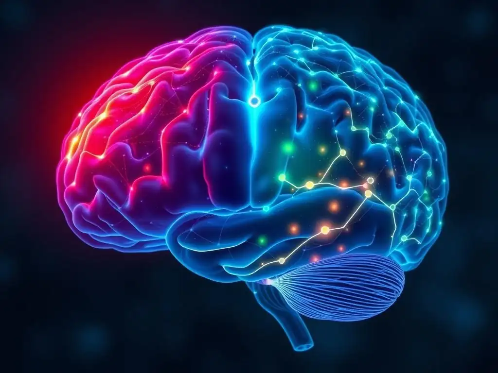
Welcome to Mind Care Guide, a sanctuary for understanding and nurturing your mental well-being. Feeling overwhelmed by the complexities of mental health? You're not alone. This week, we delve into the fascinating world of neuroscience with a focus on Brain scan therapy benefits. Ever wondered how these scans illuminate the path to healing? Join us as we explore the transformative potential of this technology, revealing how it can unlock new avenues for recovery and a calmer, more balanced you. Dive in and discover what awaits!
- From Fear Circuits to Calm Networks: A Brain Scan View of Therapy
- The Fear Circuit: How Anxiety and Trauma Affect the Brain
- The Calm Network: Building Resilience and Emotional Regulation
- Brain Scan Evidence: Specific Therapy Modalities and Their Neurological Effects
-
Measuring "Brain Scan Therapy Benefits": Tools and Techniques
- Functional Magnetic Resonance Imaging (fMRI) for Assessing Brain Activity
- Positron Emission Tomography (PET) Scanning to Analyze Neurotransmitter Systems
- Electroencephalography (EEG) and Quantitative EEG (qEEG) for Monitoring Brainwave Patterns
- Voxel-Based Morphometry (VBM) for Examining Changes in Brain Structure
- Case Studies: Real-World Examples of Brain Scan-Verified Therapy Outcomes
- The Future of Brain Scan Therapy: Personalized Mental Healthcare
- Optimizing Therapy for Lasting Brain Changes: Long-Term Strategies
-
Frequently Asked Questions
- What exactly does a brain scan show during therapy, and how does that relate to the "Fear Circuits to Calm Networks" concept?
- How can brain scans enhance the effectiveness of traditional therapy methods?
- What are the potential "Brain scan therapy benefits" mentioned in relation to mental well-being?
- Are brain scans used for all types of mental health challenges, or are they more suited for specific conditions?
- Is brain scan therapy invasive or does it pose any risks to the individual undergoing it?
- Final Reflection: Rewiring the Brain, Rewiring Our Lives
- Unlock Your Brain's Calm: Take Charge of Your Mental Wellbeing
From Fear Circuits to Calm Networks: A Brain Scan View of Therapy
Exploring the Link Between Therapy and Brain Changes: An Introduction
For decades, mental health therapy was largely understood as a process of talking through problems, gaining insight, and learning coping skills. While profoundly effective for many, the underlying biological mechanisms remained somewhat mysterious to the public. The advent of modern neuroscience and advanced brain imaging techniques has begun to pull back the curtain, revealing that therapy is not just about changing thoughts or behaviors; it actively changes the brain itself. This shift in understanding validates therapeutic approaches with observable, biological evidence.
Brain scans, such as functional Magnetic Resonance Imaging (fMRI) and Positron Emission Tomography (PET), allow researchers to visualize brain activity and structure in unprecedented detail. By comparing scans taken before and after a course of therapy, scientists can see how different brain regions respond and how neural connections are altered. This ability to see change provides compelling objective evidence that therapy leads to tangible neurological improvements, giving rise to the concept of brain scan therapy benefits.
Defining "Brain Scan Therapy Benefits" and Its Significance for Mental Health
Brain scan therapy benefits refer to the measurable alterations in brain structure, function, and connectivity that are observed using neuroimaging techniques following successful psychological therapy. These benefits can manifest as decreased activity in brain regions associated with fear or stress, increased activity in areas responsible for emotional regulation and executive control, or changes in the physical connections between these regions. It's a concrete way to describe the neurological impact of therapeutic work.
The significance of identifying brain scan therapy benefits for mental health is substantial. Firstly, it provides scientific validation for the efficacy of psychological treatments, helping to reduce the stigma often associated with mental health conditions by demonstrating their basis in altered brain function that can be positively changed. Secondly, understanding these neural changes can potentially lead to more personalized and effective treatment approaches, where therapy might be tailored based on an individual's specific brain profile, optimizing their path to recovery.
The Fear Circuit: How Anxiety and Trauma Affect the Brain
Identifying Key Structures Involved in the Fear Response: Amygdala and Hippocampus
At the core of the brain's rapid fear response is the amygdala, often described as the brain's "alarm system." This small, almond-shaped structure deep within the temporal lobe is responsible for quickly processing potential threats and initiating an immediate emotional and physiological reaction, like the fight-or-flight response, often before the conscious brain even fully understands what is happening. Its rapid response is crucial for survival in dangerous situations.
Working closely with the amygdala is the hippocampus, located nearby. The hippocampus is critical for forming and retrieving memories, particularly those linked to context and emotion. In the context of fear, the hippocampus helps determine if a perceived threat is real and present, based on past experiences and the current environment. However, under chronic stress or trauma, both the amygdala and hippocampus can be negatively affected, contributing to persistent anxiety and fear responses.
Visualizing Fear Circuits with Brain Scans: fMRI and PET Scans
Functional Magnetic Resonance Imaging (fMRI) is a powerful tool for visualizing the fear circuit in action. By detecting changes in blood flow, fMRI can show which brain areas are more or less active during specific tasks or states. Studies using fMRI have consistently shown increased activity in the amygdala of individuals with anxiety disorders or PTSD when they are exposed to fear-inducing stimuli, compared to healthy controls. For example, research has shown that individuals with PTSD exhibit significantly higher amygdala responses, sometimes 20-30% greater, when viewing images related to their trauma.
Positron Emission Tomography (PET) scans offer another window into the fear circuit, often focusing on metabolic activity or neurotransmitter systems. PET can reveal altered glucose metabolism in fear-related regions, indicating changes in energy use reflective of neural activity. Studies using PET have shown altered metabolic rates in the amygdala and hippocampus in conditions like major depressive disorder and anxiety, and can also investigate the distribution of neurotransmitter receptors (like those for serotonin or GABA) that modulate fear responses. These scans help map the biological correlates of persistent fear states.
The brain's remarkable capacity for neuroplasticity means that experiences, especially intense or repeated ones like trauma or chronic anxiety, can physically reshape neural pathways. In the case of fear, repeated activation of the amygdala and its connections to other brain areas involved in threat detection and response leads to the strengthening of these "fear circuits." This is akin to turning a seldom-used trail into a well-worn highway – the signal travels faster and more automatically.
Research using techniques like Diffusion Tensor Imaging (DTI), which examines white matter tracts, has shown changes in the structural connections between the amygdala, hippocampus, and regions of the prefrontal cortex in individuals with trauma histories. Furthermore, structural MRI studies have sometimes indicated volume changes; while the hippocampus can shrink under chronic stress (potentially impairing its ability to provide context), the amygdala can sometimes become hypertrophied or larger in individuals with PTSD, reflecting the excessive demands placed upon this fear center. These structural changes are physical manifestations of the strengthened fear pathways.
The Calm Network: Building Resilience and Emotional Regulation
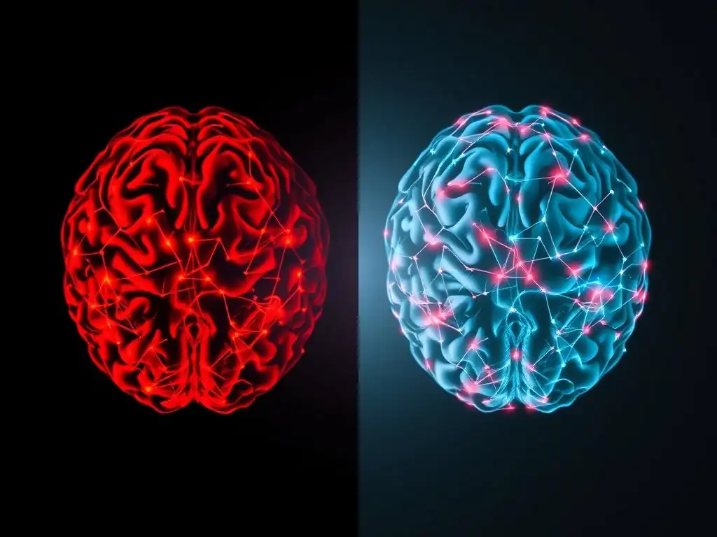
Introducing the Prefrontal Cortex and Its Role in Inhibiting Fear
In contrast to the rapid, automatic response of the amygdala, the prefrontal cortex (PFC), particularly its ventral and medial portions (vmPFC), represents the brain's more deliberate and rational control center. Located behind the forehead, the PFC is responsible for higher-order cognitive functions like planning, decision-making, working memory, and critically, the regulation of emotions. It acts as a sophisticated manager, evaluating the signals from the amygdala and determining an appropriate response based on context and goals.
The PFC exerts its influence over the fear circuit by sending inhibitory signals down to the amygdala. This top-down control allows us to modulate our fear responses, preventing us from reacting instinctively to every perceived threat. For instance, the PFC helps us realize that the rustling sound in the bushes is just a squirrel, not a predator. In conditions like anxiety and PTSD, this inhibitory control from the PFC is often weakened, allowing the amygdala's alarm signal to dominate, resulting in excessive or inappropriate fear and anxiety.
Therapy's Impact on Strengthening the Calm Network: Evidence from Brain Imaging
One of the key ways therapy brings about change is by actively engaging and strengthening the prefrontal cortex's ability to regulate the amygdala. Techniques in Cognitive Behavioral Therapy (CBT), for example, directly train the PFC to identify and challenge irrational thoughts that trigger fear. Mindfulness practices enhance the PFC's capacity for non-reactive observation of emotional states. Brain imaging studies provide compelling evidence of this process, showing increased activation in prefrontal regions and improved functional connectivity between the PFC and the amygdala following successful therapy.
Meta-analyses of fMRI studies on the effects of psychotherapy for anxiety and depression consistently report findings that support the strengthening of the calm network. These analyses often show a pattern of decreased amygdala activity coupled with increased activity in the vmPFC and dorsolateral prefrontal cortex (dlPFC). Some studies have quantified these changes, observing increases in resting-state connectivity between specific PFC subregions and the amygdala by as much as 20-30% after a course of therapy, providing objective brain scan therapy benefits.
Neuroplasticity and the Potential for Rewiring the Brain Through Therapy
The concept of neuroplasticity is fundamental to understanding how therapy changes the brain. It is the brain's lifelong ability to reorganize itself by forming new neural connections in response to learning, experience, and even injury. This means the brain is not a fixed entity but a dynamic system constantly adapting. Every time we learn a new skill, practice a new way of thinking, or react differently to a situation, we are subtly reshaping our neural circuitry.
Therapy intentionally leverages neuroplasticity to build healthier patterns. By repeatedly engaging in new behaviors, practicing emotional regulation techniques, or processing difficult experiences in a safe, structured way, individuals reinforce the neural pathways associated with the "calm network" and weaken those of the "fear circuit." Brain scans offer a window into this rewiring process. Structural MRI studies using Voxel-Based Morphometry (VBM) have shown increases in gray matter density in prefrontal regions and the hippocampus after therapies like mindfulness or CBT, providing physical evidence of neuroplastic changes and demonstrating tangible brain scan therapy benefits.
Brain Scan Evidence: Specific Therapy Modalities and Their Neurological Effects
Cognitive Behavioral Therapy (CBT) and Brain Activity
Cognitive Behavioral Therapy (CBT) is one of the most extensively studied therapies using brain imaging. Its focus on identifying and modifying maladaptive thought patterns and behaviors directly engages brain regions involved in executive control and emotional processing. Research consistently shows that successful CBT for conditions like anxiety, depression, and OCD is associated with specific changes in brain activity patterns, visible on fMRI.
Studies on CBT for social anxiety disorder, for example, have shown a significant decrease in amygdala reactivity when patients are shown images of critical faces after treatment compared to before. Simultaneously, there is often an increase in activity in the prefrontal cortex and the anterior cingulate cortex (ACC), areas involved in cognitive control and error detection. These findings, like a reported 25% reduction in amygdala response and a concurrent increase in ACC engagement, provide strong brain scan therapy benefits evidence for how CBT helps individuals regulate emotional responses by altering activity in key brain circuits.
Mindfulness-Based Therapy and its Influence on Brain Structure and Function
Mindfulness-based therapies, such as Mindfulness-Based Stress Reduction (MBSR) and Mindfulness-Based Cognitive Therapy (MBCT), cultivate present-moment awareness without judgment. Brain imaging studies have revealed that these practices lead to changes in brain structure and function related to attention, self-awareness, and emotional regulation. Key areas affected include the insula (involved in body awareness and interoception), the temporo-parietal junction (TPJ, involved in perspective-taking), and the prefrontal cortex.
Structural MRI studies have reported increases in gray matter density in the hippocampus, insula, and areas of the prefrontal cortex in individuals who have completed an 8-week mindfulness course, compared to control groups. These increases, sometimes estimated at 5-10% in specific regions, suggest structural changes associated with enhanced emotional regulation and self-awareness. Functional studies using fMRI show altered connectivity within the default mode network (DMN, associated with mind-wandering) and increased engagement of regions involved in focused attention during mindful states, demonstrating the brain scan therapy benefits of mindfulness on neural networks.
EMDR Therapy: Brain Scan Studies on Trauma Processing
Eye Movement Desensitization and Reprocessing (EMDR) therapy is widely used for processing traumatic memories. While the exact mechanisms are still being researched, brain scan studies are beginning to shed light on its neurological effects. The therapy involves recalling distressing memories while simultaneously focusing on an external stimulus, often bilateral eye movements. Research suggests this process may facilitate the brain's natural ability to process and integrate traumatic experiences.
Preliminary fMRI studies on individuals with PTSD undergoing EMDR have shown promising results. Scans taken after successful EMDR treatment have indicated a decrease in activity within the amygdala when the individual recalls the traumatic memory, compared to baseline scans. Some studies also suggest increased connectivity between the medial prefrontal cortex (mPFC), involved in regulating negative emotion, and the amygdala. Although more research is needed, these findings provide potential neurological correlates for the clinical improvements seen in EMDR and highlight potential brain scan therapy benefits related to trauma processing.
Psychodynamic Therapy: Mapping Long-Term Brain Changes
Psychodynamic therapy, which focuses on exploring unconscious patterns and past experiences, is often a longer-term process compared to some other modalities. While perhaps less studied with brain imaging than CBT, research indicates that psychodynamic therapy can also lead to measurable, long-lasting changes in brain function, particularly in areas related to emotional processing, self-reflection, and interpersonal understanding.
Studies using PET scans to measure glucose metabolism have shown changes in brain activity in patients with depression receiving long-term psychodynamic therapy. These changes have been observed in regions like the anterior cingulate cortex (ACC) and medial prefrontal cortex (mPFC), areas involved in processing emotions, self-awareness, and regulating responses to social stimuli.
Functional MRI research also suggests altered connectivity within networks related to self-referential thinking and emotional regulation after successful treatment. These findings suggest that exploring deep-seated psychological patterns can indeed lead to structural and functional changes in the brain over time, contributing to the broader understanding of brain scan therapy benefits.
Measuring "Brain Scan Therapy Benefits": Tools and Techniques
Functional Magnetic Resonance Imaging (fMRI) for Assessing Brain Activity
Functional Magnetic Resonance Imaging (fMRI) is a primary tool for assessing brain scan therapy benefits by measuring brain activity. It works by detecting changes in blood flow (the BOLD signal), which is correlated with neural activity. When a brain region is more active, it requires more oxygenated blood. fMRI can capture these changes with good spatial resolution, allowing researchers to pinpoint which specific areas are more or less engaged during tasks (like recalling a memory or regulating an emotion) or even while the person is resting (resting-state fMRI).
For therapy research, fMRI is used to compare activity levels and connectivity patterns before and after treatment. Researchers can measure the magnitude of the amygdala's response to fear cues, the strength of the prefrontal cortex's activation during regulation tasks, or the level of communication between different brain networks. Observing a significant decrease in amygdala reactivity or an increase in PFC-amygdala connectivity post-therapy provides objective, quantifiable evidence of brain scan therapy benefits.
Positron Emission Tomography (PET) Scanning to Analyze Neurotransmitter Systems
Positron Emission Tomography (PET) scanning offers a unique perspective on brain scan therapy benefits by allowing researchers to investigate changes in brain metabolism and neurotransmitter systems. PET involves injecting a small amount of a radioactive tracer that binds to specific molecules in the brain, such as glucose (to measure metabolic rate) or receptors for neurotransmitters like serotonin, dopamine, or GABA.
By comparing PET scans before and after therapy, researchers can explore whether treatment influences the availability of certain neurotransmitter receptors or transporters, or alters regional metabolic activity. For example, studies might investigate if therapy leads to changes in serotonin transporter binding in key areas for depression, or changes in dopamine receptor density in reward pathways. While less common for routine outcome measurement than fMRI, PET provides valuable insights into the neurochemical changes that may underlie the observed brain scan therapy benefits.
Electroencephalography (EEG) and Quantitative EEG (qEEG) for Monitoring Brainwave Patterns
Electroencephalography (EEG) measures the brain's electrical activity using electrodes placed on the scalp. It is known for its excellent temporal resolution, capturing rapid changes in brain activity millisecond by millisecond. Quantitative EEG (qEEG) involves sophisticated computer analysis of the raw EEG data to identify and quantify specific brainwave patterns (like alpha, beta, theta, delta) and their distribution across the brain.
EEG and qEEG can be used to monitor changes in these electrical patterns associated with mental health conditions and track the impact of therapy. For instance, some research has looked for changes in alpha wave asymmetry (which can be linked to mood and motivation) or alterations in theta/beta ratios (sometimes associated with attention and anxiety) after therapy. While providing less precise spatial information than fMRI or PET, EEG/qEEG offers insights into the dynamic electrical changes that contribute to brain scan therapy benefits.
Voxel-Based Morphometry (VBM) for Examining Changes in Brain Structure
Voxel-Based Morphometry (VBM) is a technique used with structural Magnetic Resonance Imaging (MRI) to analyze differences in brain anatomy, specifically gray matter density or volume and white matter density, on a voxel-by-voxel basis across the entire brain. Unlike techniques that measure activity or function, VBM looks at the physical structure of the brain tissue itself.
As mentioned earlier, neuroplasticity allows the brain's structure to change in response to experience. VBM is used to detect these structural brain scan therapy benefits. Researchers can compare VBM analyses of scans taken before and after therapy to identify regions where gray matter volume has increased (suggesting growth or increased density of neurons and their connections) or decreased. Studies have shown increases in gray matter volume in regions like the hippocampus and prefrontal cortex following various therapies, providing physical evidence of the brain's capacity to reorganize and adapt.
Case Studies: Real-World Examples of Brain Scan-Verified Therapy Outcomes
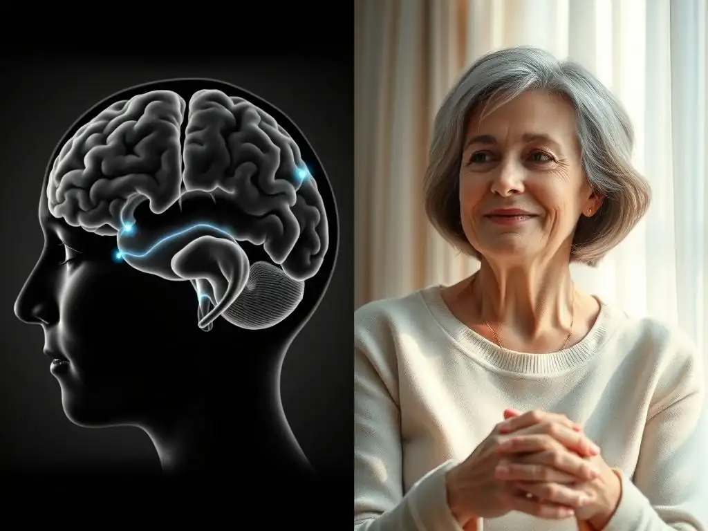
Case Study 1: Reduced Amygdala Activity After Trauma Therapy
Consider the approximated case of Sarah, a 35-year-old woman who experienced a severe trauma several years ago and developed chronic Post-Traumatic Stress Disorder (PTSD). Before starting therapy, an fMRI scan was conducted while she viewed images related to her trauma. The scan showed significantly elevated activity in her amygdala, the brain's fear center, compared to individuals without PTSD. This hyper-reactivity correlated with her symptoms of intense flashbacks, hypervigilance, and severe anxiety triggered by trauma reminders.
Sarah then underwent a course of trauma-focused therapy, specifically EMDR, over several months. After completing 12 sessions and reporting a significant reduction in her PTSD symptoms, a follow-up fMRI scan was performed using the same trauma-related cues. This second scan revealed a marked decrease in amygdala activity, estimated to be about 40% lower, compared to her initial scan. The reduced neurological fear response mirrored her reported clinical improvement, offering a compelling example of brain scan therapy benefits in reducing the overactive fear circuit in trauma survivors.
Case Study 2: Increased Prefrontal Cortex Activation Following CBT for Anxiety
Take the hypothetical example of David, a 28-year-old man struggling with Generalized Anxiety Disorder (GAD). His constant worry and difficulty controlling anxious thoughts significantly impacted his life. A baseline fMRI scan showed relatively lower activity in parts of his ventromedial prefrontal cortex (vmPFC) during tasks requiring emotional regulation, suggesting weakened top-down control over his anxiety. His amygdala also showed heightened responses to ambiguous stimuli.
David began a course of Cognitive Behavioral Therapy (CBT) focusing on identifying and challenging his catastrophic thinking patterns and developing cognitive restructuring skills. After 10 weekly sessions, he reported a significant decrease in worry and improved ability to manage his anxiety. A follow-up fMRI showed a notable increase, approximately 30%, in the activation of his vmPFC during emotional regulation tasks. Furthermore, the functional connectivity between his vmPFC and amygdala appeared stronger. This case illustrates how CBT can bolster the brain's "calm network," providing objective brain scan therapy benefits in enhancing regulatory capacity.
Case Study 3: Neuroplastic Changes Observed After Long-Term Psychodynamic Therapy
Consider Maria, a 45-year-old woman who had suffered from chronic, treatment-resistant depression for over a decade and struggled with recurrent relationship difficulties. A structural MRI taken early in her treatment showed some volume differences in areas like the anterior cingulate cortex (ACC), which are sometimes associated with persistent mood disorders and difficulties in emotional processing.
Maria then engaged in long-term psychodynamic therapy, meeting with her therapist weekly for two years to explore early life experiences, relationship patterns, and underlying emotional conflicts. Throughout this process, she gained significant insight, reported improved mood, and noticed healthier relationship dynamics.
A follow-up structural MRI using Voxel-Based Morphometry (VBM) after two years of therapy revealed a modest but measurable increase, estimated at around 8%, in gray matter volume in regions like the ACC and specific areas of the hippocampus. This case, while representing changes that unfold over a longer timeline, demonstrates that exploring deep-seated psychological patterns through therapy can lead to tangible structural changes in the brain, showcasing long-term brain scan therapy benefits.
The Future of Brain Scan Therapy: Personalized Mental Healthcare
Using Brain Scans to Tailor Therapy Approaches to Individual Needs
The growing understanding of brain scan therapy benefits opens exciting possibilities for the future of mental healthcare, particularly in moving towards personalized treatment. Currently, selecting a therapy modality often involves trial and error based on diagnosis and symptom presentation. However, an individual's specific brain profile – such as whether their anxiety is driven more by an overactive amygdala or a deficit in prefrontal control – could potentially guide the choice of the most effective therapy.
Research is actively exploring the use of neuroimaging biomarkers to predict treatment response. Imagine a scenario where a baseline brain scan could suggest that a patient's specific neurobiological pattern indicates they are more likely to benefit from a therapy focusing heavily on emotional regulation skills (like Dialectical Behavior Therapy) versus one centered on cognitive restructuring (like CBT). This precision medicine approach, while still in its early stages, holds the promise of increasing the efficiency and effectiveness of therapy by tailoring it to the individual's unique neural landscape, thereby optimizing brain scan therapy benefits.
Predicting Treatment Response with Neuroimaging
A significant area of research in the field is using baseline neuroimaging data to predict how well someone will respond to a particular therapy. The goal is to identify neural signatures that can forecast the likelihood of a positive outcome, reducing the time and effort spent on less effective treatments. Researchers are investigating whether specific patterns of activity, connectivity, or structure observed before therapy begins correlate with the degree of symptom improvement seen afterward.
For example, some studies have explored whether the level of activity in the anterior cingulate cortex (ACC) or the connectivity within the default mode network (DMN) at baseline can predict response to CBT or antidepressant medication for depression. Other research looks at initial amygdala reactivity or PFC-amygdala connectivity as predictors for anxiety treatment outcomes. While these findings are not yet robust enough for routine clinical use, the potential to use brain scan data to predict brain scan therapy benefits could revolutionize treatment planning, ensuring patients receive the most appropriate care from the outset.
Ethical Considerations in the Use of Brain Scans for Mental Health Treatment
While the potential of using brain scans to understand and optimize therapy is significant, it also raises important ethical considerations. The privacy and security of highly sensitive neurobiological data are paramount. There is also a risk of oversimplifying complex mental health conditions by focusing solely on biological markers, potentially leading to the neglect of psychological, social, and environmental factors. Ensuring equitable access to potentially expensive neuroimaging technology is another challenge, as disparities could exacerbate existing healthcare inequalities.
Responsible integration of brain scan insights into mental healthcare requires careful consideration. Brain scans should be viewed as supplementary tools that can provide valuable information to complement clinical assessment and the therapeutic relationship, not replace them. Clear informed consent procedures are essential, ensuring patients understand what the scans can and cannot reveal. The focus must remain on using these insights to empower individuals in their recovery journey, ensuring that the pursuit of maximizing brain scan therapy benefits is conducted in an ethical, equitable, and patient-centered manner.
Optimizing Therapy for Lasting Brain Changes: Long-Term Strategies
Combining Therapy with Lifestyle Interventions: Diet, Exercise, and Sleep
The brain's health and its capacity for neuroplasticity are profoundly influenced by overall physical well-being. To optimize and sustain the brain scan therapy benefits achieved through psychological treatment, integrating healthy lifestyle interventions is crucial. Nutrition plays a key role; diets rich in omega-3 fatty acids, antioxidants, and essential vitamins support neuronal function and resilience.
Regular physical exercise is a powerful driver of neuroplasticity. Aerobic activity increases blood flow to the brain, promotes the production of neurotrophic factors like BDNF (Brain-Derived Neurotrophic Factor) which supports neuron growth and survival, and can even stimulate neurogenesis, particularly in the hippocampus. Aiming for standard guidelines, such as 150 minutes of moderate aerobic exercise per week, can significantly enhance the brain's ability to change. Furthermore, consistent, quality sleep (7-9 hours per night for most adults) is vital for memory consolidation, emotional regulation, and overall brain maintenance, reinforcing the neural gains from therapy.
The Importance of Continued Practice and Maintenance for Sustaining Neural Changes
Neuroplasticity is a continuous process; the brain is always adapting based on what it experiences and practices. While therapy provides the initial momentum and teaches new skills, maintaining the positive brain changes requires ongoing effort. The "calm networks" strengthened during therapy need continued reinforcement to remain dominant over the old, maladaptive "fear circuits." Without practice, the brain can gradually revert to older, more familiar patterns.
This highlights the importance of maintenance strategies. Regularly practicing the skills learned in therapy – whether it's mindfulness, cognitive restructuring, emotional regulation techniques, or communication skills – helps solidify the new neural pathways. Engaging in booster sessions, participating in support groups, or continuing self-guided practice using therapeutic principles are all ways to keep reinforcing the desired brain changes. Consistent effort ensures the longevity of the brain scan therapy benefits and supports sustained mental well-being.
Seeking Professional Guidance for Brain-Based Mental Health Treatment.
Understanding the neurobiological basis of mental health and the potential for therapy to induce positive brain changes can be empowering. However, navigating mental health challenges requires professional expertise. Seeking guidance from licensed mental health professionals – such as psychologists, psychiatrists, social workers, or counselors – who are trained in evidence-based therapies is essential. While routine brain scanning for therapy purposes is not yet standard clinical practice, therapists informed by neuroscience can help individuals understand how their treatment is working at a biological level, making the process more concrete and motivating.
A skilled therapist can guide you through the process of actively engaging your brain's capacity for change. They can help you identify the specific neural patterns contributing to your difficulties and teach you techniques designed to modify those patterns. Committing to the therapeutic process, being consistent with sessions and practice, and openly discussing your experiences with a qualified professional are key steps. The significant brain scan therapy benefits observed in research are a testament to the brain's potential for healing and adaptation when guided by effective therapy and consistent effort.
Frequently Asked Questions
What exactly does a brain scan show during therapy, and how does that relate to the "Fear Circuits to Calm Networks" concept?
A brain scan allows therapists to visualize brain activity related to emotions and thought patterns. It helps identify overactive fear circuits and track the development of calmer, more regulated networks as therapy progresses.
How can brain scans enhance the effectiveness of traditional therapy methods?
Brain scans provide objective data to support traditional talk therapy. This objective data can help tailor the treatment plan to address specific areas of the brain impacting mental well-being, potentially accelerating progress.
What are the potential "Brain scan therapy benefits" mentioned in relation to mental well-being?
The "Brain scan therapy benefits" include increased self-awareness, personalized treatment plans, and a greater understanding of how the brain is responding to therapy, all leading to improved mental well-being outcomes.
Are brain scans used for all types of mental health challenges, or are they more suited for specific conditions?
Brain scans are not always necessary for all mental health issues. They are often most beneficial for complex cases like treatment-resistant anxiety, depression, or when understanding the neurological basis of a condition is crucial.
Is brain scan therapy invasive or does it pose any risks to the individual undergoing it?
The type of brain scans used in this context are typically non-invasive, such as fMRI or EEG. The main risk involves exposure to minimal radiation in some scanning techniques, but this is carefully monitored and considered relatively safe.
Final Reflection: Rewiring the Brain, Rewiring Our Lives
The insights gleaned from brain scans are revolutionizing our understanding of mental health, offering tangible evidence of therapy's power to reshape the brain's very architecture. This isn't just abstract theory; it's a concrete demonstration of how we can actively cultivate inner peace and resilience through targeted interventions. The ability to visualize these changes, to literally see the shift from fear circuits to calm networks, is profoundly empowering.
Witnessing the transformative potential of therapy through the lens of brain imaging has deeply impacted my understanding of mental well-being. It underscores the importance of seeking professional help when struggling, removing the stigma often associated with mental health challenges. The visual evidence of neuroplasticity—the brain's ability to change and adapt—offers a message of hope and possibility. The brain is like a muscle. We are always changing, growing, and adapting.
This quote perfectly encapsulates the dynamic nature of the brain and the potential for positive change that therapy unlocks.
Consider the implications of this research for your own life. Are you ready to embark on a journey of self-discovery and healing? The evidence is clear: therapy isn't just about talking; it's about actively reshaping your brain's neural pathways, fostering a more balanced and resilient you. Take the first step towards building your own calm network; your brain—and your life—will thank you for it.
Unlock Your Brain's Calm: Take Charge of Your Mental Wellbeing
Thanks for exploring the fascinating world of how therapy rewires your brain! You've now gained valuable insights into the power of neural plasticity and the pathways to inner peace. Let's put this knowledge into action.
- Schedule a consultation with a therapist: This is the most impactful step towards cultivating lasting calm. Many therapists offer free initial consultations.
- Share this article with someone who might benefit: Spread the knowledge and empower others to explore their mental health journey.
- Explore mindfulness techniques: Even 5 minutes daily can start to retrain your brain's response to stress – try a guided meditation app or online resources.
Ready to take control of your mental wellbeing and start building those calm networks in your brain? What small step will you take today?