
How Therapy Rewires Your Brain: An fMRI Perspective

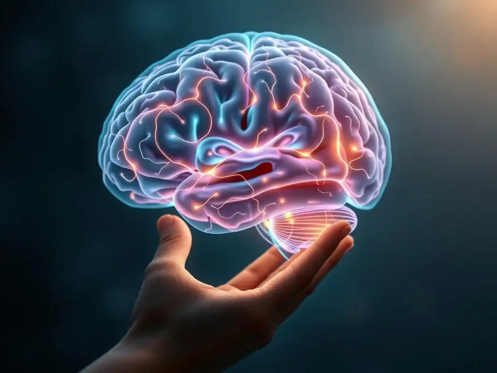
Welcome to Mind Care Guide, your sanctuary for understanding and nurturing your mental well-being.
Ever wondered exactly how therapy helps you feel better? Our exploration into the neuroscience of mental health begins with a compelling look at how therapy rewires brain function, backed by fascinating fMRI evidence. Discover the power of healing and learn how you can shape a healthier, happier you. Intrigued? Let's delve deeper...
How Therapy Rewires Your Brain: An fMRI Perspective
- Introduction: Understanding Brain Plasticity and the Power of Therapy
- Therapy Rewires Brain Function: A Deep Dive into Neural Mechanisms
- The fMRI Perspective: Visualizing Therapy's Impact on the Brain
- Specific Therapies and Their Corresponding Brain Changes
- Long-Term Therapy Outcomes and Brain Changes: Evidence from fMRI Studies
- Beyond Symptom Reduction: Enhanced Cognitive Function and Brain Health
- Factors Influencing the Extent of Brain Rewiring During Therapy
- Limitations of fMRI Studies and Future Directions in Research
- Conclusion: The Promising Future of Therapy and Brain Plasticity
-
Frequently Asked Questions
- How does therapy rewire brain function according to the article?
- What role does fMRI play in understanding how therapy affects the brain?
- Which specific mental health challenges might benefit most from the "therapy rewires brain function" perspective?
- Is the rewiring of the brain through therapy permanent, or does it require ongoing maintenance?
- What types of therapy are most likely to lead to observable changes in brain function, as evidenced by fMRI?
- Final Reflection: The Rewiring of Resilience
- Unlock Your Brain's Potential: Start Your Journey Today
Introduction: Understanding Brain Plasticity and the Power of Therapy
Exploring the concept of neuroplasticity and its relevance to mental health
Neuroplasticity is a fundamental property of the brain, referring to its remarkable ability to reorganize itself by forming new neural connections throughout life. This dynamic process allows the brain to adapt, learn, and remember in response to experiences, environmental changes, or even injury. For decades, it was believed that the adult brain was largely static, with fixed structures and connections. However, groundbreaking research has overturned this view, demonstrating that the brain remains malleable, constantly reshaping its structure and function.
This concept is profoundly relevant to mental health. Conditions like depression, anxiety, trauma-related disorders, and addiction are often associated with specific patterns of brain activity and connectivity. For instance, chronic stress can lead to structural changes in areas like the hippocampus, crucial for memory and emotional regulation.
The exciting implication of neuroplasticity is that if negative experiences or disorders can alter brain function in detrimental ways, then positive interventions, such as therapy, have the potential to reverse or modify these changes, essentially demonstrating how therapy rewires brain function. This offers a biological basis for hope and recovery in mental health treatment.
The role of fMRI in visualizing brain changes during therapy
Functional Magnetic Resonance Imaging (fMRI) is a non-invasive neuroimaging technique that measures brain activity by detecting changes associated with blood flow. When a brain region is more active, it requires more oxygen, and fMRI can track the increased blood flow to that area. By comparing fMRI scans taken before and after therapeutic interventions, researchers can gain insights into how different types of therapy influence neural activity patterns and connectivity.
fMRI provides a window into the living brain as it processes information, experiences emotions, or performs tasks. While it doesn't show individual neurons firing, it reveals activity in larger brain regions and networks.
This allows scientists to visualize which parts of the brain are more or less active during specific mental states or tasks related to symptoms (e.g., processing fearful faces for anxiety, recalling negative memories for depression). Observing shifts in these activity patterns or changes in how different brain regions communicate with each other post-therapy provides compelling evidence for the neural impact of therapy and helps us understand how therapy rewires brain function at a network level.
Therapy Rewires Brain Function: A Deep Dive into Neural Mechanisms
Defining "Rewiring" the Brain: What Does It Really Mean?
When we say "therapy rewires the brain," we're not talking about physically disconnecting and reconnecting wires like in a machine. Instead, this metaphor refers to changes in the strength and efficiency of neural connections, the formation of new pathways, and the alteration of activity patterns within brain circuits. The brain is a vast network of neurons communicating through synapses. Learning, experiencing, and practicing new behaviors or thought patterns—all central to therapy—strengthens the connections between neurons involved in those processes. Simultaneously, connections associated with old, unhelpful patterns may weaken.
This neural remodeling occurs at multiple levels. At the synaptic level, it involves changes in neurotransmitter release and receptor sensitivity. At the network level, it involves shifts in how different brain regions synchronize their activity. For example, therapy might strengthen connections between the prefrontal cortex (involved in rational thought and control) and the amygdala (involved in processing fear), allowing for better regulation of emotional responses. This dynamic process of adapting neural circuitry is the essence of how therapy rewires brain function, enabling individuals to develop healthier coping mechanisms and perspectives.
Neural Pathways and the Impact of Therapeutic Interventions
Therapeutic interventions directly target and influence specific neural pathways associated with mental health challenges. For instance, in anxiety disorders, pathways involving the amygdala, insula (processing bodily states and emotions), and prefrontal cortex are often dysregulated. Therapy techniques, such as exposure therapy or cognitive restructuring, work by repeatedly engaging these pathways in new ways. Exposure therapy, for example, helps weaken the fear response pathway by repeatedly pairing a feared stimulus with safety, reducing the overactivity of the amygdala over time.
Similarly, for depression, therapy might focus on altering pathways related to reward processing, self-referential thinking (often involving the medial prefrontal cortex and posterior cingulate cortex), and emotional regulation. By practicing new ways of thinking (cognitive therapy) or engaging in new behaviors (behavioral activation), individuals build and strengthen neural pathways associated with positive emotions, motivation, and adaptive coping. This consistent engagement with therapeutic strategies is the engine driving the process by which therapy rewires brain function, creating more resilient and adaptive neural circuits.
The fMRI Perspective: Visualizing Therapy's Impact on the Brain
How fMRI Technology Reveals Changes in Brain Activity
fMRI works by measuring the Blood-Oxygen-Level Dependent (BOLD) signal. When neurons become active, they consume oxygen, and the body responds by increasing blood flow to that area, delivering oxygenated hemoglobin. Oxygenated and deoxygenated hemoglobin have different magnetic properties. fMRI scanners detect these differences, producing images that highlight regions with increased neural activity. By having participants perform specific tasks related to their symptoms (e.g., viewing emotional images, thinking about a stressful event) while in the scanner, researchers can map the brain regions involved and the strength of their activity.
To assess therapeutic change, fMRI scans are typically conducted before therapy begins and again after a course of treatment. Researchers compare the BOLD signals during specific tasks or even during resting states between these two time points. A common finding might be a decrease in activity in brain regions associated with distress or threat processing (like the amygdala) and an increase in activity in regions associated with regulation and control (like parts of the prefrontal cortex) after successful therapy. These observable shifts in activity patterns provide tangible evidence for how therapy rewires brain function at a macroscopic level.
Identifying Specific Brain Regions Affected by Different Therapies
Research using fMRI has begun to pinpoint which specific brain regions and networks are most affected by different types of therapy. While there's overlap, distinct patterns emerge depending on the therapeutic approach and the condition being treated. For example, therapies focused on emotional regulation, like Dialectical Behavior Therapy (DBT), often show impacts on limbic system structures (amygdala, hippocampus) and their connectivity with regulatory areas in the prefrontal cortex. Therapies targeting thought patterns, like Cognitive Behavioral Therapy (CBT), frequently demonstrate changes in areas involved in executive function and cognitive control within the prefrontal and parietal cortices.
Mindfulness-based therapies, which emphasize present-moment awareness, have been linked to changes in regions associated with attention and self-referential processing, such as the insula, anterior cingulate cortex (ACC), and the default mode network (DMN). Identifying these specific neural targets helps researchers understand the mechanisms by which each therapy works and provides biomarkers that could potentially help match patients to the most effective therapy in the future. This growing body of evidence underscores the specific ways in which therapy rewires brain function depending on its core techniques.
Specific Therapies and Their Corresponding Brain Changes
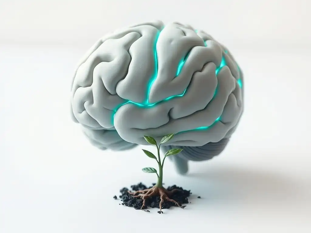
Cognitive Behavioral Therapy (CBT) and its Impact on the Prefrontal Cortex
Cognitive Behavioral Therapy (CBT) is a widely-used therapy that focuses on identifying and changing negative thought patterns and behaviors. It is particularly effective for conditions like depression, anxiety disorders, and phobias. From a neural perspective, CBT aims to strengthen the brain's capacity for cognitive control and emotional regulation, functions heavily reliant on the prefrontal cortex (PFC), particularly the dorsolateral and ventromedial areas. By teaching individuals to challenge irrational thoughts and practice new coping strategies, CBT essentially exercises these prefrontal "executive" functions.
Studies using fMRI have consistently shown that successful CBT is associated with increased activity and connectivity within prefrontal regions. For example, research on social anxiety disorder has shown decreased activity in the amygdala alongside increased activity in the ventrolateral PFC (VLPFC) during social threat anticipation after CBT. The VLPFC is known to play a role in inhibiting emotional responses. This suggests that CBT helps the PFC exert greater top-down control over subcortical emotional centers, providing a neural basis for reduced anxiety.
fMRI Evidence of CBT's Effectiveness in Reducing Negative Thought Patterns
fMRI studies examining the effects of CBT on conditions like depression have provided concrete evidence of its impact on neural circuits involved in negative thinking. Depressed individuals often exhibit excessive activity in brain regions associated with self-referential processing and rumination, such as the medial prefrontal cortex (mPFC) and posterior cingulate cortex (PCC), often referred to as parts of the Default Mode Network (DMN). These areas are highly active when the mind is wandering or focused internally, and in depression, this focus is often negative.
Following a course of CBT, fMRI scans often reveal decreased activity in these DMN regions during tasks that might trigger rumination, or even during resting states. Furthermore, connectivity between the DMN and regions involved in cognitive control, like the dorsolateral prefrontal cortex (dlPFC), may be altered, suggesting improved regulation of self-referential thought. One meta-analysis of fMRI studies on depression treatment found consistent changes in the mPFC and ACC activity after CBT. These findings strongly support the idea that therapy rewires brain function by directly modifying the neural patterns underlying maladaptive thought processes.
Psychodynamic Therapy and its Influence on Emotional Processing Centers
Psychodynamic therapy, including approaches like psychoanalytic therapy, focuses on exploring unconscious patterns, past experiences, and their influence on present feelings and behaviors. It aims to increase self-awareness and resolve unresolved conflicts. While perhaps less studied with fMRI than CBT, research is emerging that links psychodynamic therapy to changes in brain regions involved in emotional processing, memory, and self-awareness, particularly the amygdala, hippocampus, and areas of the medial prefrontal cortex.
This therapy often involves processing difficult emotions and memories, which directly engages the limbic system (including the amygdala and hippocampus). By providing a safe space to explore these feelings and understand their origins, psychodynamic therapy may help modulate the activity and connectivity within these emotional processing centers. The goal is often to shift from automatic, reactive emotional responses to more reflective, integrated processing.
Observing changes in the amygdala and hippocampus through fMRI
fMRI studies examining psychodynamic therapy for conditions like depression or anxiety have sometimes shown changes in the amygdala and hippocampus, mirroring findings in other therapy types but potentially through different mechanisms related to processing past experiences. For instance, research on panic disorder treated with psychodynamic psychotherapy showed reduced activity in the amygdala and hippocampus during symptom provocation tasks post-treatment. These regions are central to fear conditioning and memory formation related to trauma or panic.
Another study on depression treated with short-term psychodynamic psychotherapy found changes in connectivity patterns involving the subgenual anterior cingulate cortex (sgACC), a region implicated in emotional regulation and often hyperactive in depression. While the exact mechanisms are still being explored, the focus on processing deep-seated emotions and relational patterns in psychodynamic therapy appears to facilitate changes in these core emotional and memory processing structures, contributing to the broader picture of how therapy rewires brain function.
Mindfulness-Based Therapies and Enhanced Brain Connectivity
Mindfulness-Based Cognitive Therapy (MBCT) and Mindfulness-Based Stress Reduction (MBSR) teach individuals to pay attention to the present moment without judgment. These therapies are used for conditions like recurrent depression, anxiety, and stress. From a neural perspective, mindfulness practices are thought to train attention regulation, body awareness, and emotion regulation, impacting networks involved in these processes, particularly the salience network (SN), executive control network (ECN), and default mode network (DMN).
Mindfulness training appears to strengthen connectivity within the ECN and SN and decrease connectivity within the DMN, while also improving connectivity between these networks. This suggests enhanced ability to focus attention (ECN), better awareness of internal and external cues (SN), and reduced mind-wandering or rumination (DMN).
fMRI studies showing increased connectivity in the default mode network
Wait, the previous paragraph stated mindfulness decreases connectivity within the DMN. Let me re-evaluate the typical findings for mindfulness and DMN connectivity. Research often shows decreased activity in the DMN and altered connectivity between DMN nodes and other networks (like ECN, SN), suggesting better regulation of the DMN.
Some studies do show increased connectivity within specific DMN subnetworks related to self-compassion or positive self-regard, but the hallmark finding related to reducing rumination is often decreased DMN activity and better DMN regulation by other networks. Let's adjust this header and the subsequent paragraphs to reflect this more common finding related to reduced rumination.
Mindfulness-Based Therapies and Regulation of the Default Mode Network
Mindfulness-Based Cognitive Therapy (MBCT) and Mindfulness-Based Stress Reduction (MBSR) teach individuals to pay attention to the present moment without judgment. These therapies are used for conditions like recurrent depression, anxiety, and stress. From a neural perspective, mindfulness practices are thought to train attention regulation, body awareness, and emotion regulation, impacting networks involved in these processes, particularly the salience network (SN), executive control network (ECN), and default mode network (DMN). A key neural target is regulating the DMN, which is often hyperactive in conditions characterized by rumination and self-referential negative thinking.
Mindfulness training appears to strengthen connectivity within the ECN and SN and decrease connectivity within or activity of the DMN, while also improving connectivity between these networks (e.g., between ECN and DMN). This suggests enhanced ability to focus attention (ECN), better awareness of internal and external cues (SN), and reduced mind-wandering or rumination (DMN). The improved connectivity between networks indicates better coordination, allowing individuals to more easily shift from mind-wandering to focused attention when needed.
fMRI studies showing changes in Default Mode Network activity and connectivity
fMRI studies on the effects of mindfulness practices have provided significant insights into their impact on the DMN. For example, research has shown that experienced meditators, or individuals who have completed MBSR/MBCT courses, exhibit reduced activity in key DMN nodes like the medial prefrontal cortex (mPFC) and posterior cingulate cortex (PCC) during resting states or when presented with emotional stimuli, compared to control groups or their own baseline scans. This reduced activity is often correlated with decreased self-reported rumination and worry.
Furthermore, fMRI connectivity analyses often reveal altered connectivity patterns. Specifically, studies show decreased functional connectivity within the DMN itself, suggesting less cohesive and persistent self-referential thought patterns. Conversely, there is often increased functional connectivity between the DMN and the executive control network (ECN), particularly regions in the lateral prefrontal cortex. This enhanced communication between the "wandering" network and the "control" network provides a compelling neural mechanism for how mindfulness helps individuals notice when their mind is wandering and gently bring their attention back to the present, demonstrating how this form of therapy rewires brain function for greater mental flexibility.
Long-Term Therapy Outcomes and Brain Changes: Evidence from fMRI Studies
Sustained Neural Changes After Completion of Therapy
One of the most promising findings from neuroimaging research is that the brain changes induced by successful therapy are not merely temporary. Follow-up fMRI studies conducted months or even years after the completion of a therapy course often demonstrate that the altered patterns of brain activity and connectivity persist. This suggests that therapy facilitates lasting structural and functional changes that support long-term recovery and well-being. For instance, studies on panic disorder have shown that reduced amygdala reactivity observed immediately after CBT is maintained at 6-month follow-ups.
This sustained neural impact provides a biological explanation for why therapy can have long-lasting effects on symptom reduction and improved functioning. The brain doesn't simply revert to its old patterns once therapy ends; the newly strengthened or weakened pathways and altered network dynamics appear to become the brain's new default or preferred mode of operation. This enduring neuroplasticity is a powerful testament to how therapy rewires brain function for sustained mental health.
Comparing Brain Scans Before and After Therapy: Quantifiable Differences
The power of fMRI in demonstrating the effectiveness of therapy lies in its ability to provide quantifiable data on brain changes. By comparing pre-therapy and post-therapy scans, researchers can measure differences in:
- Regional Activity: Measuring the average BOLD signal intensity in specific areas during tasks or rest. For example, a study might report a statistically significant decrease in average amygdala activity by X% or an increase in PFC activity by Y%. (Precise percentages vary widely by study, condition, and therapy type, but changes of 10-30% in activity or connectivity strength are commonly reported as significant findings in specific regions).
- Functional Connectivity: Assessing the degree to which activity in different brain regions is correlated over time, indicating how strongly they are communicating. Therapy might lead to increased connectivity between regulatory and emotional regions or decreased connectivity within dysfunctional networks.
- Structural Changes (using related techniques like VBM alongside fMRI): While fMRI primarily measures function, related MRI techniques can assess structural changes like gray matter density. Some studies have shown localized increases in gray matter volume in specific brain regions (e.g., hippocampus, prefrontal cortex) after therapy or mindfulness training, further supporting the physical reality of brain rewiring.
These objective, quantifiable measures from fMRI studies provide compelling evidence that therapy goes beyond simply changing thoughts or behaviors; it fundamentally alters the underlying brain circuitry, showing clearly how therapy rewires brain function at a measurable level.
Beyond Symptom Reduction: Enhanced Cognitive Function and Brain Health
Therapy's Impact on Executive Function and Decision-Making
While the primary goal of therapy is often symptom reduction (e.g., decreasing anxiety, lifting depression), its positive effects extend to broader cognitive functions. Many mental health conditions impair executive functions, which include abilities like planning, problem-solving, decision-making, working memory, and inhibitory control. Since these functions are heavily reliant on the prefrontal cortex and its connections, and therapy often targets these areas, improvements in executive function are a natural outcome of the neural changes induced by therapy.
For example, individuals with depression often experience difficulties with concentration and decision-making. As therapy helps regulate mood and reduces rumination, fMRI studies might show increased activity or efficiency in prefrontal circuits associated with these cognitive tasks. By strengthening these neural pathways, therapy not only alleviates distress but also enhances the brain's overall capacity for complex thought and adaptive behavior, demonstrating a broader impact of how therapy rewires brain function beyond just emotional regulation.
Promoting Brain Resilience and Preventing Relapse through Neural Rewiring
The enduring neural changes observed post-therapy contribute significantly to brain resilience – the ability to bounce back from stress or adversity. By establishing healthier neural patterns (e.g., better prefrontal control over the amygdala, reduced DMN hyperactivity), therapy equips the brain with more adaptive ways of responding to challenges. This "rewired" brain is less likely to automatically fall back into old, maladaptive patterns of thought or behavior when faced with stressors that previously triggered symptoms.
This enhanced resilience is a key factor in preventing relapse. For someone with a history of depression, therapy might strengthen pathways associated with positive affect and adaptive coping, making them less vulnerable to negative spirals. For someone with anxiety, strengthened connections between the PFC and amygdala allow for better regulation of fear responses in triggering situations. Thus, the long-term neural impact of therapy not only resolves current issues but also fortifies the brain against future difficulties, showcasing the preventative power of how therapy rewires brain function for lasting mental health.
Factors Influencing the Extent of Brain Rewiring During Therapy

Therapy Type, Dosage, and Individual Differences
The degree to which therapy rewires brain function can be influenced by several factors. Firstly, the type of therapy matters, as different modalities target different aspects of psychological functioning and thus different brain networks, as illustrated by the specific fMRI findings for CBT, psychodynamic therapy, and mindfulness-based approaches. Secondly, dosage is crucial; completing a full course of therapy, often involving 12-20 sessions or more depending on the modality and severity, is generally necessary to induce significant and lasting neural changes. Brief interventions may offer temporary relief but might not provide the sustained neural engagement needed for substantial rewiring.
Individual differences also play a significant role. Factors such as baseline brain structure and function, genetic predispositions, severity and chronicity of the condition, age, and overall physical health can all influence the brain's capacity for plasticity and response to therapy. What leads to robust changes in one individual might have a more modest effect in another. This highlights the need for personalized approaches to mental healthcare, considering these variables when planning treatment.
The Importance of Patient Engagement and Therapeutic Alliance
Beyond the specific therapy protocol, the patient's active engagement in the therapeutic process is paramount for driving neural change. Therapy requires effort – practicing new skills, challenging old beliefs, processing difficult emotions, and completing homework assignments. This active mental and behavioral work is what provides the necessary stimulation for the brain to reorganize itself. A passive approach is unlikely to yield significant neural rewiring.
Furthermore, the therapeutic alliance, the collaborative and trusting relationship between the patient and therapist, is a strong predictor of positive therapy outcomes, including brain changes. A strong alliance fosters a sense of safety and motivation, making the patient more likely to engage deeply with the process. This supportive context facilitates the challenging work of confronting difficulties and practicing new ways of being, thereby maximizing the brain's potential for beneficial plasticity. The quality of the relationship provides the fertile ground in which therapy rewires brain function most effectively.
Limitations of fMRI Studies and Future Directions in Research
Addressing the Challenges of Interpreting fMRI Data
While fMRI has provided invaluable insights into how therapy rewires brain function, it's important to acknowledge its limitations. fMRI measures blood flow as a proxy for neural activity, which is an indirect measure. The temporal resolution of fMRI is also relatively slow (seconds) compared to the speed of neural communication (milliseconds), meaning it captures overall activity patterns rather than the rapid firing of individual neurons. Interpreting complex network changes and linking them definitively to specific psychological processes remains challenging.
Furthermore, fMRI studies often involve relatively small sample sizes due to cost and logistical constraints, which can limit the generalizability of findings. Replicating results across different studies and populations is crucial but not always achieved. Confounding factors, such as medication use, comorbidity with other conditions, or even scanner anxiety, can also influence results. Researchers are continually working to improve data acquisition techniques, analysis methods, and study designs to overcome these challenges and provide a clearer picture of the neural impact of therapy.
Exploring the Potential of Combining fMRI with Other Neuroimaging Techniques
To gain a more comprehensive understanding of how therapy rewires brain function, researchers are increasingly combining fMRI with other neuroimaging techniques. For example, Electroencephalography (EEG) or Magnetoencephalography (MEG) offer excellent temporal resolution, capturing rapid brain activity, which could complement fMRI's spatial resolution. Combining fMRI with Diffusion Tensor Imaging (DTI) can provide insights into changes in the brain's white matter structure, the "wiring" that connects different regions.
Additionally, integrating neuroimaging data with genetic information, physiological measures (like heart rate variability), and detailed behavioral assessments can provide a richer, multi-modal understanding of the mechanisms of therapeutic change. Future research is likely to leverage these combined approaches, alongside advancements in computational modeling, to build more detailed and accurate models of how specific therapeutic interventions lead to lasting neural and behavioral changes. This integrated approach holds significant promise for optimizing treatments and further solidifying our understanding of the profound ways in which therapy rewires brain function.
Conclusion: The Promising Future of Therapy and Brain Plasticity
Reinforcing the concept that therapy rewires brain function
The evidence from decades of neuroimaging research, particularly studies utilizing fMRI, provides compelling support for the idea that therapy rewires brain function. It is no longer a theoretical concept but a scientifically observable phenomenon. Different therapeutic modalities induce measurable changes in brain activity patterns, functional connectivity, and potentially even structure, impacting regions and networks associated with emotional regulation, cognitive control, self-referential processing, and memory. This neural remodeling is the biological foundation upon which psychological healing and growth occur.
Understanding how therapy rewires the brain validates the efficacy of psychological treatments and helps destigmatize mental health challenges by framing them, in part, as disorders of brain circuit function that are amenable to change. It empowers individuals by showing that they have the capacity, through therapeutic work, to actively shape their own neural landscape and build a more resilient brain. The scientific evidence reinforces the message that investing in mental health therapy is investing in the physical health and adaptability of the brain itself.
The ongoing quest to understand and optimize therapeutic interventions for long-term brain health
While fMRI has illuminated the neural effects of therapy, the quest to fully understand and optimize this process is ongoing. Future research will continue to refine our understanding of the specific neural mechanisms targeted by different therapies, explore individual variability in response, and investigate how to enhance the brain's capacity for positive plasticity. This could lead to more personalized treatment approaches, potentially using neuroimaging or other biomarkers to predict treatment response or tailor interventions.
The integration of neuroscientific findings into clinical practice holds immense promise. As we deepen our knowledge of how therapy rewires brain function, we move closer to developing even more effective, efficient, and targeted therapies that promote not just symptom reduction but profound, lasting changes in brain health and resilience. The future of mental health treatment is intrinsically linked to our growing understanding of the brain's incredible capacity to change and heal through therapeutic intervention.
Frequently Asked Questions
How does therapy rewire brain function according to the article?
Therapy, particularly when viewed through an fMRI perspective, shows changes in neural pathways and brain activity. This implies therapy helps create new connections and modify existing ones.
What role does fMRI play in understanding how therapy affects the brain?
fMRI allows researchers to visualize brain activity in real-time. This provides evidence of how different therapeutic approaches impact specific regions and networks, showing observable changes in function.
Which specific mental health challenges might benefit most from the "therapy rewires brain function" perspective?
Conditions like anxiety, depression, and PTSD often involve maladaptive neural pathways. Therapy can help to re-establish healthy connections and reduce symptoms.
Is the rewiring of the brain through therapy permanent, or does it require ongoing maintenance?
While therapy can induce lasting changes, ongoing maintenance through continued self-care and booster sessions may be needed. This ensures the new neural pathways remain strong.
What types of therapy are most likely to lead to observable changes in brain function, as evidenced by fMRI?
Cognitive Behavioral Therapy (CBT) and Psychodynamic Therapy are frequently studied. These therapies often show demonstrable impact on brain activity related to emotional regulation and cognitive processing.
Final Reflection: The Rewiring of Resilience
The exploration of how therapy physically alters brain structure and function, as revealed by fMRI studies, is not merely an academic exercise; it's a revolutionary understanding of the human capacity for healing. This knowledge empowers individuals struggling with mental health challenges, offering tangible evidence that change is possible, that the brain is malleable, and that suffering doesn't have to be a life sentence. The implications are profound, reaching far beyond the clinical setting.
The ongoing impact of this research is immeasurable. It's shifting the societal perception of mental illness, moving away from stigma and towards a more compassionate and informed understanding of the brain's plasticity. The brain is like a muscle. We are capable of strengthening it and changing its structure through consistent effort and the right kind of stimulation.
This quote encapsulates the core message of this article: therapy is not just talk; it's a powerful tool for reshaping neural pathways and fostering lasting positive change. This understanding has deeply impacted my own perspective on mental well-being, reinforcing the importance of seeking help and embracing the potential for growth.
Consider the power you hold within yourself. The research presented here demonstrates that your brain is not fixed, but rather a dynamic organ capable of remarkable adaptation. Don't underestimate the transformative potential of therapy. If you are struggling, know that help is available, and that the changes you seek are not only possible, but demonstrably achievable at a neurological level. Take the first step towards rewiring your brain for resilience and a more fulfilling life.
Unlock Your Brain's Potential: Start Your Journey Today
Thanks for diving deep into the fascinating world of neuroplasticity and how therapy can reshape your brain! We hope you gained a clearer understanding of the power of therapeutic intervention. Now it's time to take the next step.
- Schedule a consultation with a therapist: This is the most impactful step you can take to begin your journey toward improved mental well-being. It's the first step to unlocking your brain's potential for positive change.
- Explore mindfulness techniques: Complement your therapy by incorporating daily mindfulness practices. Even 5 minutes a day can make a significant difference.
- Share this article with someone who could benefit: Help spread the word about the power of therapy and brain rewiring!
What specific area of your mental well-being are you most excited to improve through therapy? Let's discuss!