
Mapping the Mind: How Therapy Reshapes Brain Networks Seen in Scans


Welcome to Mind Care Guide, a space dedicated to understanding and nurturing your mental well-being. Ever wondered how talking can actually change your brain? In this article, we delve into the fascinating world of neuroscience to explore how therapy rewires brain networks 🧠, revealing the tangible impact of healing on your neural pathways. Join us as we uncover the science behind transformation and explore the incredible potential within your own mind.
Mapping the Mind: How Therapy Reshapes Brain Networks Seen in Scans
- Introduction: Unveiling the Brain's Plasticity Through Therapy
- The Neuroscience of Therapy: How Does Therapy Change the Brain?
- Brain Scans: A Window into Therapy's Impact
- Long-Term Therapy Outcomes: Evidence from Brain Scan Studies
- Factors Influencing Brain Changes During Therapy
- The Future of Therapy and Brain Imaging
- Conclusion: Therapy's Transformative Power on the Brain
-
Frequently Asked Questions
- ### How does therapy actually change the brain?
- ### Can brain scans really show the effects of therapy?
- ### Which types of therapy are most effective at rewiring brain networks?
- ### How long does it take to see changes in brain networks through therapy?
- ### Is rewiring brain networks through therapy a permanent change?
- Final Reflection: The Rewiring of Resilience
- Unlock Your Brain's Potential: Start Your Journey Today!
Introduction: Unveiling the Brain's Plasticity Through Therapy
Understanding Brain Networks and Mental Health
The human brain is a complex web of interconnected regions, forming intricate networks that govern everything from our thoughts and emotions to our behaviors. These networks are not static; they are dynamic systems that constantly adapt based on our experiences. For individuals facing mental health challenges, specific disruptions or imbalances within these networks can contribute significantly to symptoms. For instance, in depression, there's often observed altered activity in the network involving the prefrontal cortex (responsible for decision-making and emotional regulation) and the amygdala (involved in processing fear and emotion), suggesting a potential biological basis for the condition.
Understanding these brain networks is crucial because it moves beyond viewing mental health solely as a psychological or behavioral issue. It highlights the physical substrate of our emotional and cognitive lives. Research using advanced imaging techniques has shown that conditions like anxiety disorders, PTSD, and even substance use disorders are associated with specific patterns of connectivity and activity in distinct brain circuits. By mapping these networks, scientists and clinicians gain deeper insights into the mechanisms underlying mental illness and how interventions might target these biological underpinnings.
The Promise of Brain Scans in Assessing Therapy Outcomes
For decades, the effectiveness of therapy was primarily assessed through subjective reports from patients and observations by clinicians. While invaluable, these methods provide only one perspective. The advent of advanced brain imaging technologies has opened a new window into objectively measuring the impact of therapy on the brain itself. Techniques like fMRI and EEG allow researchers to observe brain activity and connectivity in real-time or shortly after therapeutic interventions.
These brain scans hold the promise of providing tangible evidence for the changes that occur within the brain as a result of therapy. They can potentially show how therapy works at a biological level, revealing shifts in neural activity patterns or the strength of connections between different brain regions. This not only helps validate the efficacy of various therapeutic approaches but also aids in understanding which types of therapy might be most effective for specific conditions or even individual patients, paving the way for more personalized mental healthcare.
Therapy's Impact on Brain Networks: An Emerging Reality
The idea that talking therapy, without medication, can induce physical changes in the brain might seem counterintuitive to some, but it is increasingly supported by scientific evidence. This emerging reality is rooted in the concept of neuroplasticity, the brain's inherent ability to reorganize itself by forming new neural connections throughout life. Therapy, through structured conversations, skill-building, and emotional processing, provides experiences that can actively stimulate this neuroplastic process.
Studies using brain imaging before and after courses of therapy are beginning to demonstrate these changes. For example, research on successful treatment for depression often shows normalization of activity in brain areas involved in emotional processing. Similarly, therapy for anxiety can lead to reduced reactivity in fear-related circuits. This body of evidence strongly suggests that therapy doesn't just change minds; therapy rewires brain networks, leading to observable and measurable alterations in how the brain functions.
The Neuroscience of Therapy: How Does Therapy Change the Brain?
Neuroplasticity: The Brain's Capacity for Change
Neuroplasticity is the biological foundation upon which therapy exerts its effects. Far from being a fixed organ after childhood, the adult brain possesses a remarkable capacity to change its structure and function in response to experience, learning, and even injury. This involves various mechanisms, such as synaptogenesis (the formation of new synapses or connections between neurons), dendritic branching (neurons growing new extensions to connect with more cells), and even neurogenesis (the birth of new neurons in certain brain regions, like the hippocampus).
Therapeutic interventions leverage this plasticity by providing new experiences, perspectives, and coping strategies. For instance, practicing mindfulness in therapy can strengthen connections in brain areas associated with attention and emotional regulation. Learning to challenge negative thought patterns in CBT can create new neural pathways for processing information. These repeated mental exercises, guided by a therapist, act as powerful stimuli that encourage the brain to adapt and reorganize its networks in healthier ways. Essentially, through consistent engagement, therapy helps the brain build and reinforce pathways associated with desired thoughts, emotions, and behaviors, while potentially weakening those linked to maladaptive patterns.
How Different Therapy Types Impact Brain Structure and Function
While the core principle of neuroplasticity underpins all effective therapy, different therapeutic modalities may influence brain networks through distinct mechanisms, reflecting their unique approaches and targets.
Cognitive Behavioral Therapy (CBT) and Brain Network Modulation
CBT is a goal-oriented therapy that focuses on identifying and challenging distorted thinking patterns and changing problematic behaviors. Studies using fMRI have shown that successful CBT for conditions like depression and anxiety is associated with changes in the connectivity between the prefrontal cortex and the amygdala. For example, research might show increased top-down regulation from the prefrontal cortex over the amygdala's activity, suggesting improved emotional control.
A study on social anxiety disorder treated with CBT showed decreased activity in the amygdala and hippocampus during social situations, areas typically hyperactive in this condition. Furthermore, connectivity changes were observed in networks related to attention and self-referential processing. These findings indicate that CBT helps modulate activity within key emotional and cognitive networks, demonstrating how therapy rewires brain networks by restructuring how these areas interact.
Psychodynamic Therapy and its Effects on Brain Connectivity
Psychodynamic therapy explores how unconscious processes, past experiences, and relational patterns influence current behavior and emotional states. While perhaps less studied with neuroimaging than CBT, research suggests it can impact brain regions involved in self-awareness, emotional processing, and interpersonal understanding. This type of therapy often involves processing difficult emotions and memories, which can influence areas like the insula (involved in bodily awareness and feeling) and the medial prefrontal cortex (involved in self-reflection).
Studies have shown that psychodynamic therapy for depression can lead to changes in the connectivity within the default mode network (DMN), a network active during introspection and mind-wandering, which is often dysregulated in depression. For instance, some findings suggest a normalization of connectivity within the DMN or between the DMN and areas like the subgenual cingulate cortex, a region implicated in mood regulation. This suggests that exploring the roots of emotional distress can lead to a re-organization of brain networks involved in self-perception and emotional processing.
Mindfulness-Based Therapies: Enhancing Brain Regulation
Mindfulness-based therapies, such as MBSR (Mindfulness-Based Stress Reduction) and MBCT (Mindfulness-Based Cognitive Therapy), train individuals to pay attention to the present moment without judgment. This practice is believed to enhance emotional regulation and reduce reactivity. Neuroimaging studies consistently show that mindfulness training impacts brain regions associated with attention, self-awareness, and emotion regulation, including the prefrontal cortex, anterior cingulate cortex (ACC), and insula.
For example, research has demonstrated that mindfulness training can increase gray matter density in the hippocampus and ACC, areas linked to learning, memory, and self-regulation. Functionally, fMRI studies often show decreased activity in the amygdala during emotional tasks and increased connectivity between the prefrontal cortex and the amygdala, indicating better top-down control over emotional responses. These findings highlight how cultivating present-moment awareness actively shapes brain networks involved in crucial regulatory functions.
Brain Scans: A Window into Therapy's Impact
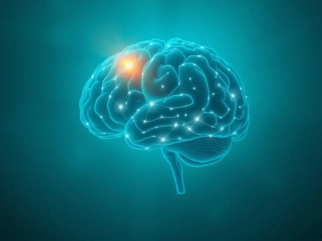
Types of Brain Scans Used to Study Therapy Effects
Neuroimaging plays a vital role in understanding the biological mechanisms of therapy. Different types of scans offer unique insights into various aspects of brain function and structure.
Functional Magnetic Resonance Imaging (fMRI): Mapping Brain Activity
fMRI is one of the most widely used techniques to study the brain's response to therapy. It measures brain activity by detecting changes in blood flow, as active brain regions require more oxygenated blood. By observing which areas are more or less active during specific tasks (like processing emotional images) or at rest (measuring resting-state connectivity), researchers can infer how therapy alters neural function.
For instance, an fMRI study might compare brain activity patterns in individuals with PTSD while recalling a traumatic memory before and after trauma-focused therapy. A common finding is reduced activity in the amygdala (the fear center) and increased activity in the medial prefrontal cortex (involved in emotional regulation) after successful treatment. This provides visual evidence of how therapy helps the brain process and regulate distressing experiences differently.
Electroencephalography (EEG): Measuring Brainwave Patterns
EEG measures the brain's electrical activity using electrodes placed on the scalp. It is excellent for capturing the timing of neural events with millisecond precision, although its spatial resolution is lower than fMRI. EEG can reveal changes in brainwave patterns (such as alpha, beta, theta, or delta waves) associated with different states of consciousness or cognitive processes.
Studies using EEG might examine how therapy affects brainwave coherence (synchronization between different brain regions) or event-related potentials (ERPs), which are brain responses to specific stimuli. For example, research on mindfulness meditation, often incorporated into therapy, has shown changes in alpha wave activity associated with relaxed alertness and increased gamma wave activity linked to enhanced cognitive processing. These electrical signatures provide another layer of data on how therapy influences the brain's dynamic electrical communication.
Diffusion Tensor Imaging (DTI): Examining Brain White Matter Integrity
DTI is an MRI technique that measures the diffusion of water molecules in the brain's white matter. White matter consists of bundles of nerve fibers (axons) covered in myelin, which act as the brain's communication highways. The way water diffuses along these fibers provides information about their directionality and integrity. Changes in DTI metrics can indicate alterations in the structure or health of these connections.
While less common in therapy outcome studies than fMRI or EEG, DTI can offer insights into how therapy might affect the physical infrastructure of brain networks. For example, some research has explored whether DTI can detect changes in the white matter tracts connecting the amygdala and prefrontal cortex after therapy for anxiety, potentially indicating strengthened connections that support better emotional regulation. This technique provides a structural perspective on how therapy rewires brain networks at the level of their physical connections.
Interpreting Brain Scan Results: What Do Changes Signify?
Interpreting changes seen in brain scans after therapy requires careful consideration. A change in activity or connectivity in a specific brain region doesn't automatically mean the therapy was successful or caused the change in isolation. Researchers compare scans taken before and after therapy, often including control groups (e.g., patients on waiting lists or receiving different treatments) to isolate the effects attributable to the specific therapy.
Observed changes typically signify alterations in the functional or structural organization of brain networks associated with the targeted symptoms. For instance, decreased activity in the amygdala during exposure to fear-inducing stimuli after therapy for phobia suggests a reduction in the brain's fear response. Increased connectivity between the prefrontal cortex and emotion centers might indicate improved emotional regulation capabilities.
These findings provide objective, biological correlates to the subjective improvements reported by patients, strengthening the evidence for therapy's profound impact on the physical brain. However, it's crucial to remember that brain scan findings are complex and often represent statistical averages across groups, and individual responses can vary.
Long-Term Therapy Outcomes: Evidence from Brain Scan Studies
The most compelling evidence for therapy's lasting impact comes from studies that not only show immediate post-treatment changes but also demonstrate that these brain alterations are sustained over time or correlate with long-term clinical improvements. These studies reinforce the idea that therapy rewires brain networks for enduring change.
Specific Mental Health Conditions and Observed Brain Network Changes
Neuroimaging research has begun to map specific brain network changes associated with successful therapy across various conditions.
Depression: Therapy's Influence on the Amygdala and Prefrontal Cortex
Major Depressive Disorder (MDD) is often associated with hyperactivity in the amygdala and reduced activity in the prefrontal cortex, particularly areas involved in cognitive control and reward processing. Studies of various psychotherapies for depression, including CBT and Interpersonal Therapy (IPT), frequently report changes in these regions and their connectivity.
One meta-analysis of fMRI studies on psychotherapy for depression found consistent evidence of altered activity in the prefrontal cortex and limbic system (including the amygdala) post-treatment. For example, some studies show increased activity in the dorsolateral prefrontal cortex (involved in cognitive control) and decreased activity in the subgenual anterior cingulate cortex (often hyperactive in depression) after successful therapy. These changes suggest that therapy helps restore balance in the brain circuits governing mood and emotional regulation, providing biological support for the clinical remission observed in patients.
Anxiety Disorders: Restructuring Fear Circuits in the Brain
Anxiety disorders, such as specific phobias, social anxiety disorder, and generalized anxiety disorder, are characterized by excessive fear and worry. Neuroimaging often reveals heightened activity in the amygdala and insula, brain regions critical for processing fear and bodily sensations, and sometimes reduced activity in prefrontal areas responsible for modulating these responses. Exposure therapy, a common and effective treatment, involves gradually confronting feared situations.
Research using fMRI on individuals with specific phobias treated with exposure therapy has shown significant reductions in amygdala activity when exposed to the feared stimulus post-treatment compared to before. Simultaneously, increased activation or connectivity is often observed in prefrontal regions, suggesting enhanced top-down control over the fear response. This demonstrates how therapy helps restructure the brain's fear circuits, enabling individuals to respond to previously terrifying stimuli with reduced fear and anxiety.
Post-Traumatic Stress Disorder (PTSD): Healing Trauma-Related Brain Alterations
PTSD is linked to alterations in a network of brain regions involved in fear processing, memory, and emotional regulation, including the amygdala, hippocampus, and medial prefrontal cortex. Trauma-focused therapies, such as Prolonged Exposure (PE) and Cognitive Processing Therapy (CPT), aim to help individuals process traumatic memories and reduce associated distress.
Neuroimaging studies of successful trauma-focused therapy for PTSD have reported several consistent findings. These include decreased amygdala reactivity to trauma-related cues, suggesting a reduction in the automatic fear response. Often, studies also find increased activation or volume in the hippocampus, a region crucial for memory processing that is sometimes smaller in individuals with chronic PTSD. Furthermore, changes in the connectivity between the medial prefrontal cortex and the amygdala are frequently observed, indicating improved regulation of emotional responses to trauma reminders. These findings provide powerful biological evidence that therapy can help heal the brain's response to trauma.
Case Studies: Real-World Examples of Therapy-Induced Brain Changes
While large group studies provide statistical power, individual case studies or smaller group studies can offer compelling illustrations of how therapy rewires brain networks in specific instances. For example, a study might follow a small group of individuals with Obsessive-Compulsive Disorder (OCD) undergoing Exposure and Response Prevention (ERP) therapy, a highly effective behavioral treatment.
Before therapy, fMRI scans might show heightened activity in brain areas like the orbitofrontal cortex and caudate nucleus, implicated in repetitive thoughts and behaviors. After a successful course of ERP, follow-up scans could reveal a significant reduction in activity within these very same circuits when the individual is presented with stimuli that would previously trigger compulsions. While not every individual will show the exact same pattern, such case studies help to concretely illustrate the principle that behavioral interventions can lead to measurable biological changes, reinforcing the potential for recovery and change.
Factors Influencing Brain Changes During Therapy
Not everyone experiences the same degree of change in therapy, and similarly, the observed brain changes can vary between individuals. Several factors can influence the extent and nature of brain network remodeling during treatment.
Therapy Duration and Intensity: Optimizing Brain Network Remodeling
The principle of "practice makes perfect" applies to the brain's capacity for change. Neuroplasticity is often stimulated by repeated, consistent experiences. Therefore, the duration and intensity of therapy are significant factors in promoting observable brain changes. Shorter, less intense interventions might lead to subtle shifts, while longer-term, more intensive therapy is often associated with more robust and lasting alterations in brain networks.
For instance, studies comparing brief interventions to longer courses of therapy for depression or anxiety often find more pronounced and stable changes in brain connectivity patterns (like those involving the prefrontal cortex and amygdala) following extended treatment. A typical course of CBT might involve 12-20 sessions, and research suggests that significant brain changes become more evident as individuals progress through treatment, particularly during later sessions when new skills are being consolidated. This highlights the importance of completing a full course of therapy as recommended by a clinician to maximize the potential for brain network remodeling.
The Role of the Therapeutic Relationship in Brain Plasticity
Beyond the specific techniques used, the quality of the relationship between the therapist and the client is a crucial, non-specific factor influencing therapy outcomes and, potentially, brain changes. A strong therapeutic alliance characterized by trust, empathy, and collaboration provides a safe environment for exploring difficult emotions and practicing new behaviors.
This safe and supportive relationship may influence brain regions involved in social bonding, trust, and emotional regulation, such as the ventral striatum and areas within the prefrontal cortex. While direct neuroimaging studies specifically isolating the impact of the therapeutic alliance on brain plasticity are complex to design, the clinical evidence is strong: a positive therapeutic relationship is a consistent predictor of positive outcomes across various therapies. It is plausible that this relational safety facilitates the brain's openness to new learning and emotional processing, thereby indirectly supporting the neuroplastic changes targeted by the therapeutic techniques themselves.
Individual Differences: Genetics, Lifestyle, and Brain Response to Therapy
Each person's brain is unique, shaped by a combination of genetics, life experiences, and current lifestyle factors. These individual differences can significantly influence how the brain responds to therapeutic interventions. Genetic variations may affect the expression of neurochemicals or the structure of neural pathways, potentially influencing the baseline state of brain networks and their capacity for change.
Lifestyle factors such as sleep quality, diet, physical exercise, and stress levels also play a profound role in brain health and plasticity. For example, regular aerobic exercise has been shown to promote neurogenesis in the hippocampus and improve connectivity in various brain regions, potentially enhancing the brain's receptiveness to therapeutic change. Conversely, chronic stress can impair neuroplasticity. Understanding these individual factors can help clinicians tailor therapy approaches and potentially predict which individuals might benefit most from specific types of interventions, though this area requires much more research.
The Future of Therapy and Brain Imaging
The intersection of therapy and brain imaging is a rapidly evolving field with exciting potential to transform mental healthcare.
Personalized Therapy: Tailoring Treatment Based on Brain Scan Data
Currently, selecting the most appropriate therapy for an individual often relies on diagnostic criteria, symptom presentation, and therapist expertise. However, brain imaging offers the tantalizing possibility of a more personalized approach. Imagine using brain scans to identify specific patterns of network dysfunction associated with a patient's symptoms and then selecting the therapy modality most likely to normalize those particular patterns.
While still largely in the research phase, studies are exploring whether baseline brain activity or connectivity patterns can predict response to different types of therapy. For instance, preliminary research might investigate if individuals with a specific pattern of prefrontal-amygdala connectivity are more likely to benefit from CBT than psychodynamic therapy.
If successful, this could lead to a future where brain scans help guide treatment decisions, making therapy more efficient and effective by matching the right intervention to the individual's unique brain profile. This would be a significant step in truly leveraging the understanding that therapy rewires brain networks in a targeted way.
Combining Brain Stimulation Techniques with Traditional Therapy
Another area of future potential involves combining traditional talk therapy with non-invasive brain stimulation techniques, such as Transcranial Magnetic Stimulation (TMS) or Transcranial Direct Current Stimulation (tDCS). These techniques can directly modulate activity in specific brain regions. The idea is that stimulating relevant brain areas might enhance the brain's plasticity and make it more receptive to the changes promoted by psychotherapy.
For example, research might explore using TMS to stimulate the dorsolateral prefrontal cortex, a region often underactive in depression, while a patient simultaneously engages in therapy sessions focused on cognitive restructuring. The hope is that this combined approach could accelerate or amplify the positive brain changes observed with therapy alone, leading to faster or more complete symptom remission. While still experimental, this approach represents a fascinating frontier in enhancing the brain's capacity for therapeutic change.
Ethical Considerations: Using Brain Scans Responsibly in Mental Healthcare
As brain imaging becomes more integrated into understanding and potentially guiding mental health treatment, important ethical considerations arise. Issues of privacy and data security surrounding sensitive brain imaging data are paramount. Ensuring that scan data is used responsibly and does not lead to stigmatization or discrimination based on brain characteristics is critical.
Furthermore, it is essential to manage expectations about what brain scans can and cannot tell us. While they provide valuable insights into group-level brain changes associated with therapy, interpreting individual scans in a clinical context is complex and not yet a standard practice for diagnosis or treatment planning. Future guidelines and ethical frameworks will be necessary to ensure that the use of brain imaging in mental healthcare is beneficial, equitable, and respects patient autonomy and privacy.
Conclusion: Therapy's Transformative Power on the Brain
Reinforcing Therapy's Impact on Brain Networks
The accumulating evidence from neuroimaging studies provides compelling biological support for what clinicians and patients have long observed: therapy works. These studies move beyond subjective reports to show objective, measurable changes in the brain's structure and function. They demonstrate that engaging in therapeutic processes like challenging negative thoughts, processing difficult emotions, or practicing mindfulness actively stimulates neuroplasticity, leading to the reorganization and strengthening of healthier brain networks.
From modulating activity in the amygdala in anxiety to normalizing connectivity in the prefrontal cortex in depression, research consistently shows that therapy rewires brain networks in ways that correlate with symptom reduction and improved well-being. This understanding reinforces the value of therapy as a powerful intervention that can induce profound and lasting changes at the most fundamental level of our being – our brain.
The Potential of Brain Imaging to Revolutionize Mental Health Treatment
Brain imaging techniques offer incredible potential to revolutionize the field of mental health. By providing a window into the brain's response to therapy, they can help us better understand how different treatments work, paving the way for more targeted and potentially more effective interventions.
The ability to visualize and measure therapy-induced brain changes opens doors for refining existing therapies, developing new ones, and potentially personalizing treatment based on an individual's unique neural profile. While challenges remain in translating research findings into clinical practice, the promise of using brain insights to improve mental healthcare is immense.
Further Research and the Ongoing Quest to Understand the Brain.
Despite the significant progress made, our understanding of how therapy changes the brain is still in its early stages. Much more research is needed to fully unravel the complex mechanisms of neuroplasticity engaged by different therapeutic approaches, to identify reliable neural biomarkers that predict treatment response, and to explore how individual differences influence brain changes.
The ongoing quest to map and understand the brain is a vast and challenging endeavor. As neuroimaging technology continues to advance and researchers conduct more sophisticated studies, we can anticipate gaining even deeper insights into therapy's transformative power. This knowledge will not only enhance our ability to treat mental health conditions but will also deepen our appreciation for the incredible adaptability and resilience of the human brain.
Frequently Asked Questions
### How does therapy actually change the brain?
Therapy, particularly talk therapy, stimulates neuroplasticity, allowing the brain to form new connections and weaken old ones. This process effectively rewires brain networks.
### Can brain scans really show the effects of therapy?
Yes, advanced brain imaging techniques like fMRI can reveal changes in brain activity and connectivity after therapy. These scans show how therapy impacts specific brain regions.
### Which types of therapy are most effective at rewiring brain networks?
While effectiveness varies, therapies like Cognitive Behavioral Therapy (CBT) and Dialectical Behavior Therapy (DBT) are often cited for their ability to restructure thought patterns and related neural circuits.
### How long does it take to see changes in brain networks through therapy?
The timeline varies depending on the individual, the type of therapy, and the severity of the mental health challenge. Noticeable changes in brain network activity can sometimes be observed after several weeks or months of consistent therapy.
### Is rewiring brain networks through therapy a permanent change?
While therapy can lead to significant and lasting changes, maintaining those changes often requires ongoing effort and continued engagement with healthy coping mechanisms. Relapse is possible, highlighting the importance of consistent mental health care.
Final Reflection: The Rewiring of Resilience
The brain's plasticity, once a theoretical concept, is now vividly demonstrable through neuroimaging. This means that therapy, far from being a mere talking cure, is a powerful tool for actively reshaping the neural pathways that underpin our mental health. We've seen how different therapeutic approaches, from Cognitive Behavioral Therapy to mindfulness practices, leave distinct marks on brain networks, fostering resilience and emotional regulation where once there was struggle.
The implications of this are profound, extending far beyond the individual. Understanding how therapy physically alters the brain challenges the stigma surrounding mental illness, reframing it as a condition amenable to change and healing. The mind is not a vessel to be filled, but a fire to be kindled.
- Plutarch. This quote resonates deeply, as therapy acts as that very kindling, igniting the inherent capacity for growth and change within each of us. The more we understand the neurological mechanisms at play, the more effectively we can support individuals on their journeys to well-being, fostering a culture of empathy and understanding.
Consider the power you now hold. You have witnessed the tangible effects of therapy on the brain, the literal rewiring of neural pathways towards greater mental health. This knowledge is not just information; it's a call to action. Are you ready to explore the potential within yourself or to support others in their journey of self-discovery and healing? Embrace the plasticity of your own mind, and remember that the path to resilience is paved with understanding and the courage to seek help when needed.
Unlock Your Brain's Potential: Start Your Journey Today!
Thanks for exploring the fascinating world of neuroplasticity with us! You've learned how therapy can literally reshape your brain for the better – empowering knowledge, right? Now it's time to take the next step.
- Schedule a consultation with a therapist: This is your key to unlocking personalized strategies for improved mental well-being. Let's work together to map your unique brain pathways.
- Explore our resource library: Discover additional articles and guides designed to support your mental health journey. Find the tools and insights you need to thrive.
- Share this article with someone who could benefit: Let's spread the word about the incredible power of the human brain and the transformative effects of therapy.
What step will you take today to invest in your mental well-being and start reshaping your brain's networks?