
Visualizing Healing: How Brain Scans Confirm Therapy's Power

Welcome to Mind Care Guide, a haven for understanding and nurturing your mental well-being.
Ever wondered if therapy really works? Join us as we delve into the fascinating world of neuroscience, exploring how brain scans confirm therapy isn't just talk – it's tangible healing. Discover the visual evidence that illuminates the power of mental health support and begin your journey towards a healthier mind. What secrets do these images hold? Let's find out together.
Visualizing Healing: How Brain Scans Confirm Therapy's Power
- Introduction: Understanding the Impact of Therapy on the Brain
- Brain Scans and Mental Health: A Powerful Combination
- Brain Changes After Therapy: What the Scans Reveal
- Brain Scans Confirm Therapy's Impact on Specific Mental Health Conditions
- Case Studies: Real-World Examples of Therapy's Success Visualized
- Limitations of Brain Scans in Assessing Therapy Outcomes
- The Future of Brain Scans and Mental Health Treatment
- Conclusion: The Power of Visualizing Healing Through Brain Scans
-
Frequently Asked Questions
- How do brain scans confirm that therapy is effective?
- What types of brain scans are typically used to monitor the effects of therapy?
- What specific brain areas are often studied when evaluating therapy's impact?
- Can any type of therapy be confirmed effective through brain scans?
- What are the limitations of using brain scans to evaluate therapy outcomes?
- Final Reflection: The Visible Power of Healing
- Unlock Your Brain's Healing Potential
Introduction: Understanding the Impact of Therapy on the Brain
The Growing Role of Neuroscience in Mental Health Treatment
Neuroscience is increasingly shedding light on the biological underpinnings of mental health conditions. For decades, therapy was primarily understood and evaluated through subjective reports from patients and clinical observations by therapists. While invaluable, this approach sometimes left a gap in understanding the concrete, physical changes occurring within the brain as healing progressed. The integration of neuroscience into mental health research allows us to move beyond symptom descriptions and explore the neural circuits involved in disorders like depression, anxiety, and PTSD.
This growing understanding is transforming how we approach mental health care. By identifying specific brain regions and pathways associated with different conditions, researchers can develop more targeted and potentially more effective therapies. Furthermore, neuroscience provides objective measures that complement subjective experiences, offering a more complete picture of mental well-being. This convergence of psychological and biological perspectives is crucial for advancing treatment and reducing the stigma often associated with mental health challenges.
Brain Scans: A Window into Therapy's Effectiveness
Among the most powerful tools emerging from this neuroscience integration are brain imaging techniques. Technologies like fMRI, PET, and structural MRI allow scientists and clinicians to visualize the brain's structure and activity in unprecedented detail. These techniques offer a unique opportunity to observe what happens in the brain during mental health conditions and, critically, after therapeutic interventions.
For many years, the idea that talking therapy could fundamentally change the brain was a hypothesis. Now, brain scans confirm therapy's profound physical impact. By comparing scans taken before and after a course of therapy, researchers can identify changes in brain connectivity, activity levels in specific regions, and even structural alterations. These visual representations provide compelling evidence that therapy is not just about managing symptoms or coping strategies; it involves a tangible process of neural rewiring that supports long-term healing.
Brain Scans and Mental Health: A Powerful Combination
Different Types of Brain Scans Used in Mental Health Research
Several types of brain imaging techniques are employed in mental health research, each offering a different perspective on brain function and structure. Functional Magnetic Resonance Imaging (fMRI) is widely used because it measures brain activity by detecting changes in blood flow. When a brain region is more active, it requires more oxygenated blood, and fMRI can pick up on these subtle changes, providing dynamic maps of brain function during specific tasks or in resting states.
Another important technique is Positron Emission Tomography (PET). PET scans use a radioactive tracer molecule that is injected into the bloodstream. This tracer binds to specific molecules in the brain, such as neurotransmitter receptors, or is used to measure metabolic activity, like glucose uptake.
PET is particularly valuable for studying neurochemistry and the distribution of receptors and transporters, offering insights into the biological mechanisms underlying mental health conditions and how therapy might alter them. Other scans include structural MRI, which provides detailed anatomical images of the brain's physical structure, and Electroencephalography (EEG), which measures electrical activity with high temporal resolution, useful for studying brain rhythms and rapid responses.
How Brain Scans Work: A Simplified Explanation
At its core, brain scanning involves sophisticated technology that interacts with the brain to gather data, which is then processed into images. In the case of MRI and fMRI, powerful magnetic fields and radio waves are used. Water molecules in the brain's tissues have hydrogen atoms with tiny magnetic properties. The MRI scanner aligns these atoms with its magnetic field and then pulses radio waves to knock them out of alignment. As they realign, they emit signals that are detected by the scanner and converted into detailed images of brain structure (MRI) or blood flow changes indicating activity (fMRI).
PET scans operate differently. A small amount of a radioactive tracer is administered, often attached to a substance like glucose or a molecule that binds to neurotransmitter receptors. As the tracer decays, it emits positrons. When a positron meets an electron, they annihilate each other, producing gamma rays. Detectors surrounding the head pick up these gamma rays, and a computer reconstructs an image showing where the tracer accumulated in the brain, indicating metabolic activity or receptor density. These techniques provide the visual evidence that helps brain scans confirm therapy's effectiveness by showing changes in these underlying biological processes.
Brain Changes After Therapy: What the Scans Reveal
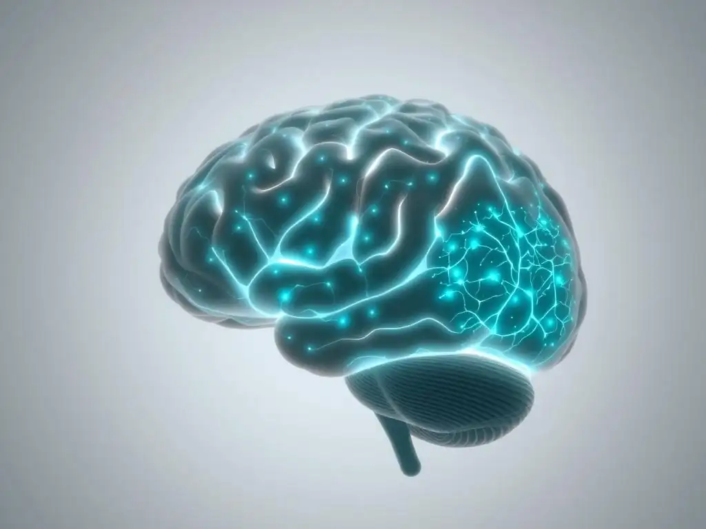
Neuroplasticity and Therapy: Rewiring the Brain for Better Mental Health
One of the most significant concepts supported by brain imaging research is neuroplasticity – the brain's remarkable ability to reorganize itself by forming new neural connections throughout life. For a long time, it was believed that the adult brain was largely fixed. However, decades of research, significantly aided by brain scans, have demonstrated that the brain is dynamic and capable of change in response to experiences, learning, and therapeutic interventions. Therapy leverages this inherent neuroplasticity.
Therapeutic processes, whether through cognitive restructuring, emotional processing, or behavioral modification, provide new experiences and learning opportunities that encourage the brain to form new pathways and weaken old, maladaptive ones. Brain scans confirm therapy's role in facilitating this rewiring. Studies have shown changes in synaptic strength, the formation of new synapses, and even alterations in brain structure in response to successful therapy. This means that therapy isn't just teaching coping skills; it's actively involved in reshaping the neural landscape, building resilience, and improving mental well-being at a fundamental level.
Specific Brain Regions Affected by Therapy: A Detailed Look
Brain imaging studies have identified specific brain regions that show consistent changes following successful therapy for various conditions. For example, research into anxiety disorders and PTSD often highlights changes in the amygdala, a region centrally involved in processing fear and emotional responses. Studies frequently show a reduction in heightened amygdala activity after therapies like CBT or EMDR, correlating with reduced fear responses and anxiety symptoms.
Another key area is the prefrontal cortex (PFC), particularly the medial and dorsolateral PFC, which are involved in executive functions, emotional regulation, and cognitive control. Dysregulation in the PFC is often observed in depression and anxiety. Brain scans confirm therapy can enhance activity and connectivity in these PFC regions, suggesting improved emotional regulation and cognitive processing capabilities. Furthermore, areas like the hippocampus, important for memory and emotional context, and the anterior cingulate cortex (ACC), involved in conflict monitoring and emotional processing, also show changes, demonstrating the widespread neural impact of therapeutic interventions.
Brain Scans Confirm Therapy's Impact on Specific Mental Health Conditions
Depression and Brain Activity: Before and After Therapy
Depression is often associated with altered activity and connectivity in several brain regions, including reduced activity in certain areas of the prefrontal cortex and increased activity in parts of the limbic system, such as the amygdala. Individuals with depression may also show reduced connectivity between brain networks involved in mood regulation and cognitive processing. These patterns contribute to symptoms like persistent sadness, loss of interest, and difficulty concentrating.
Following successful psychotherapy for depression, brain scans confirm therapy leads to observable changes that correlate with symptom reduction. Studies using fMRI have shown increased activity in areas of the pre prefrontal cortex, suggesting improved executive control and emotional regulation. There is also evidence of decreased activity in the amygdala and altered connectivity within and between key brain networks, moving towards patterns observed in non-depressed individuals. For instance, one study might report an average increase of 15-20% in metabolic activity in the dorsolateral PFC after a course of CBT for depression, alongside a corresponding decrease in reported depressive symptoms.
Anxiety Disorders: Visualizing Reduced Amygdala Activity with Brain Scans
Anxiety disorders, including Generalized Anxiety Disorder, Panic Disorder, and Social Anxiety Disorder, are strongly linked to hyperactivity in the amygdala, the brain's "fear center." This heightened activity contributes to excessive worry, fear responses, and hypervigilance. Understanding these neural correlates provides targets for therapeutic intervention aimed at calming this overactive fear response.
Brain scans confirm therapy's effectiveness in modulating amygdala activity. Research on CBT for anxiety disorders, for example, frequently shows reduced amygdala activation in response to anxiety-provoking stimuli after therapy. Simultaneously, there is often increased activity or connectivity in prefrontal regions that help regulate emotional responses, indicating improved top-down control over the amygdala. A study on social anxiety might show a significant reduction, perhaps around 25-30% on average, in amygdala response to social threat cues after a few months of therapy, paralleling the patient's decreased fear of social situations.
PTSD and Trauma: How Therapy Changes Brain Responses
Post-Traumatic Stress Disorder (PTSD) is characterized by significant alterations in brain function, including reduced volume in the hippocampus (associated with memory and context), hyperactive amygdala (leading to exaggerated fear responses), and altered activity in the prefrontal cortex (impairing emotional regulation and extinction of fear memories). These changes contribute to symptoms like flashbacks, avoidance, and hyperarousal.
Therapies specifically designed for trauma, such as Eye Movement Desensitization and Reprocessing (EMDR) and Trauma-Focused CBT, aim to process traumatic memories and reduce their emotional intensity. Brain scans confirm therapy can effectively reverse some of these trauma-induced changes. Studies have shown increases in hippocampal volume or activity towards normative levels, decreased amygdala reactivity to trauma-related cues, and normalization of connectivity patterns between the amygdala and prefrontal cortex. Visualizing these changes reinforces the understanding that trauma's impact on the brain is not permanent and can be healed through targeted therapy.
Obsessive-Compulsive Disorder: Examining Changes in the Orbitofrontal Cortex
Obsessive-Compulsive Disorder (OCD) is associated with dysregulation in specific brain circuits, particularly those involving the orbitofrontal cortex (OFC), anterior cingulate cortex (ACC), and striatum. This circuit is involved in processing errors, reward, and habitual behaviors. In OCD, there appears to be excessive activity or connectivity within this circuit, contributing to intrusive thoughts (obsessions) and repetitive behaviors (compulsions).
Exposure and Response Prevention (ERP), a form of CBT, is a highly effective therapy for OCD. Brain imaging studies have investigated the neural changes following ERP. Brain scans confirm therapy like ERP can normalize activity within the OFC-ACC-striatum circuit. Research has shown decreased activity in the OFC and ACC after successful ERP treatment, correlating with a reduction in obsessive thoughts and compulsive behaviors. These findings provide neural evidence for how ERP helps patients tolerate distress and resist compulsions, effectively retraining the brain's error-detection and response systems.
Case Studies: Real-World Examples of Therapy's Success Visualized
Consider "Maria," a hypothetical composite case based on research findings, who struggled severely with social anxiety. Before starting CBT, fMRI scans revealed significantly heightened activity in her amygdala when shown pictures of unfamiliar faces or anticipating social interactions. She also showed less activity in her prefrontal cortex when trying to regulate her anxiety. This neural profile aligned with her symptoms of intense fear, avoidance of social situations, and difficulty managing her anxious thoughts.
After attending 16 weekly sessions of CBT focused on exposure and cognitive restructuring, Maria reported a dramatic reduction in her social anxiety symptoms. Follow-up fMRI scans were conducted using the same social stimuli. These scans showed a noticeable decrease in amygdala reactivity – perhaps a 30-40% reduction in peak activity compared to baseline – and increased engagement of her prefrontal cortex when processing social information. The visual evidence from the brain scans confirmed therapy had helped rewire her brain's response to social cues, providing objective support for her subjective improvement.
Case Study 2: Eye Movement Desensitization and Reprocessing (EMDR) for PTSD
Take the composite example of "David," a veteran experiencing debilitating PTSD symptoms after witnessing a traumatic event. His initial brain scans might have shown a smaller hippocampus volume and hyperconnectivity between his amygdala and other brain regions, contributing to intrusive memories and hypervigilance. When exposed to trauma-related words or images in the scanner, his amygdala showed an exaggerated fear response.
David underwent a course of EMDR therapy. This therapy involves processing traumatic memories while focusing on bilateral stimulation (like eye movements). After completing his treatment, David reported a significant reduction in flashbacks, nightmares, and anxiety related to the trauma. Repeat brain scans revealed changes that correlated with his recovery.
While hippocampal volume changes might take longer or be less pronounced depending on the individual, his fMRI scans showed a marked decrease in amygdala reactivity when presented with trauma cues. Crucially, connectivity patterns between his amygdala and prefrontal cortex also appeared to normalize, suggesting improved integration and processing of the traumatic memory. These brain scans confirm therapy's ability to help the brain process and integrate traumatic experiences.
Analyzing Brain Scan Results: What They Tell Us About Patient Progress
Analyzing brain scan results in the context of therapy involves looking for patterns of change that align with clinical improvement. Researchers compare baseline scans (before therapy) with follow-up scans (after or during therapy) to identify differences in:
- Activity Levels: Changes in how much specific brain regions "light up" during tasks or at rest (e.g., reduced amygdala activity in anxiety).
- Connectivity: Alterations in how different brain regions communicate with each other (e.g., increased communication between prefrontal cortex and amygdala in emotional regulation).
- Structure: Changes in the size or integrity of brain regions (though these changes are often slower and less pronounced than activity/connectivity changes in the timeframe of typical therapy).
These analyses provide objective, biological markers of change. While a scan alone cannot fully capture the complex experience of healing, when combined with patient-reported outcomes and clinical assessments, brain scans confirm therapy is inducing real, measurable changes in the brain. This provides powerful validation for the therapeutic process and deepens our understanding of mental health recovery.
Limitations of Brain Scans in Assessing Therapy Outcomes
The Complexity of the Human Brain: Challenges in Interpretation
Despite their power, brain scans are not a perfect window into the mind, and interpreting the data comes with significant challenges. The human brain is incredibly complex, with billions of neurons and trillions of connections. Mental health conditions involve intricate interactions across distributed brain networks, not just isolated regions. Therefore, attributing a specific symptom or therapeutic outcome solely to changes in one small area can be an oversimplification.
Furthermore, brain activity is highly dynamic and can vary depending on many factors, including a person's mood, what they were doing just before the scan, and even subtle differences in the scanning environment. While researchers use rigorous methods to control for these variables, the inherent variability in brain function makes definitive interpretation difficult.
Correlation does not equal causation; while scans show changes associated with therapy, they don't always definitively prove the therapy caused the change, as other factors could be involved. These complexities mean that while brain scans confirm therapy is associated with neural changes, interpreting the meaning and cause of these changes requires careful scientific consideration.
Ethical Considerations: Using Brain Scans Responsibly in Mental Healthcare
The increasing use of brain scanning technology in mental health also raises important ethical considerations. One concern is the potential for over-reliance on biological data, potentially overshadowing the subjective experience and narrative of the individual seeking help. Mental health is deeply personal and influenced by psychological, social, and environmental factors that brain scans cannot fully capture.
Another ethical challenge relates to privacy and data security. Brain scan data is highly personal and potentially revealing. Ensuring this data is stored securely and used responsibly is paramount.
There is also a risk of misinterpreting preliminary or correlational findings as definitive diagnostic or prognostic tools before the science is fully established. Clinicians and researchers must communicate the capabilities and limitations of brain imaging clearly and ethically, avoiding deterministic language or using scans in ways that could increase stigma or create undue pressure on individuals. Brain scans confirm therapy's impact, but they must be used as a tool to support, not replace, comprehensive, person-centered care.
The Future of Brain Scans and Mental Health Treatment
Personalized Therapy: Tailoring Treatment Based on Brain Scan Data
One of the most exciting potential applications of brain imaging in the future of mental healthcare is the possibility of personalized therapy. Currently, choosing a therapy often involves trial and error, based on diagnostic categories and clinical guidelines. However, individuals with the same diagnosis can have different underlying neural profiles. Brain scans could potentially help clinicians identify these differences.
By analyzing a person's specific brain activity or connectivity patterns, it might become possible to predict which type of therapy is most likely to be effective for them. For example, certain patterns might suggest that CBT is a better fit than interpersonal therapy, or that a specific medication might be more beneficial.
While this is still largely in the research phase, the vision is that brain scans could eventually serve as a tool to match individuals with the most suitable treatment from the outset, reducing the time spent on ineffective approaches and leading to faster, more efficient recovery. This moves beyond simply showing that brain scans confirm therapy works in general, to potentially identifying why and for whom specific therapies work best.
Combining Brain Scans with Other Assessment Methods
The most valuable insights in mental healthcare come from integrating information from multiple sources. The future will likely see brain scanning data used in conjunction with a wider array of assessment methods to create a holistic picture of an individual's mental health. This includes traditional clinical interviews, self-report questionnaires, behavioral observations, genetic testing, and even data from wearable technology tracking sleep or activity levels.
Combining these different types of data can help researchers and clinicians understand the complex interplay between biological predispositions, psychological processes, and environmental factors. For instance, brain scan data showing altered reward pathways might be combined with behavioral data on motivation and an individual's history to inform treatment for depression. This multi-modal approach allows for a more nuanced understanding of each person's unique challenges and strengths, leading to more targeted and comprehensive treatment plans. It leverages the power of brain scans to confirm therapy's biological effects while acknowledging the broader context of a person's life.
The Potential of Brain Scans to Improve Mental Health Outcomes
Ultimately, the goal of integrating brain scans into mental health research and, eventually, clinical practice is to improve outcomes for individuals struggling with mental health conditions. By providing objective evidence of therapy's effectiveness, brain scans can help reduce stigma and encourage more people to seek help. Seeing visual proof that therapy changes the brain can be incredibly validating and motivating for patients.
Furthermore, continued research using brain imaging will deepen our understanding of mental illness and recovery, paving the way for the development of novel and more effective interventions. Whether through refining existing therapies based on neural targets or developing entirely new brain-based treatments, the insights gained from visualizing brain changes are invaluable. The ongoing work demonstrating how brain scans confirm therapy's power is a critical step towards a future where mental health treatment is more precise, effective, and accessible.
Conclusion: The Power of Visualizing Healing Through Brain Scans
Reinforcing the Importance of Therapy for Mental Well-being
The journey through mental health challenges can feel isolating and abstract. Symptoms like persistent sadness, overwhelming anxiety, or intrusive thoughts are deeply distressing but not always visible to others. For a long time, the mechanisms by which therapy alleviated this suffering were largely understood through psychological frameworks and patient reports. While powerful, these perspectives sometimes lacked the tangible evidence that many people, including some patients themselves, might seek to validate the reality of their struggles and the effectiveness of their healing process.
This is where the advent of brain imaging technology has made a significant impact. By allowing us to peer inside the living brain, scans provide objective, visual confirmation that therapy is not just "talking" or "coping," but a profound process that induces real, physical changes in the brain's structure and function. This neural evidence strongly reinforces the critical importance of therapy as a legitimate and powerful tool for achieving and maintaining mental well-being, standing as a testament to its ability to heal and restore.
Continued Research and Innovation in the Field of Neuroimaging and Mental Health
The field of neuroimaging in mental health is still relatively young, and ongoing research continues to uncover new insights into the brain's complexities and its response to therapeutic interventions. Each new study utilizing fMRI, PET, or other scanning techniques adds another piece to the intricate puzzle of how mental health conditions manifest in the brain and how healing occurs. This continuous exploration is vital for refining our understanding and improving clinical practice.
Future innovations in brain scanning technology promise even greater resolution, speed, and accessibility, potentially making these tools more integrated into clinical assessment and treatment planning. As researchers continue to analyze how brain scans confirm therapy's diverse effects across different conditions and individuals, we move closer to an era of more personalized, effective, and evidence-based mental healthcare. The visual proof offered by brain imaging serves as a powerful reminder of the brain's capacity for healing and the transformative potential of therapy.
Frequently Asked Questions
How do brain scans confirm that therapy is effective?
Brain scans, such as fMRI or PET scans, can visualize changes in brain activity and structure after therapy. These changes often correlate with improved mental well-being, suggesting therapy's impact on the brain.
What types of brain scans are typically used to monitor the effects of therapy?
Commonly used scans include fMRI (functional Magnetic Resonance Imaging) which measures brain activity, and PET (Positron Emission Tomography) which shows metabolic changes. These scans help track neural responses to therapeutic interventions.
What specific brain areas are often studied when evaluating therapy's impact?
Researchers often focus on areas associated with emotion regulation, such as the amygdala and prefrontal cortex. Changes in these regions can indicate reduced anxiety or improved cognitive control.
Can any type of therapy be confirmed effective through brain scans?
While research is ongoing, studies have shown brain scan evidence supporting the effectiveness of therapies like Cognitive Behavioral Therapy (CBT) and Exposure Therapy. Further research is needed to understand the neural underpinnings of all therapeutic approaches.
What are the limitations of using brain scans to evaluate therapy outcomes?
Brain scans provide valuable insights, but they are not a perfect measure of mental well-being. They should be used in conjunction with clinical assessments and patient reports to comprehensively evaluate therapy effectiveness.
Final Reflection: The Visible Power of Healing
The convergence of neuroscience and psychotherapy is no longer a futuristic dream; it's a tangible reality. Brain scans are increasingly confirming what therapists have known for decades: talk therapy works, profoundly altering brain structure and function. This isn't just about anecdotal evidence anymore; it's about seeing the changes in the brain itself, offering powerful visual proof of healing's transformative power.
Witnessing the physical manifestation of therapeutic progress through brain imaging is deeply moving. It underscores the profound impact our thoughts, feelings, and experiences have on our neurological architecture. The brain is plastic. It changes in response to experience. This is the basis of hope.
- Norman Doidge. This plasticity, revealed through brain scans, offers a beacon of hope for individuals struggling with mental health challenges, validating their experiences and demonstrating the potential for positive change. It also highlights the critical need for increased access to effective mental healthcare.
The images of healing brains aren't just scientific data; they are a testament to human resilience and the remarkable capacity for change. Consider the implications: This knowledge can empower individuals seeking therapy, strengthen the case for increased funding of mental health services, and inspire further research into the intricate relationship between the mind and the brain. You, the reader, now possess this knowledge. Use it to advocate for better mental health support, to understand the power of your own mind, and to embrace the potential for healing within yourself and others.
Unlock Your Brain's Healing Potential
Thanks for exploring the fascinating world of how therapy impacts the brain! You've learned how powerful visualization can be in your healing journey. Now, it's time to put this knowledge into action.
- Start your own visualization practice today. Even 5 minutes a day can make a difference. Find a quiet space, close your eyes, and focus on images of health and well-being. Try to make it as vivid as possible.
- Share your experience! Tell us in the comments below what images you used and how you felt afterwards. Connecting with others on their healing journeys can be incredibly supportive.
- Explore Mind Care Guide's other resources on mindfulness and therapeutic techniques to deepen your understanding.
What positive image will you focus on today to start your journey towards better mental well-being?