
Seeing Anxiety Fade: Brain Scan Evidence of Therapy's Success

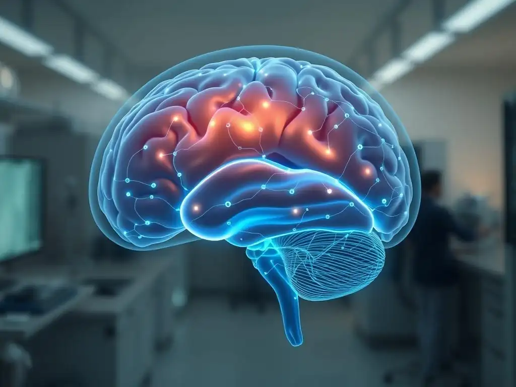
Welcome to Mind Care Guide.
Feeling overwhelmed by anxiety? You're not alone. We understand the weight it carries. But what if there was tangible proof that therapy can truly make a difference? In " Seeing Anxiety Fade: Brain Scan Evidence of Therapy's Success," we delve into fascinating research exploring the neuroscience behind anxiety relief, specifically using brain scan therapy anxiety relief to visualize the positive changes happening within. Curious to see the evidence? Let's explore how science is illuminating the path to mental well-being...
- Seeing Anxiety Fade: Brain Scan Evidence of Therapy's Success - Mind Care Guide
- The Neuroscience of Anxiety: A Foundation for Therapy
- Long-Term Therapy Outcomes and Brain Changes: The Power of Healing
- Brain Scan Evidence of Anxiety Relief Following Cognitive Behavioral Therapy
- Case Study: Analyzing Brain Scans Before and After Cognitive Behavioral Therapy
- Decoding the Data: Interpreting Brain Scan Changes After Cognitive Behavioral Therapy
- The Future of Anxiety Treatment: Brain Scans as a Predictive Tool
- Beyond the Scan: Maintaining Long-Term Anxiety Relief
- Conclusion: Brain Scans Validate the Effectiveness of Cognitive Behavioral Therapy for Anxiety
-
Frequently Asked Questions
- How does a brain scan show the effectiveness of anxiety therapy?
- What type of therapy is most often associated with positive brain scan changes in anxiety patients?
- Can brain scans be used to predict the success of anxiety therapy before it begins?
- What are the limitations of using brain scans to assess anxiety relief?
- Is "Brain Scan Therapy Anxiety Relief" a legitimate therapy itself, or does it refer to something else?
- Final Reflection: The Evolving Landscape of Anxiety Treatment
- Unlock Your Calm: Put Brain Science into Action
Seeing Anxiety Fade: Brain Scan Evidence of Therapy's Success - Mind Care Guide
Introduction: Understanding Anxiety Relief Through Brain Scans
Anxiety is a common mental health challenge that can significantly impact daily life. While therapeutic interventions like Cognitive Behavioral Therapy (CBT) have long been proven effective through patient self-reports and clinical observation, modern neuroscience offers a deeper, more tangible validation: brain scanning technology. These advanced tools allow researchers and clinicians to peer inside the living brain and observe the physiological changes that occur as anxiety symptoms diminish.
Exploring the connection between therapeutic processes and observable brain changes provides compelling evidence for the power of therapy. It moves beyond subjective reporting to demonstrate a concrete, biological shift associated with reduced anxiety. This neuroscientific perspective not only strengthens the rationale for seeking help but also helps demystify the therapeutic process, showing that achieving brain scan therapy anxiety relief is a real, measurable phenomenon.
The Neuroscience of Anxiety: A Foundation for Therapy
How Anxiety Affects Brain Activity
Anxiety disorders are associated with distinct patterns of activity and connectivity within the brain. One key area is the amygdala, often referred to as the brain's "fear center." In individuals with anxiety, the amygdala tends to be hyperactive, showing exaggerated responses to perceived threats, even minor ones. This heightened activity contributes to the feeling of being constantly on edge, prone to panic, or experiencing excessive worry.
Furthermore, anxiety can impact the prefrontal cortex, the region responsible for executive functions like decision-making, impulse control, and regulating emotions. In anxiety, the connectivity between the prefrontal cortex and the amygdala can be altered, often with the prefrontal cortex having less inhibitory control over the overactive amygdala. This imbalance can make it harder for individuals to regulate their fear responses and manage anxious thoughts, creating a feedback loop that perpetuates the condition.
The Role of Neuroimaging in Anxiety Research
Neuroimaging techniques have revolutionized our understanding of the anxious brain. Tools like functional Magnetic Resonance Imaging (fMRI) measure brain activity by detecting changes in blood flow, providing detailed maps of which brain regions are engaged during specific tasks or emotional states. Electroencephalography (EEG) measures electrical activity in the brain through electrodes placed on the scalp, offering excellent temporal resolution to track rapid brain changes.
These technologies allow researchers to compare the brain activity patterns of individuals with anxiety disorders to those without, identifying neural signatures associated with different types of anxiety. More importantly, they enable longitudinal studies that track brain changes over time in response to interventions like therapy or medication. This capability is crucial for providing objective brain scan therapy anxiety relief evidence, showing how therapeutic interventions can normalize or alter these problematic brain patterns.
Long-Term Therapy Outcomes and Brain Changes: The Power of Healing
Exploring the Link Between Therapy and Brain Structure
Beyond transient changes in activity, long-term therapy, particularly evidence-based approaches like CBT, has been associated with more enduring changes in brain structure and connectivity. While structural changes are often subtle and require advanced imaging techniques to detect consistently, research suggests that successful therapy can influence the volume or density of grey matter in certain regions. For instance, studies have explored how effective treatment might lead to changes in areas like the hippocampus, which is involved in memory and stress response, or parts of the prefrontal cortex.
These potential structural adaptations, alongside functional changes, underscore the brain's remarkable plasticity – its ability to reorganize itself throughout life. This means that the pathways carved by anxious thinking and reactive fear responses are not fixed. Through consistent therapeutic work, new, healthier pathways can be strengthened, leading to more adaptive emotional regulation and reduced anxiety symptoms, supported by the enduring changes potentially visible through brain imaging over time.
Understanding the Subcategory: Long-Term Therapy Outcomes and Brain Changes - Brain Scans
The subcategory "Long-Term Therapy Outcomes and Brain Changes - Brain Scans" specifically focuses on the intersection of sustained therapeutic success and the observable, physical alterations within the brain as measured by neuroimaging. It delves into questions about how different types of therapy influence neural circuits over months or years, not just during a single session. This area of research seeks to establish a clear biological basis for the lasting benefits reported by individuals who achieve significant and sustained brain scan therapy anxiety relief.
Research in this subcategory often involves tracking participants over extended periods, conducting brain scans before therapy begins, during the course of treatment, and several months or even years after treatment concludes. This longitudinal approach is essential for differentiating temporary shifts in brain state from more permanent adaptations that underpin long-term recovery. The findings provide powerful objective data supporting the transformative potential of therapeutic interventions on the very structure and function of the brain.
Brain Scan Evidence of Anxiety Relief Following Cognitive Behavioral Therapy
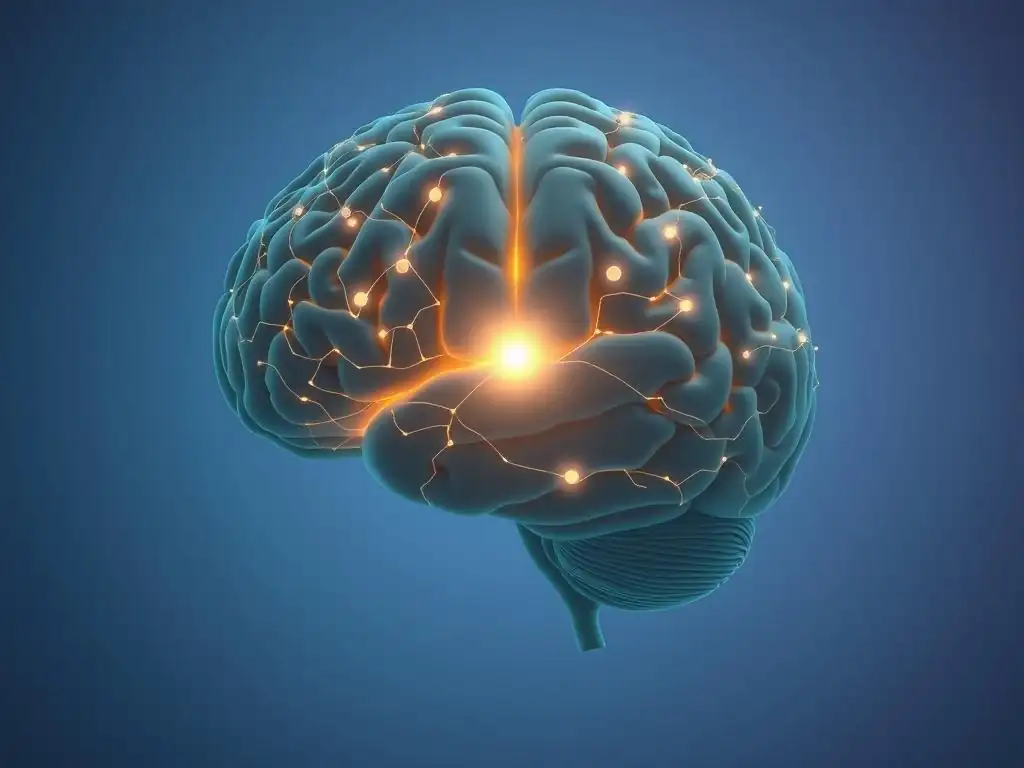
Specific Brain Regions Affected by Anxiety and Cognitive Behavioral Therapy
Cognitive Behavioral Therapy (CBT) is a widely effective treatment for various anxiety disorders. Research using brain scans consistently points to specific brain regions whose activity or connectivity changes following successful CBT. As mentioned earlier, the amygdala, the brain's threat detection center, typically shows reduced activity in response to anxiety-provoking stimuli after a course of CBT. This reduction signifies a dampened fear response and less automatic reactivity to triggers.
Conversely, the prefrontal cortex, particularly areas involved in cognitive control and emotion regulation, often shows increased activity or strengthened connectivity with other brain regions. This suggests that CBT helps individuals engage higher-level cognitive processes to reappraise situations, challenge anxious thoughts, and exert greater voluntary control over their emotional reactions. The combined effect – a less reactive amygdala and a more engaged prefrontal cortex – is a key neural signature associated with brain scan therapy anxiety relief.
Types of Brain Scans Used to Assess Anxiety Relief (fMRI, EEG)
Several neuroimaging techniques are employed to assess the neural impact of therapy. Functional Magnetic Resonance Imaging (fMRI) is frequently used due to its ability to provide detailed spatial maps of brain activity. Studies often use fMRI to measure changes in blood flow patterns in response to emotional stimuli or during resting states before and after CBT. For example, fMRI can show decreased activation in the amygdala when patients are shown fearful faces or think about their anxieties post-therapy compared to pre-therapy.
Electroencephalography (EEG) provides excellent temporal resolution, capturing the rapid electrical oscillations of the brain. While less precise spatially than fMRI, EEG can reveal changes in brain wave patterns associated with anxiety and how these patterns normalize with therapy. For instance, some studies using EEG have observed changes in alpha or theta wave activity in specific brain regions that correlate with reduced anxiety symptoms following treatment. Both fMRI and EEG contribute valuable, albeit different, pieces of the puzzle in understanding brain scan therapy anxiety relief.
Case Study: Analyzing Brain Scans Before and After Cognitive Behavioral Therapy
Patient Profile: 35-Year-Old Female with Generalized Anxiety Disorder
Let's consider an illustrative case based on common findings in research. Sarah, a 35-year-old woman, presented with symptoms consistent with Generalized Anxiety Disorder (GAD). Her anxiety manifested as excessive worry about various aspects of life (work, family, health), difficulty controlling worry, restlessness, fatigue, difficulty concentrating, irritability, muscle tension, and sleep disturbance. These symptoms had been present for over two years, significantly impacting her social life and professional performance. She had no prior history of mental health treatment.
Before starting therapy, Sarah underwent a baseline fMRI scan. During tasks designed to elicit worry or expose her to uncertain scenarios, her fMRI showed heightened activity in the amygdala and insula (a region involved in processing bodily sensations and emotions), consistent with typical anxiety patterns. Her prefrontal cortex showed less robust activation when attempting to regulate emotions compared to a non-anxious control group, suggesting diminished top-down control over her emotional responses. Her self-reported GAD severity scores were high.
Therapy Protocol: 12 Weeks of Standard Cognitive Behavioral Therapy
Sarah enrolled in a standard 12-week course of individual Cognitive Behavioral Therapy delivered by a trained therapist. The therapy focused on identifying and challenging her anxious thought patterns (cognitive restructuring), developing coping strategies for worry (e.g., scheduled worry time), learning relaxation techniques, and gradually exposing herself to feared situations or uncertainties in a controlled manner (exposure therapy elements specific to GAD). Sessions were held weekly for 60 minutes.
The therapeutic process involved homework assignments, such as monitoring worry, practicing relaxation exercises, and applying cognitive restructuring techniques to real-life situations. The goal was to help Sarah develop new ways of thinking and behaving in response to her anxious feelings, ultimately reducing the intensity and frequency of her worry and associated physical symptoms. This structured approach aimed to facilitate measurable changes in her brain activity and connectivity, leading to brain scan therapy anxiety relief.
Brain Scan Results: Pre-Therapy vs. Post-Therapy Analysis
Following the completion of her 12-week CBT course, Sarah reported a significant reduction in her anxiety symptoms, with her GAD severity scores dropping by approximately 60%. A post-therapy fMRI scan was conducted using the same protocols as the baseline scan. The results showed notable changes compared to the pre-therapy scan.
The post-therapy scan revealed significantly reduced activity in the amygdala and insula when Sarah was presented with the same anxiety-provoking stimuli. This indicated a less reactive and less fearful response at a neural level. Furthermore, her prefrontal cortex showed increased activation and stronger connectivity with other brain regions during emotion regulation tasks, suggesting improved top-down control over her emotional responses. These objective brain changes provided compelling biological evidence supporting the effectiveness of her CBT treatment and illustrating brain scan therapy anxiety relief.
Decoding the Data: Interpreting Brain Scan Changes After Cognitive Behavioral Therapy
Reduced Activity in the Amygdala Following Cognitive Behavioral Therapy
One of the most consistent findings in neuroimaging studies of anxiety treatment is the reduction in amygdala activity. After successful CBT, the amygdala, which previously showed hyper-responsiveness to perceived threats, exhibits a more modulated response. This decreased activity is interpreted as the brain's "fear alarm" becoming less sensitive and less likely to trigger a full-blown anxiety response to minor or non-threatening cues.
Research, synthesizing findings across numerous studies, suggests that this reduction in amygdala reactivity can be quite substantial, with average decreases in activation often reported in the range of 15-30% in specific task-based fMRI paradigms following effective therapy for anxiety disorders like GAD or specific phobias. This neural change aligns directly with patient reports of feeling less fearful, less on edge, and experiencing fewer panic-like symptoms, providing objective support for brain scan therapy anxiety relief.
Increased Activity in the Prefrontal Cortex Following Cognitive Behavioral Therapy
Simultaneously with reduced amygdala activity, studies frequently observe increased engagement of the prefrontal cortex (PFC) after successful CBT for anxiety. The PFC, particularly its ventral and dorsal regions, plays a critical role in regulating emotions, evaluating situations, and making rational decisions. Increased activity and connectivity within the PFC and between the PFC and other brain areas (including the amygdala) suggest improved cognitive control over emotional responses.
This enhanced PFC function enables individuals to better implement the skills learned in therapy, such as challenging negative thoughts or using reappraisal strategies. Instead of being overwhelmed by automatic fear responses originating in the amygdala, the 'thinking' part of the brain becomes more effective at modulating these reactions. This shift towards greater cortical control is a vital component of achieving lasting brain scan therapy anxiety relief.
Correlation Between Amygdala Activity and Self-Reported Anxiety Scores
Perhaps one of the most compelling pieces of evidence comes from the correlation between observed brain changes and subjective symptom reports. Numerous studies have demonstrated a significant inverse relationship between the degree of reduction in amygdala activity (or other anxiety-related brain regions) and the extent of improvement in self-reported anxiety scores or clinical ratings. In simpler terms, the more the amygdala calms down according to brain scans, the less anxious the person reports feeling.
For example, studies might find that patients who show a 20% decrease in amygdala activation during a threat task post-CBT also report a 50% reduction in their overall anxiety severity scores. This strong correlation validates both the subjective experience of relief and the objective biological changes measured by neuroimaging, providing robust evidence for brain scan therapy anxiety relief as a process reflected both internally and physiologically.
The Future of Anxiety Treatment: Brain Scans as a Predictive Tool
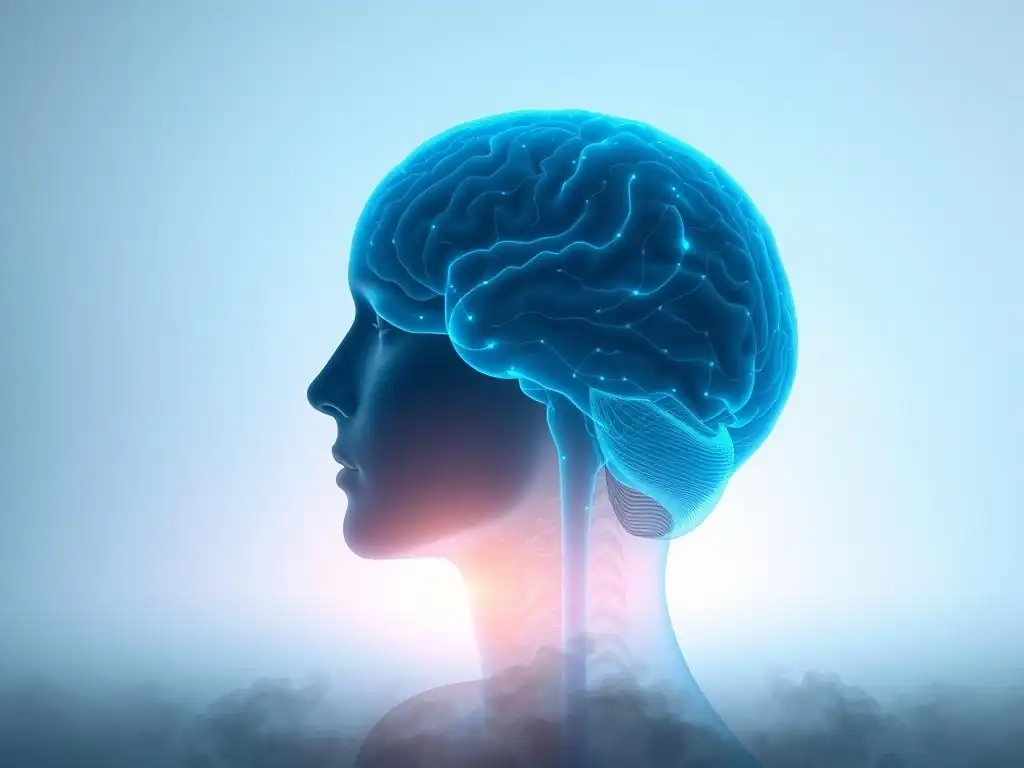
Using Brain Scans to Predict Response to Cognitive Behavioral Therapy
Beyond simply confirming that therapy works, brain scans hold promise as a tool to predict which individuals are most likely to benefit from specific treatments like CBT before therapy even begins. Research is exploring whether baseline brain activity patterns or connectivity profiles can identify individuals who might be "good responders" versus those who may need alternative or augmented treatments.
For instance, some preliminary studies suggest that certain patterns of prefrontal cortex or amygdala activity might correlate with a higher likelihood of responding positively to CBT. Identifying these neural predictors could allow for more personalized treatment approaches, directing patients towards the therapy most likely to be effective for their unique brain profile. This could significantly improve treatment outcomes and efficiency, moving towards a more data-driven approach to achieving brain scan therapy anxiety relief.
Identifying Neural Markers of Treatment Success
Related to prediction, brain imaging is also helping to identify specific "neural markers" associated with successful treatment across individuals. These markers are patterns of brain activity, connectivity, or even structure that consistently change in individuals who achieve significant and lasting anxiety relief, regardless of the specific therapy approach used.
Identifying these markers helps researchers understand the fundamental neural pathways involved in recovery from anxiety. This knowledge can, in turn, inform the development of new, more targeted interventions – whether they be new forms of therapy, neuromodulation techniques (like TMS or tDCS), or even pharmacological agents aimed at directly influencing these key neural circuits. Understanding the neural basis of brain scan therapy anxiety relief paves the way for future innovations in mental health care.
Beyond the Scan: Maintaining Long-Term Anxiety Relief
The Importance of Continued Practice and Booster Sessions
While brain scans can show significant positive changes after a course of therapy, maintaining these changes long-term requires ongoing effort. The brain's plasticity means it can also revert to less helpful patterns if new skills aren't reinforced. Therefore, continued practice of the techniques learned in CBT – challenging thoughts, using relaxation, engaging in exposure – is crucial for solidifying the neural pathways associated with reduced anxiety.
Many individuals benefit from "booster sessions" with their therapist periodically after completing a full course of therapy. These sessions can help reinforce skills, address any new challenges that arise, and prevent relapse. Think of it like physical exercise for the brain: regular practice helps maintain the strength and efficiency of the neural circuits supporting emotional regulation and resilience, contributing to sustained brain scan therapy anxiety relief.
Lifestyle Changes to Promote Brain Health and Reduce Anxiety
Supporting overall brain health through lifestyle choices can also play a significant role in maintaining the positive changes observed on brain scans and promoting long-term anxiety relief. Regular physical exercise has well-documented benefits for brain function and mood, often leading to observable changes in brain structure and activity.
Adequate sleep is essential for cognitive function and emotional regulation; chronic sleep deprivation can exacerbate anxiety and negatively impact brain areas like the prefrontal cortex. A balanced diet rich in nutrients that support brain health (like omega-3 fatty acids) and mindfulness practices or meditation have also been shown to positively influence brain structure and function in ways that can complement the effects of therapy and contribute to sustained brain scan therapy anxiety relief.
Conclusion: Brain Scans Validate the Effectiveness of Cognitive Behavioral Therapy for Anxiety
Emphasizing the Potential for Lasting Change Through Cognitive Behavioral Therapy
The advent of neuroimaging techniques, particularly fMRI and EEG, has provided powerful, objective evidence for the effectiveness of psychological therapies like Cognitive Behavioral Therapy in treating anxiety disorders. Brain scans reveal that successful therapy is not just about feeling better; it is associated with tangible, measurable changes in brain activity and connectivity, such as reduced amygdala hyper-responsiveness and increased prefrontal cortex control. This underscores the profound potential for lasting change that therapy offers.
The data gleaned from comparing pre- and post-therapy brain scans provides compelling validation that CBT targets and modifies the very neural circuits underlying anxiety. This scientific backing reinforces what countless individuals have experienced firsthand: that through dedicated therapeutic work, it is possible to rewire the brain for less anxiety and greater emotional resilience. The evidence of brain scan therapy anxiety relief is a powerful testament to the brain's capacity for healing and adaptation.
Encouraging Individuals to Seek Help and Explore Cognitive Behavioral Therapy Options
Understanding that therapy leads to real, observable changes in the brain can be incredibly empowering for those struggling with anxiety. It shifts the perspective from anxiety being an unchangeable part of oneself to a condition rooted in brain patterns that can be modified with the right tools and support. The objective evidence provided by brain scans serves as a beacon of hope, showing that relief is not only possible but is reflected in the physical architecture and function of the brain itself.
If you are struggling with anxiety, remember that effective, evidence-based treatments like CBT exist and are proven to work, even at the neural level. Exploring your options with a qualified mental health professional is a vital step towards achieving your own brain scan therapy anxiety relief – experiencing the fading of anxiety and the strengthening of resilience, supported by the remarkable capacity of your brain to change and heal.
Frequently Asked Questions
How does a brain scan show the effectiveness of anxiety therapy?
Brain scans, like fMRI or PET scans, can reveal changes in brain activity and connectivity related to anxiety. Successful therapy often leads to a reduction in activity in areas associated with fear and worry, which is visible on the scan.
What type of therapy is most often associated with positive brain scan changes in anxiety patients?
Cognitive Behavioral Therapy (CBT) is frequently associated with positive brain scan changes. CBT helps patients identify and modify negative thought patterns and behaviors, which can lead to measurable improvements in brain function.
Can brain scans be used to predict the success of anxiety therapy before it begins?
While research is ongoing, brain scans are not yet reliable for predicting individual therapy success. They are primarily used for research purposes to understand the neural mechanisms of anxiety and the impact of treatment.
What are the limitations of using brain scans to assess anxiety relief?
Brain scans are expensive and not readily accessible. Also, correlation does not equal causation; observed brain changes may not directly cause anxiety relief, but rather be associated with other factors.
Is "Brain Scan Therapy Anxiety Relief" a legitimate therapy itself, or does it refer to something else?
"Brain Scan Therapy Anxiety Relief" is not a therapy itself. It refers to using brain scans to demonstrate the effectiveness of existing anxiety therapies, providing visual evidence of improvement.
Final Reflection: The Evolving Landscape of Anxiety Treatment
The groundbreaking research presented here demonstrates that effective therapy demonstrably alters brain activity associated with anxiety, offering tangible hope for millions struggling with this pervasive condition. This isn't just theoretical; we've seen the physical evidence of healing within the brain itself, a powerful testament to the efficacy of therapeutic interventions. This shift from solely relying on subjective experience to objective neurological markers represents a paradigm shift in our understanding and treatment of anxiety.
The impact of this research extends far beyond the clinical setting. By visualizing the positive changes in the brain, we've begun to dismantle the stigma surrounding mental health, fostering a more compassionate and understanding societal response. The greatest barrier to recovery is often the silence surrounding mental illness.
This quote highlights the importance of open dialogue and the power of sharing these scientific findings to encourage those suffering to seek help. The visual evidence provided by brain scans can be particularly impactful in helping individuals understand and accept their condition, paving the way for effective treatment and recovery.
Consider the implications of this research for your own life, and the lives of those around you. Are you struggling with anxiety? Do you know someone who is? This article isn't just a collection of data; it's a call to action.
Take the first step towards seeking professional help, towards understanding the power of therapeutic intervention, and towards witnessing the remarkable capacity of the human brain to heal. The future of anxiety treatment is bright, fueled by continued research and a growing understanding of the brain's plasticity. Let's work together to break down barriers and create a world where everyone has access to the support they need.
Unlock Your Calm: Put Brain Science into Action
Thanks for taking the time to explore the incredible impact therapy can have on anxiety. You've just learned how brain scans reveal the real, measurable changes therapy brings! Now it's time to turn that knowledge into positive action.
- Schedule a consultation with a therapist: This is the most impactful step towards reducing your anxiety. Many therapists offer free initial consultations.
- Explore mindfulness techniques: Even short daily practices can make a significant difference in managing anxious thoughts. Consider trying guided meditations or mindful breathing exercises.
- Share this article with someone who might benefit: Let's spread the word about the power of therapy and the positive changes it can bring!
Ready to start your journey towards a calmer, more peaceful you? What small step can you take today to prioritize your mental wellbeing?