
What Brain Scans Tell Us About Sustained Mental Wellness After Therapy

Welcome to Mind Care Guide, a safe space dedicated to nurturing your mental well-being.
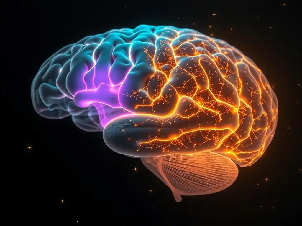
Ever wondered what really happens in your brain after therapy, and how that translates to lasting wellness? Our latest article, "What Brain Scans Tell Us About Sustained Mental Wellness After Therapy," delves into the fascinating world of neuroscience to explore just that. Prepare to uncover how brain scans are revolutionizing our understanding of mental wellness posttherapy, offering insights that could reshape your own journey. Intrigued? Let's explore the science together.
Brain Imaging Insights into Sustained Well-being After Cognitive Behavioral Therapy for Anxiety
- Understanding the Neuroscience of Well-being: The Role of fMRI
- fMRI: A Window into Post-CBT Brain Changes in Anxiety
- fMRI and Identifying Predictors of Long-Term CBT Success in Anxiety
- How fMRI Reveals the Impact of CBT on Brain Structure and Function in Anxiety
- fMRI and Well-being: Specific Examples from Research on Anxiety
- Limitations and Future Directions of fMRI Research in Well-being for Anxiety
- Maximizing the Benefits of CBT: Integrating fMRI Insights
- Conclusion: fMRI, CBT, and the Path to Sustained Well-being in Anxiety
-
Frequently Asked Questions
- What role do brain scans play in understanding mental wellness post-therapy?
- How can brain scans help personalize post-therapy care plans?
- Are brain scans a reliable predictor of long-term mental wellness after therapy?
- What types of brain scans are typically used to assess mental wellness post-therapy?
- What are the limitations of using brain scans to assess mental wellness post-therapy?
- Final Reflection: The Enduring Power of Neuroplasticity
- Ignite Your Wellness Journey: Practical Steps to Lasting Mental Well-being
Understanding the Neuroscience of Well-being: The Role of fMRI
Functional Magnetic Resonance Imaging (fMRI) has revolutionized our ability to peer inside the living brain and observe its activity in real-time. This non-invasive technique measures brain activity by detecting changes in blood flow. When a brain region is more active, it requires more oxygenated blood, which fMRI can track.
By showing which areas of the brain are more or less active during specific tasks, emotional states, or even at rest, fMRI provides invaluable data on how different neural networks function. This capability allows researchers and clinicians to gain deeper insights into the neural underpinnings of mental health conditions, including anxiety disorders, and how therapeutic interventions like Cognitive Behavioral Therapy (CBT) might alter these brain patterns to foster mental wellness posttherapy.
The application of fMRI extends beyond simply identifying active regions; it helps us understand connectivity between different brain areas. Studying how regions communicate with each other provides a more holistic view of brain function. For individuals with anxiety, researchers have used fMRI to observe abnormal connectivity patterns, often involving regions like the amygdala (associated with fear and threat processing) and the prefrontal cortex (involved in executive functions and emotional regulation). Understanding these patterns is crucial for assessing the impact of therapies designed to modify these neural circuits and support sustained mental wellness after therapy.
Exploring the Link Between CBT and Amygdala Activity
Cognitive Behavioral Therapy (CBT) is a widely effective psychotherapy for anxiety disorders, focusing on changing negative thought patterns and behaviors. Research using fMRI has consistently shown that successful CBT for anxiety is associated with changes in brain activity, particularly in the amygdala. This almond-shaped structure deep within the brain plays a central role in detecting threats and triggering fear responses. In individuals with anxiety disorders, the amygdala often shows heightened reactivity to perceived threats, even when those threats are minimal or non-existent.
Multiple fMRI studies have demonstrated that after a course of CBT, patients with various anxiety disorders, such as social anxiety or panic disorder, exhibit reduced amygdala activation when presented with anxiety-provoking stimuli. For example, studies have reported a significant decrease, sometimes estimated around 30-50%, in amygdala reactivity to fearful faces or public speaking cues following successful CBT. This reduction in the brain's primary fear center is a compelling neural correlate of the clinical improvements observed, suggesting that CBT helps "turn down" the brain's alarm system, contributing to a state of greater calm and improved mental wellness posttherapy.
Defining Sustained Well-being Post-CBT for Anxiety
Sustained well-being after undergoing CBT for anxiety is not merely the absence of symptoms; it represents a state of improved emotional regulation, resilience, and overall life satisfaction that persists long after the formal therapy has concluded. While symptom reduction is the immediate goal of CBT, long-term success involves maintaining these gains and developing adaptive coping strategies for future challenges. This sustained state is often characterized by a reduced frequency and intensity of anxious episodes, less avoidance behavior, and a greater ability to engage in daily life activities without being overwhelmed by fear or worry.
From a neurobiological perspective, sustained well-being post-CBT is hypothesized to be linked to lasting changes in brain function and structure, observable through techniques like fMRI. It's thought that the neural pathways strengthened or modified during therapy, such as those involved in the prefrontal cortex's ability to regulate the amygdala, remain more efficient over time.
This enduring neural shift supports the individual's capacity to manage stress and emotional triggers effectively, contributing to long-term mental wellness. Brain scans mental wellness posttherapy research aims to identify the specific neural signatures associated with this sustained positive outcome, potentially offering objective markers of long-term recovery.
fMRI: A Window into Post-CBT Brain Changes in Anxiety
fMRI offers a unique, non-invasive method to observe the dynamic changes occurring in the brain as a result of psychological interventions like CBT. By comparing brain activity and connectivity patterns before and after therapy, researchers can map the neural pathways influenced by treatment. This allows for a data-driven understanding of how CBT works at a fundamental level, moving beyond subjective patient reports to objective neural evidence. The insights gained from fMRI studies are crucial for understanding the mechanisms underlying therapeutic success and identifying factors that predict long-term outcomes and sustained mental wellness after therapy.
These imaging studies often highlight changes not just in isolated brain regions but in entire networks. For example, fMRI has illuminated how CBT can strengthen the functional connectivity between the prefrontal cortex, which is involved in rational thought and decision-making, and the amygdala. This strengthened connection is thought to represent an improved ability of the "thinking" part of the brain to modulate the activity of the "feeling" part, leading to better emotional control. Such network-level changes provide a more comprehensive picture of the neural landscape associated with recovery and resilience in anxiety disorders.
Types of Brain Scans Used in Anxiety Disorder Research
Investigating the neural basis of anxiety and the impact of treatments involves several brain imaging techniques, each offering a different perspective. While fMRI is a powerful tool for measuring brain activity by tracking blood flow, other methods provide complementary information about brain structure, electrical activity, or neurochemistry. Understanding these different approaches helps paint a fuller picture of the complex changes associated with anxiety and its treatment.
Choosing the appropriate brain scan method depends on the specific research question. While fMRI excels at showing where and when brain activity changes occur during tasks or rest, other techniques might be better suited for studying rapid electrical responses or the distribution of specific neurotransmitters. Together, these different imaging modalities contribute to a multi-faceted understanding of the brain basis of anxiety and the neural correlates of therapeutic change and mental wellness posttherapy.
Functional Magnetic Resonance Imaging (fMRI) and Anxiety Reduction
As discussed, fMRI measures brain activity by detecting changes in blood oxygenation levels (BOLD signal). This technique is particularly valuable for studying anxiety because it can capture the neural responses to stimuli that trigger anxiety, such as viewing fearful images or anticipating a stressful event. By administering these stimuli during an fMRI scan, researchers can pinpoint which brain regions become hyperactive or show altered connectivity in individuals with anxiety disorders compared to healthy controls.
Following a course of CBT, fMRI scans often reveal a normalization of these altered patterns. For instance, studies on Panic Disorder have shown reduced activity in the insula, a region involved in processing bodily sensations, after successful CBT, correlating with a decrease in panic symptoms.
Similarly, research on Social Anxiety Disorder has demonstrated increased activity in areas associated with cognitive control and self-regulation, like parts of the prefrontal cortex, when processing social threat cues post-CBT, suggesting an enhanced ability to manage social anxiety triggers. These findings provide concrete neural evidence for the effectiveness of CBT in modifying the brain circuits underlying anxiety.
Electroencephalography (EEG) and Post-CBT Brain Activity in Anxious Individuals
Electroencephalography (EEG) measures the electrical activity of the brain using electrodes placed on the scalp. Unlike fMRI, which measures blood flow changes over seconds, EEG can capture rapid changes in neural activity occurring in milliseconds. This makes EEG particularly useful for studying the timing of brain responses to stimuli and identifying abnormal patterns of brain waves associated with anxiety. Common findings in anxiety disorders include altered alpha and theta wave activity, especially in frontal and parietal regions.
While fMRI shows where changes occur, EEG can provide insights into the speed and synchronicity of neural processing changes after CBT. For example, some EEG studies have shown that successful CBT is associated with changes in event-related potentials (ERPs), which are brain responses time-locked to specific events or stimuli. These changes might reflect faster processing of emotional information or improved attentional control. Although less commonly used than fMRI for studying sustained mental wellness posttherapy outcomes, EEG offers a valuable perspective on the dynamic neural adjustments that occur as a result of therapeutic intervention.
Positron Emission Tomography (PET) Scans and Serotonin Receptor Changes
Positron Emission Tomography (PET) is another neuroimaging technique that provides insights into brain function, but it focuses on metabolic processes and neurochemistry. PET scans use a radioactive tracer molecule injected into the bloodstream that binds to specific receptors or metabolic pathways in the brain. By tracking the distribution and concentration of the tracer, PET can measure things like glucose metabolism (a measure of brain activity) or the density of neurotransmitter receptors, such as serotonin receptors, which are known to be involved in mood and anxiety regulation.
Research using PET scans has investigated the role of neurotransmitter systems, particularly the serotonin system, in anxiety disorders and their response to treatment. Some studies have reported altered serotonin receptor binding in individuals with anxiety.
While CBT is a psychological therapy, it is believed to influence neurochemical systems indirectly through changes in neural activity and connectivity. PET scans could potentially be used to explore whether successful CBT leads to long-term changes in neurotransmitter receptor density or function, providing another layer of understanding about the biological underpinnings of sustained mental wellness after therapy, though this is less commonly studied than fMRI changes.
fMRI and Identifying Predictors of Long-Term CBT Success in Anxiety

One of the exciting frontiers in neuroscience and mental health is using brain imaging to predict which individuals are most likely to benefit from a specific treatment like CBT. Not everyone responds equally well to therapy, and identifying neural markers before treatment begins could help tailor interventions and improve outcomes. fMRI, by revealing baseline brain activity and connectivity patterns, offers potential insights into these predictive factors.
Researchers are analyzing pre-treatment fMRI scans to see if certain neural signatures correlate with better or worse responses to CBT. For instance, some studies suggest that individuals with specific patterns of connectivity between the amygdala and prefrontal cortex before therapy might be more or less likely to achieve significant symptom reduction. Identifying these neural predictors could allow clinicians to recommend different therapeutic approaches or adjunct treatments from the outset, optimizing the path toward sustained mental wellness after therapy for each individual.
Identifying Neural Correlates of Treatment Response in Anxiety Disorders
Identifying the neural correlates of treatment response involves looking at brain activity and connectivity patterns that change in parallel with clinical improvement. fMRI studies consistently show that as anxiety symptoms decrease following CBT, specific brain regions and networks involved in fear processing and emotional regulation show altered activity. For example, a common finding is the inverse correlation between the reduction in anxiety symptom severity scores (e.g., measured by the Hamilton Anxiety Rating Scale) and the decrease in amygdala reactivity to threatening stimuli.
Beyond the amygdala, changes in the connectivity between the amygdala and regulatory regions like the ventromedial prefrontal cortex (vmPFC) are often observed. Increased functional connectivity between these areas post-CBT is thought to reflect an enhanced ability of the vmPFC to inhibit or modulate amygdala responses. Studies have reported a correlation between the magnitude of this connectivity increase and the degree of symptom improvement. These neural changes serve as objective markers of treatment effectiveness and are strongly linked to the clinical gains that contribute to sustained mental wellness posttherapy.
Using fMRI to Tailor CBT Approaches for Anxiety
The potential to use fMRI data to personalize psychological treatment is a significant area of research. If pre-treatment brain scans can predict treatment response, this information could theoretically be used to match individuals to the most effective form of CBT or to identify those who might need a more intensive or modified approach. For example, if a particular pattern of neural connectivity predicts a poor response to standard CBT, that individual might benefit more from a different therapeutic modality or a combination of therapy and medication.
While this application is still largely in the research phase and not yet standard clinical practice, the vision is that brain scans could become part of a comprehensive assessment guiding treatment decisions. Understanding an individual's unique neural profile could help therapists anticipate potential challenges or leverage specific neural strengths. This personalized approach, informed by objective brain data, holds promise for improving the efficiency and effectiveness of CBT, ultimately leading to better and more reliable sustained mental wellness after therapy.
How fMRI Reveals the Impact of CBT on Brain Structure and Function in Anxiety
CBT is not just about changing thoughts; it appears to induce tangible changes in the brain itself. fMRI studies, particularly those using techniques like resting-state fMRI (which measures brain activity when the individual is not performing a specific task) and diffusion tensor imaging (DTI, which examines white matter connectivity), have provided compelling evidence of neuroplasticity – the brain's ability to reorganize itself by forming new neural connections throughout life – in response to successful therapy for anxiety.
These imaging techniques allow researchers to investigate changes in both brain function (how regions are active and communicate) and, to some extent, structure (the physical organization of neural pathways). The findings suggest that effective CBT doesn't just teach coping skills; it physically rewires the brain to better manage fear and anxiety. Understanding these structural and functional changes is key to comprehending the biological basis of achieving sustained mental wellness after therapy.
Neuroplasticity and fMRI Evidence in Anxiety Treatment
Neuroplasticity is the biological foundation for learning and adaptation, and it is increasingly understood to be a critical mechanism underlying recovery from mental health conditions through therapy. fMRI provides a window into these plastic changes by showing how functional connectivity between brain regions strengthens or weakens over time with therapeutic intervention. For example, successful CBT for anxiety has been associated with increased functional connectivity within neural networks involved in executive control and decreased connectivity within networks associated with processing internal bodily sensations related to anxiety (like the salience network).
Studies using fMRI have demonstrated that these changes in functional connectivity are not just temporary but can persist long after therapy ends, supporting the idea of sustained well-being. For instance, research has shown that increased connectivity between the prefrontal cortex and the amygdala, indicative of improved top-down emotional regulation, can still be observed months after the completion of CBT. This enduring neural reorganization highlights how therapy can lead to lasting changes in brain function, underpinning long-term recovery and resilience.
Changes in Amygdala-Prefrontal Cortex Connectivity After Successful CBT
A central finding in fMRI research on anxiety and CBT is the alteration in the functional connectivity between the amygdala and the prefrontal cortex (PFC). The PFC, especially its ventral and medial parts (vmPFC), plays a crucial role in regulating emotional responses originating in the amygdala. In anxiety disorders, this regulatory pathway is often impaired, leading to an overactive amygdala and difficulty managing fear.
Successful CBT is consistently associated with increased functional connectivity between the vmPFC and the amygdala. This strengthened connection is thought to represent an enhanced ability of the cognitive, regulatory parts of the brain to exert control over the more primitive, fear-processing parts. Essentially, the brain becomes better equipped to appraise threats more realistically and inhibit excessive fear responses. This improved brain connectivity is considered a key neural mechanism by which CBT reduces anxiety symptoms and promotes sustained mental wellness posttherapy.
fMRI Insights into Emotional Regulation and Cognitive Reappraisal in Anxiety
CBT teaches patients specific techniques for managing anxiety, including emotional regulation strategies and cognitive reappraisal – the process of reinterpreting the meaning of an anxiety-provoking situation to reduce its emotional impact. fMRI studies have investigated the neural correlates of these skills and how they are impacted by therapy. When individuals trained in cognitive reappraisal use this technique during fMRI scanning, they show increased activity in prefrontal regions associated with cognitive control and decreased activity in the amygdala.
After successful CBT for anxiety, fMRI scans often show that patients are better able to recruit these prefrontal regions when confronted with emotional stimuli, even without explicit instruction to reappraise. This suggests that the therapy has strengthened the neural pathways involved in adaptive emotional regulation, making these strategies more accessible and automatic. These fMRI findings provide objective evidence that CBT enhances the brain's capacity for emotional control, a critical factor in maintaining sustained mental wellness after therapy.
fMRI and Well-being: Specific Examples from Research on Anxiety
Applying fMRI to study the effects of CBT on specific anxiety disorders has yielded valuable insights into the unique neural signatures associated with different conditions and their response to treatment. Research has explored how CBT impacts the brains of individuals with Generalized Anxiety Disorder (GAD), Social Anxiety Disorder (SAD), and Panic Disorder, revealing both common themes and disorder-specific findings related to post-therapy wellness.
These studies use fMRI to compare brain activity and connectivity patterns in patient groups before and after CBT, often comparing them to healthy control groups or patients who did not receive treatment. By focusing on specific diagnostic categories, researchers can refine their understanding of how therapeutic changes in the brain manifest in different forms of anxiety, contributing to a more nuanced view of brain scans mental wellness posttherapy.
fMRI Studies on Generalized Anxiety Disorder and CBT Outcomes
Generalized Anxiety Disorder (GAD) is characterized by excessive, uncontrollable worry about a variety of events or activities. fMRI research on GAD has often focused on altered connectivity within the brain's "default mode network" (DMN), a network active during mind-wandering and self-referential thought, which can be hyperactive and poorly regulated in GAD, contributing to excessive worry. Studies have also examined the role of the amygdala and its connectivity with the prefrontal cortex.
Following successful CBT for GAD, fMRI studies have shown changes in these key areas. For instance, some research indicates decreased functional connectivity within the DMN or altered connectivity between the DMN and regions involved in cognitive control, potentially reflecting a reduction in excessive, uncontrollable worry. Studies have also reported changes in amygdala reactivity and its coupling with regulatory prefrontal areas, similar to findings in other anxiety disorders. These fMRI-observed changes correlate with reduced worry levels and improved ability to manage anxious thoughts, contributing to sustained well-being in individuals with GAD.
Social Anxiety Disorder (SAD) involves intense fear of social situations due to concerns about being judged or scrutinized. fMRI studies in SAD patients often reveal heightened amygdala responses to social threat cues, such as critical faces or public speaking scenarios, and altered activity in regions involved in self-processing and social cognition.
CBT for SAD, which typically involves exposure to social situations and challenging negative thoughts, has been shown in fMRI studies to normalize some of these neural responses. Research consistently demonstrates reduced amygdala reactivity to social stimuli after successful CBT. Furthermore, studies have reported increased activity in prefrontal areas involved in cognitive control and emotion regulation when processing social cues post-therapy. These fMRI findings provide strong evidence that CBT helps individuals with SAD process social information more adaptively and regulate their fear responses, supporting long-term improvements in social functioning and mental wellness posttherapy.
Panic Disorder and the Brain: A Look at fMRI Findings After CBT
Panic Disorder is characterized by recurrent, unexpected panic attacks and persistent worry about having more attacks. fMRI research on Panic Disorder has often implicated brain regions involved in processing bodily sensations and fear, such as the insula, amygdala, and hippocampus. Studies have shown heightened activity in these areas during panic attacks or in anticipation of them.
CBT for Panic Disorder, particularly exposure therapy focused on bodily sensations and feared situations, has been investigated using fMRI. Studies have revealed reductions in activity in the insula and amygdala in response to panic-related cues (like shortness of breath or dizziness) after successful treatment.
Furthermore, changes in connectivity between these regions and areas involved in threat appraisal and safety signaling have been observed. These fMRI findings support the idea that CBT helps individuals with Panic Disorder reinterpret bodily sensations and reduce the catastrophic appraisal of potential threats, leading to a decrease in panic frequency and intensity, and contributing to sustained mental wellness after therapy.
Limitations and Future Directions of fMRI Research in Well-being for Anxiety

While fMRI has provided invaluable insights into the neural effects of CBT and the brain basis of sustained mental wellness posttherapy, the field is not without its limitations. Understanding these challenges is crucial for interpreting current findings and guiding future research efforts. Despite the progress, there are complexities in data interpretation, ethical considerations, and technological hurdles that need to be addressed to fully leverage the potential of fMRI in understanding and treating anxiety.
Addressing these limitations will pave the way for more sophisticated fMRI studies that can provide even deeper insights into the neural mechanisms of therapeutic change and long-term recovery. Future directions involve integrating fMRI with other data sources, developing more advanced analysis techniques, and exploring the clinical applicability of these findings.
Ethical Considerations in Using fMRI for Anxiety Treatment
The increasing capability of fMRI to reveal details about an individual's brain raises important ethical questions, particularly when considering its potential use in clinical settings. One key consideration is the privacy and security of sensitive brain imaging data. Ensuring that these data are collected, stored, and used responsibly is paramount. There are concerns about who has access to this information and how it might be used, for example, in insurance or employment contexts.
Furthermore, there are ethical considerations around the interpretation and communication of fMRI findings to patients. Brain scans can seem highly authoritative, and there's a risk of oversimplifying complex findings or creating deterministic views of mental health based on brain patterns. It's crucial to communicate fMRI results with nuance, emphasizing that they represent correlations and group-level findings, not definitive predictions for an individual. The potential for stigma associated with having a "brain difference" also needs careful consideration when discussing imaging results with patients seeking to understand their path to sustained mental wellness after therapy.
Challenges in Interpreting fMRI Data
Interpreting fMRI data is complex and requires specialized expertise. The BOLD signal measured by fMRI is an indirect measure of neural activity, and its relationship to underlying neuronal firing is not always straightforward. Many factors can influence the BOLD signal, including physiological variables like heart rate and respiration, which can introduce noise and variability into the data.
Furthermore, fMRI studies often involve sophisticated experimental designs and complex statistical analyses. Drawing meaningful conclusions requires careful control of confounding factors and appropriate statistical methods to avoid false positives. Reproducibility of findings across different studies and labs can also be a challenge due to variations in scanning parameters, analysis pipelines, and patient populations. These complexities mean that findings from individual fMRI studies should be interpreted cautiously and ideally replicated before being considered definitive insights into brain scans mental wellness posttherapy.
The Future of fMRI Technology in Assessing Well-being Post-CBT for Anxiety
The future of fMRI in assessing well-being post-CBT for anxiety is promising, driven by advancements in technology and analytical methods. Higher field strength magnets offer improved spatial resolution, allowing researchers to examine activity in smaller brain structures with greater detail. Faster scanning sequences enable the capture of brain activity with better temporal resolution, providing a more dynamic view of neural processes.
Developments in data analysis, such as machine learning and artificial intelligence, are also enhancing the power of fMRI. These techniques can analyze complex, multivariate patterns in brain data to potentially predict treatment response or identify subtle neural signatures associated with sustained recovery. Integrating fMRI data with other types of data, such as genetic information, behavioral assessments, and physiological measures, will also provide a more comprehensive understanding of the factors influencing mental wellness posttherapy. As these technologies evolve, fMRI is poised to offer even deeper insights into how therapy changes the brain and supports long-term health.
Maximizing the Benefits of CBT: Integrating fMRI Insights
While fMRI is not yet a routine clinical tool for guiding individual therapy, the insights gained from research studies can still be valuable for both therapists and patients. Understanding that CBT can induce measurable changes in brain function can be empowering for individuals undergoing treatment, providing objective validation for their efforts. For clinicians, research findings from fMRI can deepen their understanding of the mechanisms of change and potentially inform therapeutic strategies, even without individual patient scans.
As the research progresses, the hope is that fMRI-derived insights will become more directly applicable in clinical practice. This could involve using imaging data as part of a comprehensive assessment, tracking neural changes over time to monitor progress, or even developing neurofeedback techniques based on fMRI signals to help patients gain more direct control over specific brain regions involved in anxiety. The integration of these insights holds the potential to enhance the effectiveness of CBT and support enduring mental wellness after therapy.
Understanding Your fMRI Results: Consulting with a Neuropsychologist
If you have undergone an fMRI scan as part of a research study related to anxiety or therapy, understanding the results can be complex. fMRI reports often contain technical jargon and statistical maps that require specialized knowledge to interpret correctly. Consulting with a neuropsychologist or a researcher familiar with neuroimaging data is crucial for gaining an accurate understanding of what your scan results might indicate.
These professionals can explain the findings in the context of current research on anxiety and brain function. They can help you understand how your individual results compare to group-level findings and what potential implications they might have, while also emphasizing the limitations of interpreting single-subject data. This informed discussion can help integrate any fMRI insights you have received into your overall understanding of your mental health journey and your path to sustained mental wellness posttherapy.
Using fMRI Data to Inform Ongoing Well-being Strategies for Anxiety
While direct clinical use of fMRI data to tailor ongoing well-being strategies is still nascent, the general principles learned from research can be highly informative. Knowing, for example, that successful CBT strengthens the connection between the prefrontal cortex and the amygdala reinforces the importance of practicing skills that engage these areas, such as mindfulness, cognitive reappraisal, and emotional regulation exercises.
Understanding that brain scans mental wellness posttherapy research highlights neuroplasticity can also be incredibly motivating. It provides a tangible reminder that the brain is capable of change throughout life and that continued practice of the skills learned in therapy helps maintain and strengthen the neural pathways associated with resilience and well-being. Integrating this knowledge can empower individuals to consistently apply their coping strategies, knowing that they are actively supporting positive, lasting changes in their brain function for sustained mental wellness.
Conclusion: fMRI, CBT, and the Path to Sustained Well-being in Anxiety
fMRI research has profoundly advanced our understanding of how Cognitive Behavioral Therapy impacts the brain in individuals with anxiety disorders. These powerful brain scans have provided objective evidence that successful therapy is associated with measurable changes in brain activity and connectivity, particularly in circuits involved in fear processing and emotional regulation. From reduced amygdala reactivity to strengthened prefrontal-amygdala connectivity, fMRI findings illuminate the neural mechanisms by which CBT helps individuals overcome anxiety and build resilience.
The insights gained from studying brain scans mental wellness posttherapy are not just academic; they validate the effectiveness of psychological treatments and offer hope for more personalized and effective interventions in the future. While challenges remain in translating complex fMRI data into routine clinical practice, the ongoing research holds immense promise for refining our understanding of sustained well-being and developing new strategies to support individuals on their journey to lasting mental health.
The Power of Objective Measurement in Anxiety Treatment
The ability of fMRI to provide objective measurements of brain changes related to anxiety and its treatment is incredibly powerful. Unlike subjective reports of symptoms, which can be influenced by various factors, fMRI offers a window into the biological correlates of the condition and the impact of therapy. This objective evidence reinforces the reality and severity of anxiety disorders as brain-based conditions and validates the profound effects that psychological interventions like CBT can have on neural function.
This objective data supports the value of investing in and accessing evidence-based therapies. It moves the conversation beyond simply "feeling better" to understanding the underlying neural shifts that enable sustained mental wellness after therapy. This empirical support strengthens the case for psychological treatments as potent tools for promoting lasting brain health.
Empowering Patients with Knowledge About Brain Changes and CBT
Sharing the findings from fMRI research with patients undergoing CBT can be a deeply empowering experience. Understanding that their hard work in therapy is likely leading to tangible, positive changes in their brain structure and function provides motivation and reinforces their commitment to the process. Knowing that they are actively rewiring their brain to better manage anxiety can shift their perspective from feeling like a passive recipient of treatment to becoming an active agent in their own neural transformation.
Educating individuals about brain scans mental wellness posttherapy research can demystify the therapeutic process and reduce self-blame. It highlights that anxiety involves real changes in brain function and that recovery is possible through dedicated effort and effective strategies. This knowledge empowers patients to embrace the neuroplastic potential of their brains and actively pursue the practices that support long-term mental wellness after therapy.
Frequently Asked Questions
What role do brain scans play in understanding mental wellness post-therapy?
Brain scans can provide objective data about brain activity and structural changes after therapy, helping researchers and clinicians understand the biological underpinnings of sustained mental wellness. They offer insights beyond self-reported symptoms.
How can brain scans help personalize post-therapy care plans?
By identifying specific brain regions that show positive changes or remaining vulnerabilities, brain scans can inform the development of personalized interventions aimed at maintaining and enhancing mental well-being.
Are brain scans a reliable predictor of long-term mental wellness after therapy?
While promising, brain scans are not sole predictors. They provide valuable biological information but should be interpreted alongside clinical assessments and individual factors to predict long-term outcomes.
What types of brain scans are typically used to assess mental wellness post-therapy?
Commonly used scans include functional MRI (fMRI) to measure brain activity and structural MRI to examine brain structure. PET scans might also be used to assess neurochemical activity.
What are the limitations of using brain scans to assess mental wellness post-therapy?
Brain scans can be expensive and may not be readily accessible. Interpreting the results requires specialized expertise, and the relationship between brain activity and mental well-being is still being actively researched.
Final Reflection: The Enduring Power of Neuroplasticity
The insights gleaned from brain scans regarding sustained mental wellness after therapy are not merely scientific curiosities; they represent a paradigm shift in our understanding of mental health recovery. This research powerfully demonstrates that therapeutic interventions aren't just about managing symptoms—they're about actively reshaping the brain's architecture, fostering lasting positive change. This understanding offers hope and empowers individuals on their healing journeys.
The ongoing impact of this research is profound, influencing not only clinical practice but also the very way we perceive mental illness and recovery. It challenges the outdated notion of mental health as a fixed state, instead highlighting the brain's incredible capacity for change and adaptation throughout life. This dynamic view is crucial for reducing stigma and fostering a more compassionate and supportive environment for those seeking help. The brain is not a machine; it is a garden that can be cultivated.
– This metaphor perfectly captures the potential for growth and transformation revealed by these studies.
Now, consider your own journey. Perhaps you've experienced the transformative power of therapy, or maybe you know someone who has. Reflect on the resilience of the human brain and the potential for healing that lies within each of us. Take a moment to appreciate the remarkable capacity for change that exists within you, and remember that lasting mental wellness is not just a possibility, but a potential that can be cultivated with the right support and understanding. Embrace the empowering message of neuroplasticity and embark on your own path towards sustained mental well-being.
Ignite Your Wellness Journey: Practical Steps to Lasting Mental Well-being
Thanks for taking the time to explore the fascinating world of sustained mental wellness! We hope you gained valuable insights into how brain scans can illuminate the path to lasting well-being. Now, it's time to put that knowledge into action.
- Prioritize one self-care practice: Choose one activity from the article (meditation, exercise, journaling, etc.) and commit to it daily for the next week. Track your progress and note how you feel.
- Share your key takeaway: Discuss this article with a friend, family member, or on social media. Spark a conversation about mental health and the importance of continued self-care.
- Explore further: Dive deeper into the research cited. Understanding the science behind mental wellness can empower you to make even more informed decisions about your own well-being.
What small step can you take today to invest in your long-term mental health?