
Your Brain on a Scan: What to Expect During Mental Health Imaging Procedures

Welcome to Mind Care Guide, your sanctuary for navigating the complexities of mental well-being. Feeling overwhelmed by the prospect of mental health imaging? You're not alone. In our featured article, "Your Brain on a Scan: What to Expect During Mental Health Imaging Procedures," we break down the science behind these powerful tools, offering a simple explanation of mental health imaging explained simply and demystifying the process, so you can approach your journey with knowledge and confidence. Curious to see what these scans can reveal about your brain? Let's explore together.
Your Brain on a Scan: What to Expect During Mental Health Imaging Procedures
- Understanding Mental Health Imaging Procedures
- Types of Mental Health Imaging Techniques
- Preparing for Your Brain Scan
- What Happens During a Scan
- Understanding the Results
- Safety Considerations and Potential Risks
- Future Trends and Advancements
- Resources
- Frequently Asked Questions
- Final Reflection: Unveiling the Mind's Landscape Through Imaging
- Unlock Your Brain's Potential: Take the Next Step
Understanding Mental Health Imaging Procedures
Why Brain Imaging is Used in Mental Health Assessments
Brain imaging techniques offer a unique window into the structure and function of the brain, providing valuable insights that complement clinical assessments in mental health. While imaging alone rarely diagnoses a mental health condition, it can help researchers and clinicians understand the biological underpinnings of various disorders.
For instance, studies using structural MRI have shown that individuals with certain conditions like schizophrenia or major depressive disorder may exhibit subtle differences in brain volume or cortical thickness in specific regions compared to control groups. These findings, though often population-level trends rather than definitive individual markers, contribute to a more comprehensive understanding of the complex interplay between brain biology and mental well-being.
Furthermore, functional imaging, such as fMRI and PET scans, allows us to observe brain activity and connectivity patterns. This helps researchers explore how different brain areas communicate with each other and how these communication networks might be altered in conditions like anxiety disorders or PTSD.
For example, fMRI studies might reveal atypical activity in the amygdala (a region associated with fear) when individuals with anxiety are exposed to specific stimuli. These insights are crucial for advancing our knowledge about the mechanisms of mental health challenges and developing more targeted therapies, offering a deeper perspective on mental health imaging explained simply.
The Role of Neuroimaging in Understanding Mental Health Conditions
Neuroimaging plays a pivotal role in advancing our scientific understanding of mental health conditions beyond observable symptoms. By providing objective data about brain structure, function, and chemistry, these techniques help researchers test hypotheses about the biological basis of disorders. For example, research using PET scans has investigated dopamine receptor availability in individuals with addiction or schizophrenia, revealing potential differences that contribute to the symptoms experienced. Similarly, fMRI has been used to study neural circuits involved in emotional regulation in bipolar disorder, showing altered connectivity patterns during mood episodes.
This research is not just academic; it directly informs the development of new treatments and interventions. Understanding which brain regions or networks are implicated in a disorder can guide the creation of targeted therapies, such as transcranial magnetic stimulation (TMS) or deep brain stimulation (DBS), which aim to modulate activity in specific brain areas.
It also helps in evaluating the effectiveness of pharmacological and psychological treatments by observing how they impact brain function over time. Thus, neuroimaging is a cornerstone of modern mental health research, helping to make mental health imaging explained simply accessible to a wider audience interested in the biological basis of these conditions.
Types of Mental Health Imaging Techniques
MRI Scans: What You Need to Know
Magnetic Resonance Imaging (MRI) is a widely used imaging technique that uses strong magnetic fields and radio waves to create detailed images of the brain's structure. Unlike X-rays or CT scans, MRI does not use ionizing radiation, making it a safer option for repeated scans. The procedure involves lying on a table that slides into a large, tube-shaped scanner.
The scanner generates a powerful magnetic field, and radio waves are pulsed through the body. As the magnetic field is turned on and off and radio waves are emitted and received, different tissues in the brain respond uniquely, allowing the scanner to build up a detailed cross-sectional image. This process can take anywhere from 30 to 60 minutes, depending on the specific type of MRI being performed.
The experience inside an MRI scanner can be noisy, with loud knocking or thumping sounds as the magnetic gradients change. Earplugs or headphones are typically provided to help reduce the noise. It is crucial to remain very still during the scan to ensure the images are clear. For some individuals, the enclosed space of the scanner can feel confining, and strategies to manage claustrophobia, such as open MRI scanners or relaxation techniques, may be discussed with your doctor beforehand. Understanding these aspects is part of having mental health imaging explained simply before your appointment.
Functional MRI (fMRI) and its Application
Functional Magnetic Resonance Imaging (fMRI) is a specialized type of MRI that measures brain activity by detecting changes in blood flow. When a specific area of the brain is more active, it requires more oxygen, and blood flow to that region increases. fMRI detects these changes in blood oxygenation levels, providing a dynamic map of brain activity while a person is performing a task, resting, or experiencing stimuli. For example, a researcher might use fMRI to observe which brain regions are active when someone is processing emotional faces or making a decision.
fMRI is a cornerstone of modern cognitive neuroscience and mental health research. It allows scientists to study neural networks and connectivity patterns associated with various psychological processes and disorders. Studies using fMRI have identified differences in brain activation patterns in individuals with depression during emotional processing tasks, or altered connectivity in the default mode network (a brain network active during rest) in conditions like autism spectrum disorder. While fMRI is primarily a research tool and not typically used for routine clinical diagnosis, its insights are invaluable for understanding the functional basis of mental health conditions.
Structural MRI and Identifying Brain Differences
Structural MRI focuses on creating detailed images of the brain's anatomy, providing information about its shape, size, and tissue composition. This technique allows researchers and clinicians to identify any structural abnormalities or differences in brain regions. For example, structural MRI can reveal the presence of tumors, lesions, or areas of atrophy. In the context of mental health research, structural MRI studies have compared brain volumes in large populations of individuals with specific disorders versus healthy controls.
Findings from structural MRI research have shown, for instance, that individuals with schizophrenia may have slightly enlarged ventricles (fluid-filled spaces in the brain) or reduced gray matter volume in certain areas like the prefrontal cortex or hippocampus. Similarly, studies on major depressive disorder have sometimes reported reduced hippocampal volume, particularly in chronic cases.
It's important to note that these findings are statistical trends observed across groups and do not mean every individual with a condition will show these specific structural differences. However, these studies contribute significantly to our understanding of how brain structure might be related to vulnerability to or the manifestation of mental health conditions.
PET Scans: Exploring Brain Activity
Positron Emission Tomography (PET) scans are imaging tests that use a small amount of a radioactive tracer to measure metabolic activity in the body, including the brain. Before the scan, a radioactive substance (often a form of glucose or a molecule that binds to specific receptors or transporters) is injected into a vein.
This tracer travels through the bloodstream and accumulates in areas of high metabolic activity, which in the brain often correlates with neural activity. As the tracer decays, it emits positrons, which are detected by the PET scanner, allowing a computer to create detailed images of brain activity or the distribution of specific molecules like neurotransmitter receptors.
PET scans are particularly useful for studying brain function at a molecular level. In mental health research, PET is frequently used to investigate neurotransmitter systems, such as dopamine, serotonin, or opioid receptors, which are implicated in various conditions like depression, addiction, and schizophrenia.
For example, PET scans can help determine the density or availability of dopamine receptors in different brain regions, providing insights into how these systems might be altered in psychiatric disorders. The scan itself typically takes about 30-90 minutes, and there is a waiting period after the tracer injection (usually 30-60 minutes) to allow it to distribute throughout the brain.
How PET Scans Help Understand Neurotransmitter Activity
PET scans provide a unique ability to visualize and quantify the activity of specific neurotransmitter systems in the living brain. Neurotransmitters are chemical messengers that play a crucial role in regulating mood, cognition, behavior, and many other brain functions; imbalances or dysfunctions in these systems are thought to contribute to various mental health conditions. By using tracers that selectively bind to specific neurotransmitter receptors or transporters, PET can measure their density, availability, or how they are affected by medications or stimuli.
For instance, PET studies using tracers that bind to dopamine receptors have provided valuable insights into conditions like Parkinson's disease (which involves dopamine-producing neurons) and schizophrenia (where dopamine system dysregulation is hypothesized). Researchers can compare receptor availability in individuals with these conditions to healthy controls to identify potential differences. Similarly, PET has been used to study serotonin transporters in depression or opioid receptors in addiction, helping to understand the neurobiological basis of these disorders and evaluate the impact of treatments. This molecular-level view is a key aspect of mental health imaging explained simply when discussing PET technology.
CT Scans: When Are They Used?
Computed Tomography (CT) scans, also known as CAT scans, use a series of X-ray images taken from different angles around the body to create cross-sectional images. A computer then processes these images to create detailed views of bones, blood vessels, and soft tissues. While MRI provides more detailed images of soft tissues like the brain, CT scans are faster and more readily available in emergency settings.
In the context of mental health, CT scans are not typically used for diagnosing psychiatric conditions themselves, as they don't show brain activity or subtle tissue differences as clearly as MRI or PET. However, CT scans may be used in mental health assessments to rule out other potential medical causes for symptoms that might mimic psychiatric disorders.
For example, if someone presents with sudden behavioral changes, confusion, or new onset of psychosis, a CT scan might be performed to check for structural issues like a brain tumor, stroke, head injury, or hydrocephalus (excess fluid in the brain) that could be causing the symptoms. It's a quick way to get a structural overview, especially in urgent situations.
EEG (Electroencephalography): Measuring Brain Waves
Electroencephalography (EEG) is a non-invasive technique that measures the electrical activity of the brain. Small electrodes are placed on the scalp, which detect the electrical signals generated by neurons communicating with each other. These signals are then amplified and recorded as wave patterns on a computer. Different brain states (e.g., sleep, wakefulness) and activities are associated with distinct patterns of brain waves (e.g., alpha, beta, theta, delta).
In mental health, EEG is primarily used in clinical settings to diagnose or monitor conditions related to abnormal electrical activity, such as epilepsy, which can sometimes present with symptoms that might be mistaken for psychiatric issues. It can also be used in sleep studies to assess sleep disorders, which are often co-occurring with mental health conditions like depression or anxiety.
In research, EEG provides excellent temporal resolution (measuring changes in milliseconds), allowing scientists to study the timing of brain responses to stimuli. While it doesn't provide detailed spatial images like MRI or PET, EEG is valuable for understanding the dynamic nature of brain activity.
Preparing for Your Brain Scan
Before the Scan: Discussing with Your Doctor
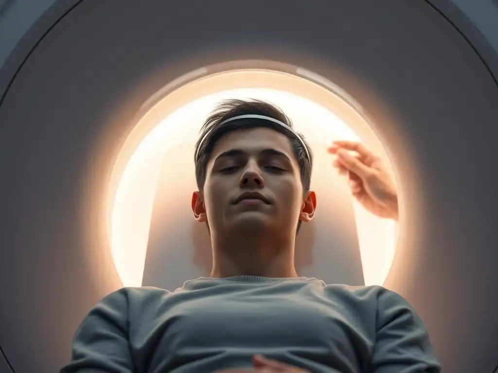
Before undergoing any brain imaging procedure, it is essential to have a thorough discussion with your doctor or mental health professional. They will explain why the scan is being recommended, what type of scan it will be, and what information they hope to gain from it.
This is your opportunity to ask questions about the procedure, any potential risks, and what the results might mean for your specific situation. Be sure to inform your doctor about your medical history, including any allergies, pre-existing conditions, medications you are taking, and whether you have any metal implants or devices in your body (this is particularly crucial for MRI).
It's also important to discuss any anxieties you might have about the scan, especially if you are claustrophobic or sensitive to loud noises. Your doctor can offer strategies or potentially prescribe a mild sedative if necessary to help you feel more comfortable during the procedure. They will also provide specific instructions regarding food and drink restrictions, medication adjustments, or other preparations required for your particular scan (e.g., for a PET scan, you might need to fast for several hours beforehand). This conversation ensures you are well-prepared and understand mental health imaging explained simply in the context of your care.
What to Wear and What to Avoid
Specific instructions regarding clothing and personal items will be provided before your scan, but general guidelines apply. For most brain imaging scans, particularly MRI, it is best to wear comfortable clothing that is free of metal zippers, buttons, snaps, or metallic threads. Sweatpants, leggings, or shorts and a t-shirt are often good choices. You will likely be asked to remove all jewelry, watches, glasses, hairpins, and any other metallic objects before entering the scanning room.
For MRI scans, you will also need to remove items containing metal like hearing aids, removable dental work, and potentially certain types of makeup or temporary tattoos that contain metallic particles. It is crucial to inform the technologist if you have any permanent metal in or on your body, such as pacemakers, cochlear implants, joint replacements, metal plates, screws, surgical clips, or even shrapnel.
For PET scans, while metal isn't a safety concern in the same way as MRI, you'll still be asked to remove jewelry and items that could interfere with image quality. Following these instructions is vital for both your safety and the clarity of the scan images.
Understanding Consent
Before any medical procedure, including brain imaging, you will be asked to provide informed consent. This means that a healthcare professional will explain the nature of the procedure, its purpose, potential benefits, risks, and alternatives. You will have the opportunity to ask questions and should feel fully informed before agreeing to proceed. For brain imaging scans, this discussion will cover details specific to the type of scan, such as the use of magnetic fields and radio waves (MRI), radioactive tracers (PET), or X-rays (CT).
You have the right to understand why the scan is being done, what the technologist will be doing, how long it will take, and what you might feel or hear. For scans involving contrast agents or radioactive tracers, the consent process will include information about the substance being administered and any associated, albeit rare, risks. Providing consent indicates that you understand the procedure and agree to undergo it voluntarily. If you are under 18 or unable to provide consent, a parent, guardian, or legally authorized representative will do so on your behalf.
What Happens During a Scan
The Scanning Environment
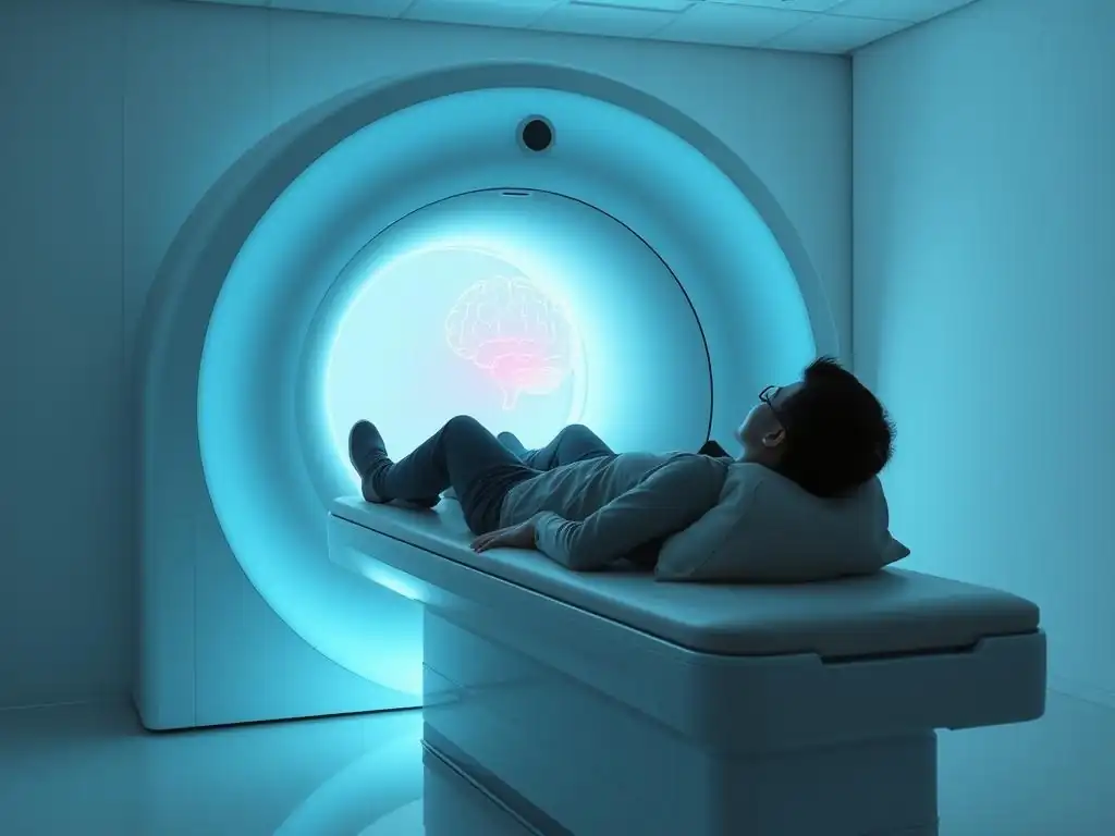
The environment for a brain imaging scan can vary slightly depending on the type of scanner. For MRI and CT scans, you will typically be in a room containing the large, donut-shaped or tube-shaped scanner machine. The room is often kept cool to ensure the equipment functions properly. For PET scans, there might be a separate room for the tracer injection and a waiting area before you proceed to the scanner room.
The scanning room is a controlled environment designed to ensure the safety and quality of the images. For MRI rooms, strict protocols are followed to prevent metal objects from entering due to the powerful magnetic field. The technologist operating the scanner will be in a separate control room, often with a window allowing them to see you during the procedure. You will be able to communicate with the technologist via an intercom system. Understanding this setting can help demystify mental health imaging explained simply.
The Procedure
Once you are positioned correctly on the scanner table, the technologist will move you into the scanner. For a brain scan, your head will be placed in a special cradle or coil that helps stabilize it and improve image quality. Depending on the scan type, you may receive an injection of a contrast agent (for some MRI or CT scans) or a radioactive tracer (for PET scans) beforehand or during the procedure.
During the scan itself, you will need to remain as still as possible. The technologist will give you instructions via the intercom, such as when to hold your breath (less common for brain scans but possible for some sequences) or if they need you to adjust your position slightly.
For MRI, you will hear loud noises; for PET and CT, the machine makes less noise but you might hear whirring or clicking sounds as the scanner rotates. The duration of the scan varies: CT scans are typically very fast (a few minutes), MRI scans usually take 30-60 minutes, and PET scans can take 30-90 minutes after the tracer has been administered and distributed.
Communicating with the Technologist
You are not alone during the scan. A technologist will be monitoring you from the control room throughout the entire procedure. Most scanning rooms are equipped with an intercom system, allowing you to speak to the technologist at any time. You will also likely be given a call button or squeeze ball to signal if you need assistance or wish to stop the scan due to discomfort or anxiety.
Do not hesitate to use the intercom or call button if you are experiencing significant discomfort, pain, or anxiety. The technologist is there to ensure your safety and comfort. They can offer reassurance, check on you, and, if necessary, pause or stop the scan. Clear communication is key to a successful and less stressful imaging experience.
Understanding the Results
How Results are Interpreted
Once the scan is complete, the raw data is processed by a computer to create detailed images. These images are then interpreted by a radiologist or a nuclear medicine physician (for PET scans), who is a medical doctor specializing in reading medical images. They will examine the images for any structural abnormalities, areas of altered activity, or other findings relevant to the reason the scan was performed. The radiologist will then generate a written report summarizing their findings.
It's important to remember that interpreting brain imaging in the context of mental health is complex. As mentioned earlier, imaging often shows group-level differences or provides insights into brain function rather than offering a definitive diagnosis for individual psychiatric conditions. The radiologist's report will describe the physical findings on the scan; correlating these findings with your clinical symptoms and history is the role of your treating physician or mental health professional.
Consulting with a Mental Health Professional
The radiologist's report will be sent to the doctor who ordered the scan, typically your psychiatrist, psychologist, or other mental health professional. They will review the imaging report in conjunction with your clinical evaluation, history, symptoms, and other test results. This integrated approach is crucial because, as mental health imaging explained simply, scan findings are just one piece of the puzzle.
Your mental health professional will discuss the results with you, explaining what the scan showed and what it means in the context of your specific situation and diagnosis (or lack thereof). They will clarify if any findings on the scan are considered normal variations or if they are potentially related to your symptoms. They will also discuss whether the scan results provide any new information that might impact your treatment plan.
Next Steps After Receiving Results
Based on the scan results and your overall clinical picture, your mental health professional will determine the appropriate next steps. If the scan revealed any significant findings unrelated to mental health (e.g., a cyst or vascular issue), you might be referred to a neurologist or other specialist for further evaluation or treatment.
If the scan results align with or provide further insight into a potential mental health condition, your doctor may adjust your treatment plan, recommend specific therapies, or suggest further assessments. Sometimes, the scan may show no significant abnormalities, which can also be valuable information, helping to rule out certain biological causes for your symptoms and focusing the treatment on other approaches. Your mental health professional will work with you to decide the best course of action moving forward, ensuring mental health imaging explained simply contributes positively to your care journey.
Safety Considerations and Potential Risks
Radiation Exposure: Minimizing Risks
For imaging techniques that use ionizing radiation, such as CT scans and PET scans, there is a small associated risk from radiation exposure. The amount of radiation used in a single diagnostic scan is generally low, comparable to the amount of natural background radiation a person is exposed to over several months or a year. Medical professionals follow strict guidelines to minimize radiation dose, using the lowest amount necessary to obtain clear images.
Radiologists and technologists adhere to the principle of "as low as reasonably achievable" (ALARA) when performing scans. This means optimizing scan parameters to reduce exposure while maintaining image quality. The risks from these low doses are considered very small compared to the potential benefits of obtaining crucial diagnostic information. However, it is important to inform your doctor if you are pregnant or suspect you might be, as radiation exposure can be harmful to a developing fetus.
MRI Safety: Screening for Metal
MRI scans do not use ionizing radiation but rely on powerful magnetic fields. The primary safety concern with MRI is the presence of metal in or on the body. The strong magnetic field can attract ferromagnetic objects (containing iron) with significant force, potentially causing injury or damaging medical devices. Therefore, a thorough screening process is mandatory before anyone enters the MRI scanning room.
You will be asked to complete a detailed questionnaire about any metal implants, devices, or foreign bodies you might have. Examples include pacemakers, defibrillators, cochlear implants, aneurysm clips, certain types of surgical clips, metal plates, screws, pins, shrapnel, and even some tattoos or permanent makeup that contain metallic pigments. Some implants are "MRI safe" or "MRI conditional," meaning they are designed to be safe in an MRI environment under specific conditions.
It is crucial to be completely honest and thorough during this screening process and provide documentation about your implants if possible. If there is any doubt about the safety of an implant or foreign body, an MRI may not be possible.
Addressing Anxiety
Undergoing a brain scan can be a source of anxiety for many people, particularly those with claustrophobia or general medical anxiety. The enclosed space of the scanner (especially traditional MRI machines), the loud noises, and the need to remain still can be challenging. Healthcare providers are aware of these concerns and have strategies to help.
Before the scan, discuss any anxieties with your doctor or the technologist. They might suggest relaxation techniques, guided imagery, or listening to music (often possible during MRI scans if headphones are provided). For significant claustrophobia, options like open MRI scanners (which are less enclosed) may be available, or your doctor might prescribe a mild anti-anxiety medication to take before the appointment. Knowing what to expect, having mental health imaging explained simply, and feeling heard about your concerns can significantly reduce anxiety surrounding the procedure.
Future Trends and Advancements
Emerging Technologies
The field of neuroimaging is constantly evolving, with researchers developing new techniques and refining existing ones to gain even deeper insights into brain function and structure. One area of advancement is ultra-high field MRI (e.g., 7T MRI), which uses even stronger magnetic fields than clinical scanners (typically 1.5T or 3T) to produce images with higher resolution and better contrast, allowing for the visualization of finer brain details.
Another area is the development of faster scanning sequences for MRI and more sensitive tracers for PET, reducing scan times and improving image quality. Hybrid imaging systems, such as integrated PET/MRI scanners, are also becoming more common, allowing simultaneous acquisition of functional and anatomical data, providing a more complete picture. Techniques like Magnetoencephalography (MEG), which measures magnetic fields produced by electrical activity in the brain, are also being used in research to study the timing and localization of brain activity with high precision.
Artificial Intelligence in Brain Scan Analysis
Artificial intelligence (AI), particularly machine learning, is playing an increasingly significant role in the analysis of brain imaging data. The sheer volume and complexity of data generated by modern scanners can be challenging for human interpretation alone. AI algorithms can be trained to identify subtle patterns, correlations, or anomalies in imaging data that might be difficult for the human eye to detect.
In mental health research, AI is being used to analyze large datasets of brain scans to identify potential biomarkers for specific conditions, predict treatment response, or differentiate between subtypes of disorders. For example, machine learning models are being developed to analyze structural MRI scans to help predict the likelihood of developing psychosis in high-risk individuals or to classify subtypes of depression based on brain connectivity patterns from fMRI.
While still largely a research tool, AI has the potential to enhance the accuracy and efficiency of brain scan interpretation in the future, contributing to a more personalized approach to mental healthcare and helping to make complex mental health imaging explained simply through data-driven insights.
Resources
Frequently Asked Questions
What is mental health imaging and why is it used?
Mental health imaging uses brain scans like MRIs or PET scans to assess brain structure and function. This helps doctors understand the biological basis of mental health conditions and aid in diagnosis and treatment planning.
What are some common types of brain scans used in mental health?
Common types include MRI (Magnetic Resonance Imaging), which shows detailed brain structure, and PET (Positron Emission Tomography), which visualizes brain activity. fMRI (functional MRI) shows brain activity in response to stimuli.
Are mental health imaging procedures painful or invasive?
Most mental health imaging procedures, like MRI and PET scans, are non-invasive and painless. You may experience some discomfort from lying still for an extended period, but there is no surgery or physical intrusion.
Can brain scans diagnose all mental health conditions definitively?
Brain scans are not a standalone diagnostic tool. They provide valuable information, but diagnoses are based on a comprehensive assessment including clinical interviews, symptom evaluation, and medical history.
How can I prepare for a mental health imaging appointment?
Preparation varies depending on the scan type, but generally involves avoiding caffeine or alcohol beforehand. You might be asked to remove metal objects and inform the technician about any medical implants or allergies.
Final Reflection: Unveiling the Mind's Landscape Through Imaging
The intricate workings of the human brain, once a complete mystery, are now increasingly accessible through the lens of advanced imaging technologies. Understanding these procedures is crucial, not only for those seeking diagnosis and treatment but also for fostering a more informed and empathetic approach to mental health. The stigma surrounding mental illness often stems from a lack of understanding; demystifying the diagnostic process is a vital step towards breaking down these barriers.
The journey into the brain, as depicted in this article, reveals a fascinating and complex world. The ability to visualize brain structures and activity offers incredible potential for improving mental healthcare. It's humbling to consider the sheer power of these technologies, and the impact they have on individuals navigating the challenges of mental illness. The greatest discovery of all time is that a person can change his future by merely changing his attitude.
– Oprah Winfrey. This sentiment rings true in the context of mental health; with better understanding and access to diagnostic tools, individuals can take control of their journey towards wellness.
Now, consider your own understanding of mental health. What preconceived notions have you held? How has this exploration of mental health imaging shifted your perspective? I urge you to share this information with others, to help spread awareness and reduce the stigma surrounding mental illness. Let us collectively work towards a future where access to advanced diagnostic tools is readily available, and where mental health is treated with the same care and understanding as physical health.
Unlock Your Brain's Potential: Take the Next Step
Thanks for exploring the fascinating world of mental health imaging with us! We hope you gained valuable insights into what these procedures entail. Now, it's time to put that knowledge to use.
- Schedule a consultation with your doctor: Discuss whether mental health imaging is right for you and explore the potential benefits. This is the most important step toward understanding your brain's unique needs.
- Share this article with someone who might find it helpful: Increase awareness and understanding of mental health imaging among your friends and family.
- Explore additional resources online about specific imaging techniques mentioned in this article; learn more about the different tools and approaches available.
What questions do you still have about mental health imaging and its role in improving your well-being? Let's start a conversation in the comments!