
Beyond the Scan: What MRI Reveals About Depression and Anxiety

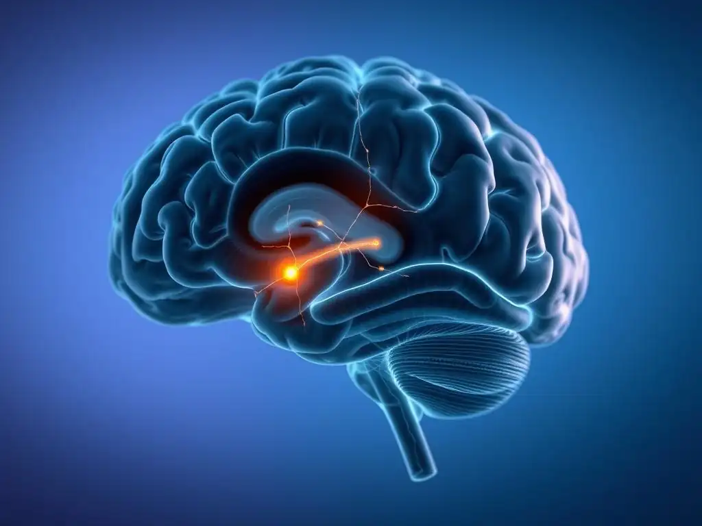
Welcome to Mind Care Guide, a space dedicated to understanding and nurturing your mental well-being. Feeling lost in the complexities of depression and anxiety? Imagine peering inside the very mechanisms of your mind. Our article, "Beyond the Scan: What MRI Reveals About Depression and Anxiety," delves into how MRI depression anxiety symptoms are increasingly understood through cutting-edge neuroscience. Curious to know what these scans are unveiling about your mental state and how it can lead to better treatments? Let's explore this fascinating frontier together.
Beyond the Scan: What MRI Reveals About Depression and Anxiety
- Neuroscience of Mental Health: Exploring Brain Imaging
- MRI and Depression: Identifying Neurological Correlates
-
MRI and Anxiety Disorders: Visualizing the Anxious Brain
- Identifying Brain Regions Involved in Anxiety: An MRI Perspective
- fMRI Studies on Anxiety: Mapping Neural Circuits of Fear and Worry
- MRI-Detected Differences in Amygdala Activity in Anxiety
- The Impact of Anxiety on Brain Structure: Insights from MRI
- Using MRI to Understand Different Types of Anxiety Disorders
- MRI and Symptom Presentation: Connecting Brain Changes to Clinical Presentation
- Limitations of MRI in Mental Health
- The Future of MRI in Mental Health Care
- Conclusion: MRI as a Tool for Understanding and Addressing Depression and Anxiety
-
Frequently Asked Questions
- What role does an MRI play in understanding depression and anxiety symptoms?
- Can an MRI scan definitively diagnose depression or anxiety?
- What specific brain areas are often examined in MRI studies related to depression and anxiety?
- How can MRI findings potentially improve treatment for depression and anxiety?
- Are MRI scans routinely recommended for individuals experiencing depression or anxiety symptoms?
- Final Reflection: Unveiling the Silent Struggle: MRI's Promise in Understanding Depression and Anxiety
- Unlock Your Mental Wellbeing: Next Steps to Understanding Anxiety & Depression
Neuroscience of Mental Health: Exploring Brain Imaging
Unveiling the Brain's Secrets: How MRI Works
Magnetic Resonance Imaging (MRI) is a non-invasive neuroimaging technique that uses powerful magnetic fields and radio waves to create detailed images of the brain's structure and function. It works by aligning the hydrogen atoms within the body's water molecules using a strong magnetic field. A radiofrequency pulse is then applied, which knocks these atoms out of alignment.
When the pulse is turned off, the atoms realign, releasing energy that is detected by the scanner. Different tissues release energy at different rates, allowing the MRI machine to differentiate between various brain structures, such as gray matter, white matter, and cerebrospinal fluid, and even observe changes in blood flow, which indicates neural activity.
The detailed images produced by MRI provide researchers and clinicians with unprecedented views of the living brain. Unlike X-rays or CT scans, MRI does not use ionizing radiation, making it safer for repeated scans, which is particularly valuable in longitudinal studies tracking changes over time. This capability allows for in-depth exploration of the complex architecture of the brain and how it might be altered in conditions like depression and anxiety disorders, offering insights into the biological underpinnings of MRI depression anxiety symptoms.
The Role of Brain Imaging in Understanding Mental Health Conditions
Brain imaging techniques, especially MRI, have revolutionized our understanding of mental health conditions by allowing us to visualize and measure structural and functional abnormalities in the brain. Before advanced imaging, understanding mental disorders relied heavily on behavioral observations, patient reports, and post-mortem studies. Now, techniques like MRI provide a window into the living brain, enabling researchers to investigate neural circuits, identify potential biomarkers, and explore the biological correlates associated with various psychiatric conditions.
By examining differences in brain structure (like volume or cortical thickness) and function (like activity levels or connectivity patterns) between individuals with and without conditions such as depression and anxiety, researchers can gain insights into the neurological basis of these disorders. This research helps to move beyond purely psychological models, providing evidence for the biological components of mental illness. Understanding these brain changes is crucial for developing more targeted and effective treatments and potentially for improving the diagnosis and prediction of MRI depression anxiety symptoms.
MRI vs. Other Brain Imaging Techniques
While MRI is a powerful tool, other brain imaging techniques also play significant roles in neuroscience and mental health research. Computed Tomography (CT) scans use X-rays to create cross-sectional images and are excellent for visualizing bone and detecting acute issues like bleeding or tumors, but they offer less detailed soft tissue contrast compared to MRI and involve radiation exposure.
Positron Emission Tomography (PET) and Single-Photon Emission Computed Tomography (SPECT) use radioactive tracers to measure metabolic activity, blood flow, or neurotransmitter distribution, providing valuable insights into brain function at a molecular level, but they have lower spatial resolution than MRI and involve radiation.
Electroencephalography (EEG) and Magnetoencephalography (MEG) measure the brain's electrical and magnetic activity directly from the scalp, offering excellent temporal resolution (capturing rapid changes in brain activity) but with lower spatial resolution compared to MRI. Functional Near-Infrared Spectroscopy (fNIRS) measures changes in blood oxygenation using infrared light, is portable and less sensitive to movement than fMRI, but is limited to studying cortical regions near the scalp.
MRI, particularly functional MRI (fMRI), strikes a balance by offering good spatial resolution to pinpoint activity in specific brain regions and reasonable temporal resolution to observe changes over seconds, making it uniquely valuable for studying the neural networks involved in the complex MRI depression anxiety symptoms.
MRI and Depression: Identifying Neurological Correlates
Structural MRI Findings in Major Depressive Disorder (MDD)
Structural MRI studies have consistently identified several brain regions that show alterations in individuals with Major Depressive Disorder (MDD). One of the most frequently reported findings is reduced volume in the hippocampus, a brain structure critical for memory and emotion regulation. Numerous studies have observed hippocampal volume reductions in depressed patients, with some meta-analyses suggesting an average reduction of around 10-15% compared to healthy controls, particularly in cases of recurrent or chronic depression. This structural change is thought to contribute to cognitive deficits often seen in depression, such as memory problems and difficulty with emotional processing.
Beyond the hippocampus, structural MRI research also points to changes in areas of the prefrontal cortex (PFC), particularly the ventromedial and dorsolateral PFC. These areas are involved in executive functions, decision-making, and mood regulation. Studies have reported decreased gray matter volume or cortical thickness in these regions in individuals with MDD. For instance, some research indicates a correlation between the severity of depressive MRI depression anxiety symptoms and the degree of volume reduction in certain PFC subregions, suggesting that structural integrity in these areas is crucial for maintaining emotional well-being.
Functional MRI (fMRI) and Brain Activity in Depression
Functional MRI (fMRI) measures brain activity by detecting changes in blood flow (specifically, the blood-oxygen-level-dependent or BOLD signal). In depression research, fMRI has revealed altered activity in several key brain networks. A common finding is dysregulation in the default mode network (DMN), a network active when the brain is at rest or engaged in internal thought processes. Studies often show increased connectivity or hyperactivity within the DMN in depressed individuals, which is hypothesized to relate to excessive rumination and self-focus, prominent MRI depression anxiety symptoms.
Furthermore, fMRI studies often highlight abnormal activity in regions associated with emotional processing and reward. The amygdala, involved in processing fear and emotional salience, is sometimes found to have heightened activity in depressed individuals, particularly when processing negative stimuli.
Conversely, areas in the reward circuit, such as the ventral striatum and orbitofrontal cortex, often show reduced activity or connectivity in response to positive stimuli, which aligns with the symptom of anhedonia (loss of interest or pleasure) frequently reported in MDD. These functional changes observed via fMRI provide a dynamic view of how the depressed brain processes information and experiences emotions.
Changes in Brain Volume and Gray Matter Density in Depression
Structural MRI allows for detailed analysis of brain volume and gray matter density, offering insights into the physical architecture of the brain in depression. As mentioned, hippocampal volume reduction is a notable finding, often linked to stress exposure and the duration of illness. Reductions in gray matter density or volume have also been reported in other areas, including parts of the anterior cingulate cortex (ACC), which plays a role in conflict monitoring and emotional regulation, and the insula, involved in interoception and emotional awareness.
For example, some studies using voxel-based morphometry (VBM), an MRI analysis technique, have identified reduced gray matter density in specific subregions of the ACC and insula in individuals with MDD compared to healthy controls. These structural alterations are not uniform across all depressed individuals, and factors like age, illness duration, and treatment history can influence the observed changes. However, the consistent findings across multiple studies regarding regions like the hippocampus and prefrontal cortex underscore the notion that depression is associated with tangible changes in brain structure, which may contribute to the range of MRI depression anxiety symptoms experienced.
MRI Biomarkers for Treatment Response in Depression
Researchers are actively investigating whether MRI findings can serve as biomarkers to predict how well an individual with depression might respond to specific treatments, such as psychotherapy or medication. For instance, some studies have explored baseline functional connectivity patterns using fMRI to predict response to antidepressant medication. Preliminary findings suggest that connectivity within certain networks, like the salience network or the cognitive control network, might predict treatment outcomes.
Specific structural features, such as the volume of certain brain regions or the integrity of white matter tracts (assessed using diffusion tensor imaging, a type of MRI), are also being studied as potential predictors. While this research is still largely experimental, the goal is to move towards personalized medicine, where an MRI scan could potentially help clinicians choose the most effective treatment for a patient based on their unique brain profile.
For example, one line of research suggests that individuals with lower baseline connectivity between the subgenual ACC and the rest of the DMN might be less likely to respond to cognitive behavioral therapy (CBT), potentially guiding treatment selection based on MRI depression anxiety symptoms and underlying neural patterns.
Exploring Brain Connectivity in Depression with MRI
Beyond looking at the activity or size of individual brain regions, MRI, particularly resting-state fMRI, is invaluable for studying functional connectivity – how different brain areas communicate with each other. Depression is increasingly understood as a disorder involving disrupted communication within and between large-scale brain networks. Studies consistently show altered connectivity in networks such as the DMN, the central executive network (CEN), and the salience network (SN).
For example, studies often report increased connectivity within the DMN and decreased connectivity between the DMN and the CEN in depressed individuals. The CEN is involved in goal-directed thought and external focus, and reduced connectivity between the DMN (internal focus) and CEN (external focus) might contribute to difficulties disengaging from negative self-referential thoughts, a core feature of MRI depression anxiety symptoms.
The SN, which detects and integrates salient internal and external stimuli, is also frequently implicated, with studies showing abnormal connectivity patterns within this network and between the SN and other networks. These findings highlight that depression is not just about problems in one brain area but involves widespread disruptions in how different parts of the brain work together.
MRI and Anxiety Disorders: Visualizing the Anxious Brain
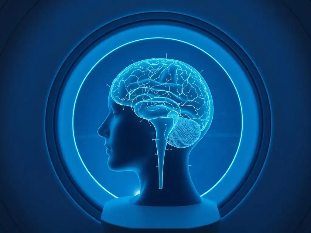
Identifying Brain Regions Involved in Anxiety: An MRI Perspective
MRI research has been crucial in identifying and characterizing the brain regions and circuits that are hyperactive or altered in various anxiety disorders, such as Generalized Anxiety Disorder (GAD), Social Anxiety Disorder (SAD), Panic Disorder, and Post-Traumatic Stress Disorder (PTSD). A central player identified by MRI studies is the amygdala, a small, almond-shaped structure deep within the temporal lobe that processes fear and threat. Studies consistently show heightened reactivity or volume changes in the amygdala in anxious individuals.
Other key regions frequently implicated in anxiety by MRI include the prefrontal cortex (PFC), particularly the medial PFC and orbitofrontal cortex, which are involved in regulating emotions and assessing threat, and the anterior cingulate cortex (ACC), involved in conflict monitoring and error detection. The insula, which processes bodily sensations and emotional awareness, also shows altered activity. These findings suggest that anxiety disorders involve a complex interplay between regions that detect threat and those that regulate emotional responses, contributing to the diverse MRI depression anxiety symptoms observed.
fMRI Studies on Anxiety: Mapping Neural Circuits of Fear and Worry
Functional MRI (fMRI) has been particularly effective in mapping the neural circuits involved in fear and worry, the hallmarks of anxiety. When individuals with anxiety disorders are exposed to anxiety-provoking stimuli (e.g., fearful faces, public speaking scenarios, unpredictable threats) during an fMRI scan, their brains often show exaggerated responses in the amygdala and insula compared to healthy controls. This hyper-reactivity is thought to represent a heightened sensitivity to potential threats in the environment.
Furthermore, fMRI studies reveal dysregulation in the connectivity between the amygdala and prefrontal control regions. In healthy individuals, the PFC typically exerts top-down control over the amygdala, helping to regulate fear responses. In anxiety, this regulatory pathway is often weakened, leading to an inability to effectively dampen down excessive fear signals. For example, studies in GAD often show increased connectivity within the "fear circuit" (amygdala, insula, ACC) and decreased connectivity between these regions and regulatory areas in the PFC, providing a neural basis for persistent worry and difficulty controlling anxious thoughts, key MRI depression anxiety symptoms.
MRI-Detected Differences in Amygdala Activity in Anxiety
Specific fMRI studies focusing on the amygdala have provided compelling evidence for its role in anxiety. Research using tasks involving the presentation of emotional faces has repeatedly shown that individuals with social anxiety disorder exhibit greater amygdala activation when viewing fearful or critical faces compared to neutral faces or compared to non-anxious individuals. This exaggerated response correlates with their heightened fear of social evaluation.
In panic disorder, fMRI studies conducted during panic attacks (sometimes induced in a controlled setting) have also shown increased activity in the amygdala, insula, and ACC. This pattern of activation aligns with the intense fear and physical sensations experienced during a panic attack. While findings can vary depending on the specific anxiety disorder and the stimuli used, the hyper-responsivity of the amygdala and its altered connectivity with other brain regions is a consistent theme in MRI research into the neural underpinnings of MRI depression anxiety symptoms related to anxiety.
The Impact of Anxiety on Brain Structure: Insights from MRI
While functional changes are prominent in anxiety research, structural MRI studies have also identified subtle but significant differences in brain structure associated with anxiety disorders. Some studies have reported increased gray matter volume in the amygdala in individuals with certain anxiety disorders, such as PTSD or social anxiety, potentially reflecting chronic hyper-activation or developmental differences. However, findings on amygdala volume are not entirely consistent across all anxiety subtypes or studies.
Beyond the amygdala, structural MRI research has also investigated areas involved in emotional regulation and cognitive control. Some studies have found altered cortical thickness or volume in regions of the prefrontal cortex or anterior cingulate cortex in anxious populations. For instance, research on GAD has sometimes indicated subtle structural differences in regions associated with cognitive control networks. These structural findings, though often less pronounced or consistent than functional findings, suggest that chronic anxiety may be associated with structural adaptations or predispositions in key emotional processing and regulatory brain areas, contributing to the persistence of MRI depression anxiety symptoms.
Using MRI to Understand Different Types of Anxiety Disorders
MRI research helps differentiate the neural correlates of various anxiety disorders, suggesting distinct patterns might underlie different clinical presentations, although there is significant overlap. For example, while amygdala hyper-reactivity is common, its specific connectivity patterns might differ. PTSD studies using fMRI often highlight altered connectivity within the fear circuit, but also between the amygdala and hippocampus, reflecting the central role of traumatic memory. Social anxiety research often focuses on responses to social stimuli and connectivity with regions involved in self-perception and social cognition.
Panic disorder studies frequently emphasize the insula and its role in processing bodily sensations, showing abnormal activity in response to interoceptive cues (sensations from within the body). By comparing the structural and functional MRI findings across different anxiety subtypes, researchers can gain a more nuanced understanding of the specific neural pathways involved in generalized worry (GAD), social fear (SAD), acute panic (Panic Disorder), and trauma responses (PTSD). This differentiation based on brain imaging helps refine our understanding of the underlying mechanisms driving the diverse MRI depression anxiety symptoms seen in clinical practice.
MRI and Symptom Presentation: Connecting Brain Changes to Clinical Presentation
How MRI Findings Correlate with Anhedonia in Depression
Anhedonia, the reduced ability to experience pleasure, is a core symptom of depression and is closely linked to dysfunction in the brain's reward system. fMRI studies investigating responses to rewarding stimuli (e.g., anticipating a monetary reward, viewing pleasant images) have consistently shown reduced activity in key reward-related brain regions, such as the ventral striatum (including the nucleus accumbens) and parts of the prefrontal cortex, in individuals with depression compared to healthy controls.
Furthermore, the degree of reduced activity or connectivity in these reward circuit areas often correlates with the self-reported severity of anhedonia. For example, studies using tasks where participants play a simple game for potential monetary rewards have shown that individuals with higher levels of anhedonia exhibit significantly blunted BOLD responses in the ventral striatum when anticipating or receiving rewards. These fMRI findings provide strong neural evidence for the biological basis of anhedonia, directly linking this specific MRI depression anxiety symptom to observable changes in brain function within the reward pathways.
Linking Hypervigilance in Anxiety to Brain Activity on MRI Scans
Hypervigilance, an exaggerated state of alertness and scanning the environment for potential threats, is a common symptom across many anxiety disorders. This symptom is thought to be mediated by altered activity and connectivity in brain regions involved in attention, threat detection, and emotional processing. fMRI studies using tasks that require attention to emotional stimuli or involve processing potential threats often show heightened activity in the amygdala, insula, and dorsal anterior cingulate cortex (dACC) in anxious individuals.
The dACC, in particular, is involved in conflict monitoring and detecting salient (important or noticeable) stimuli. Increased activity in the dACC and its connectivity with the amygdala and insula is hypothesized to contribute to the anxious individual's tendency to over-allocate attentional resources to potential threats, even ambiguous ones. This pattern of brain activity observed on fMRI scans provides a neural correlate for the subjective experience of feeling constantly on edge and scanning for danger, directly linking this MRI depression anxiety symptom to specific functional changes in the brain.
Using MRI to Predict the Severity of Depression and Anxiety
While not yet a standard clinical tool for diagnosis or severity assessment, research is exploring the potential of MRI measures to predict the severity of MRI depression anxiety symptoms. For instance, in depression, the extent of hippocampal volume reduction or the degree of abnormal connectivity within networks like the DMN has been shown in some studies to correlate with the severity of depressive symptoms as measured by clinical scales (e.g., Hamilton Depression Rating Scale).
Similarly, in anxiety disorders, the magnitude of amygdala hyper-reactivity to fearful stimuli or the strength of connectivity between the amygdala and regulatory prefrontal regions has been found to correlate with the severity of anxiety symptoms and the level of functional impairment. While these correlations are not perfect predictors and vary across individuals, the ongoing research suggests that quantitative measures derived from MRI scans could potentially contribute to a more objective assessment of symptom severity and illness burden in the future, supplementing clinical evaluations of MRI depression anxiety symptoms.
Personalized Treatment Approaches Based on MRI Results
The ultimate goal of understanding the neural basis of depression and anxiety using tools like MRI is to pave the way for more personalized and effective treatments. While still largely in the research phase, the idea is that identifying specific brain patterns associated with a patient's MRI depression anxiety symptoms might help predict which treatment is most likely to work for them. For example, if a patient's fMRI scan shows strong evidence of reward circuit dysfunction (suggesting prominent anhedonia), treatments targeting dopamine pathways or behavioral activation therapy might be prioritized.
Conversely, if a patient's scan highlights amygdala hyper-reactivity and weak prefrontal regulation (suggesting prominent fear and worry), treatments like exposure therapy or cognitive restructuring aimed at modulating these circuits might be more effective. Although complex and requiring much more research validation, the vision is that MRI could move beyond a research tool to become a precision medicine aid, helping tailor interventions based on an individual's unique neural profile underlying their specific MRI depression anxiety symptoms, leading to better outcomes.
Limitations of MRI in Mental Health
Interpreting MRI Results: Considering Individual Variability
Despite its power, interpreting MRI findings in the context of mental health is complex and comes with significant limitations. One major challenge is the considerable variability in brain structure and function across individuals, even among those without mental health conditions. Factors such as age, sex, genetics, life experiences, and even momentary states (like fatigue or mood) can influence MRI results. This means that findings observed in group studies (comparing averages between patient groups and controls) may not accurately reflect the brain of any single individual.
Therefore, identifying definitive "abnormal" patterns on an individual's MRI scan that reliably diagnose or explain their specific MRI depression anxiety symptoms is currently not possible. The field is moving towards understanding patterns and networks rather than isolated findings, but the inherent variability makes it difficult to apply group-level research findings to individual clinical cases. This underscores why MRI is currently primarily a research tool for understanding mechanisms rather than a standalone diagnostic test for depression or anxiety.
MRI as Part of a Comprehensive Diagnostic Process
Given the limitations in interpreting individual scans and the variability in findings, MRI is not used as a primary diagnostic tool for depression or anxiety disorders in current clinical practice. Diagnosis relies on a comprehensive evaluation that includes a detailed clinical interview, assessment of symptoms (including the range of MRI depression anxiety symptoms experienced), personal and family history, and consideration of other medical conditions.
MRI can be useful in a diagnostic workup if there is a suspicion of an underlying neurological condition that might be causing psychiatric symptoms (e.g., a brain tumor, stroke, or multiple sclerosis). In such cases, MRI helps rule out these organic causes. However, for diagnosing depression or anxiety themselves, MRI provides insights into potential biological correlates at a group level but does not offer the sensitivity or specificity required for individual diagnosis. Its role remains largely in research, helping us understand the why and how of MRI depression anxiety symptoms.
Cost and Accessibility of MRI Scans
Another significant limitation of using MRI more widely in mental health care is its cost and accessibility. MRI scanners are expensive pieces of equipment requiring specialized facilities and highly trained personnel to operate and interpret. This makes MRI scans relatively expensive compared to other diagnostic methods.
The high cost can be a barrier for patients, and access to MRI facilities can be limited, particularly in underserved areas. For MRI to become a more integrated tool in mental health assessment or treatment planning, issues of cost-effectiveness and equitable access would need to be addressed. Currently, these practical constraints further limit the routine use of MRI specifically for evaluating MRI depression anxiety symptoms in clinical settings.
Ethical Considerations in Using MRI for Mental Health Assessment

The increasing ability of MRI to reveal information about brain structure and function raises important ethical considerations, particularly if it were to be used for diagnosis, prognosis, or treatment selection in mental health. Issues around privacy of neural information, the potential for misinterpretation of findings, and the risk of medicalizing complex human experiences are paramount.
There is also the ethical challenge of communicating findings from research-based MRI studies to patients. Presenting correlational findings about brain differences as definitive explanations for an individual's MRI depression anxiety symptoms could be misleading and potentially harmful. Ensuring informed consent, protecting patient data, and carefully considering the implications of using brain imaging information in clinical decision-making are crucial ethical challenges that need careful navigation as MRI technology advances.
Future Directions: Improving MRI Techniques
Despite current limitations, the field of MRI technology and its application in mental health research is continuously evolving. Future directions aim to improve the sensitivity and specificity of MRI to better capture the subtle and dynamic changes associated with conditions like depression and anxiety. This includes developing higher-field strength magnets for better resolution, faster scanning sequences, and more sophisticated analysis techniques.
Advanced techniques like diffusion spectrum imaging (DSI) and quantitative MRI methods are being refined to provide more detailed information about white matter integrity and tissue properties. Machine learning and artificial intelligence are being applied to MRI data to identify complex patterns that might serve as more robust biomarkers. These advancements hold the promise of overcoming some of the current limitations, potentially making MRI a more powerful tool for understanding, and eventually helping to manage, MRI depression anxiety symptoms.
The Future of MRI in Mental Health Care
Advancements in MRI Technology
The future of MRI in mental health care will be significantly shaped by ongoing technological advancements. Higher field strength magnets (e.g., 7T and beyond) are providing unprecedented resolution, allowing visualization of smaller brain structures and finer details of connectivity. Faster scanning sequences are reducing scan times and minimizing artifacts from patient movement, improving data quality.
Simultaneously, new contrast mechanisms and quantitative MRI methods are being developed to measure specific tissue properties, such as myelination or iron content, which may be altered in mental health conditions. These technical improvements will enable researchers to probe the brain with greater precision and sensitivity, potentially revealing more subtle or specific neural correlates of MRI depression anxiety symptoms that are currently undetectable.
Integrating MRI with Other Diagnostic Tools
In the future, MRI findings are likely to be most valuable when integrated with other sources of information, rather than used in isolation. Combining MRI data with genetic information, clinical assessments, behavioral data, and potentially other neurophysiological measures (like EEG) could provide a more comprehensive picture of an individual's mental health profile.
This multimodal approach acknowledges the complex, multifactorial nature of depression and anxiety. By integrating data from various levels of analysis – from genes to circuits to behavior and subjective experience of MRI depression anxiety symptoms – researchers and clinicians can develop a more holistic understanding of the disorder in each individual, leading to more accurate characterization and potentially better-tailored interventions.
Using MRI to Develop Novel Treatments
Understanding the specific brain circuit dysfunctions associated with depression and anxiety through MRI research can directly inform the development of novel therapeutic approaches. For example, if fMRI consistently identifies underactive reward circuits in anhedonic depression, this could guide the development of treatments specifically designed to enhance activity in these pathways, perhaps through targeted neuromodulation techniques like Transcranial Magnetic Stimulation (TMS) or deep brain stimulation (DBS), or through novel pharmacological agents.
Similarly, if anxiety is linked to hyperactive fear circuits and weak regulatory control shown by MRI, treatments like exposure therapy or neurofeedback aimed at normalizing activity in these specific circuits could be refined and optimized. By providing detailed insights into the neural mechanisms underlying MRI depression anxiety symptoms, MRI research serves as a crucial bridge between basic neuroscience and clinical translation, driving the innovation of more biologically informed treatments.
The Potential of MRI to Personalize Mental Health Care
The most exciting long-term potential of MRI in mental health is its role in facilitating personalized care. As research progresses and our ability to interpret individual MRI scans improves, it may become possible to use brain imaging data to stratify patients based on their specific neural profiles and predict their likely response to different treatments.
Imagine a future where an individual presenting with MRI depression anxiety symptoms undergoes an MRI scan (potentially alongside other assessments). The patterns observed on the scan could help a clinician decide whether medication, a specific type of psychotherapy (e.g., CBT focusing on cognitive control vs. exposure therapy focusing on fear circuits), or a neuromodulation technique is most likely to be effective for that particular person. This move away from a trial-and-error approach towards precision medicine, guided partly by neuroimaging, holds significant promise for improving treatment outcomes.
MRI and Early Detection and Prevention of Mental Illness
While currently used mainly in research on established conditions, future advancements in MRI might contribute to early detection and prevention strategies. Identifying neural markers or patterns associated with increased risk for developing depression or anxiety, potentially before the full-blown clinical syndrome emerges, could allow for targeted early intervention efforts.
Longitudinal MRI studies tracking brain development and changes over time in at-risk populations (e.g., individuals with a family history of mental illness, or those exposed to significant stress) could identify early neural signatures that predict future vulnerability. This would open doors for preventative interventions aimed at building resilience or mitigating identified neural risk factors, potentially reducing the incidence or severity of MRI depression anxiety symptoms later in life. However, this application is still very much a long-term vision requiring substantial further research and ethical consideration.
Conclusion: MRI as a Tool for Understanding and Addressing Depression and Anxiety
Summarizing Insights from MRI Research
MRI has provided invaluable insights into the complex neural underpinnings of depression and anxiety disorders. Structural MRI has revealed alterations in brain volume and gray matter density in key regions like the hippocampus, prefrontal cortex, and amygdala. Functional MRI (fMRI) has illuminated dysfunctions in brain activity and connectivity within crucial networks involved in emotional processing, reward, cognitive control, and threat detection, such as the default mode network, salience network, and fear circuit. These findings have helped researchers move beyond purely psychological models to understand the biological correlates that contribute to the diverse MRI depression anxiety symptoms.
From correlating anhedonia with reduced reward circuit activity to linking hypervigilance with altered amygdala and ACC responses, MRI is helping to connect subjective symptom experiences to observable brain changes. While currently primarily a research tool, these insights are fundamental to advancing our understanding of these conditions and identifying potential targets for intervention. The picture emerging from MRI studies is one of complex network dysfunction rather than isolated regional problems, highlighting the interconnected nature of the brain's role in mental health.
Emphasizing a Holistic Approach to Mental Health Care
Despite the exciting potential of MRI and other neuroimaging techniques, it is crucial to emphasize that they are just one piece of the puzzle in understanding and addressing depression and anxiety. Mental health is influenced by a complex interplay of biological, psychological, and social factors. While MRI provides valuable biological insights into the brain's structure and function associated with MRI depression anxiety symptoms, it does not capture the full picture of a person's lived experience, environmental stressors, coping mechanisms, or personal strengths.
Therefore, the most effective approach to mental health care remains a holistic one that integrates insights from neuroscience with psychological therapies, social support, lifestyle factors, and clinical expertise. MRI findings can enhance our understanding and potentially inform future treatments, but they should always be considered within the broader context of an individual's life and unique challenges. A comprehensive approach that addresses the person as a whole, leveraging insights from various disciplines including neuroimaging research into MRI depression anxiety symptoms, offers the best path towards recovery and well-being.
Frequently Asked Questions
What role does an MRI play in understanding depression and anxiety symptoms?
MRIs can help researchers and clinicians identify structural and functional changes in the brain associated with these conditions, providing insights beyond simple symptom reporting. While not a diagnostic tool per se, it aids understanding.
Can an MRI scan definitively diagnose depression or anxiety?
No, an MRI cannot definitively diagnose depression or anxiety. Diagnosis relies on clinical assessment, including patient history and symptom evaluation by a mental health professional. MRI findings are supplementary.
Studies frequently focus on areas like the amygdala (emotional processing), hippocampus (memory), and prefrontal cortex (executive function), examining their size, activity, and connectivity.
How can MRI findings potentially improve treatment for depression and anxiety?
By identifying neural correlates of specific symptoms, MRI research can pave the way for targeted therapies and personalized treatment plans that address underlying brain mechanisms.
Are MRI scans routinely recommended for individuals experiencing depression or anxiety symptoms?
Currently, MRI scans are not routinely recommended for diagnosing or managing these conditions. They are primarily used in research settings to further understand the biological basis of depression and anxiety.
Final Reflection: Unveiling the Silent Struggle: MRI's Promise in Understanding Depression and Anxiety
The intricate dance between brain structure and mental health is no longer a mystery confined to theoretical discussions; it's a reality increasingly illuminated by advancements in neuroimaging like MRI. Understanding the neurological underpinnings of depression and anxiety is not just an academic pursuit; it's crucial for developing more effective treatments and reducing the immense suffering caused by these pervasive disorders.
The insights gained from MRI studies, as explored throughout this article, are profoundly shifting our understanding of these conditions, moving beyond simplistic diagnostic categories to a more nuanced appreciation of the individual brain's unique response to stress and trauma. The brain is not a static organ; it is constantly changing and adapting in response to our experiences.
This quote underscores the dynamic nature of mental illness and the potential for MRI to reveal the specific pathways affected in each individual, paving the way for personalized interventions. The ongoing research highlighted here is not just about identifying abnormalities; it's about fostering empathy and understanding for those struggling silently.
Consider the implications of this research for your own life, and for the lives of those you know. Are you ready to embrace a future where mental health treatment is informed by a deeper understanding of the brain's intricate workings? Take the time to learn more about the latest advancements in neuroimaging and advocate for increased funding and research in this critical area. By fostering a culture of understanding and supporting innovative research, we can move closer to a world where depression and anxiety are not insurmountable obstacles, but treatable conditions with effective, personalized solutions.
Unlock Your Mental Wellbeing: Next Steps to Understanding Anxiety & Depression
Thanks for taking the time to explore the fascinating connection between brain scans and mental health! You've gained valuable insights into how MRI technology helps us understand depression and anxiety. Now it's time to take action.
- Schedule a consultation with your doctor: This is the crucial first step to discuss your concerns and explore potential diagnostic options. Don't hesitate to address your mental health – it deserves the same attention as your physical health.
- Explore Mind Care Guide's resources on anxiety and depression: Discover additional articles and information to further your understanding.
- Share this article with someone who might find it helpful: Let's spread awareness and reduce the stigma surrounding mental health.
What initial steps will you take to prioritize your mental wellbeing today?