
Healing the Traumatized Brain: MRI Evidence of Therapy's Effects

Welcome to Mind Care Guide, a space dedicated to understanding and nurturing your mental well-being.
Are you curious about how therapy can actually reshape the brain after trauma? In this insightful article, "Healing the Traumatized Brain: MRI Evidence of Therapy's Effects," we delve into the fascinating world of trauma therapy brain changes, exploring the tangible neurological impact of healing. Discover how neuroscience illuminates the path to recovery and what MRI scans reveal about the power of therapeutic intervention. Ready to explore the science behind healing?
Healing the Traumatized Brain: MRI Evidence of Therapy's Effects
- Introduction: Understanding Trauma's Impact on the Brain
- Trauma Therapy Brain Changes: An Overview of MRI Research
-
Types of Trauma Therapy and Their Effects on Brain Structure
- Cognitive Behavioral Therapy (CBT) for Trauma: MRI Evidence of Brain Reorganization
- Eye Movement Desensitization and Reprocessing (EMDR): Exploring Brain Activity Changes
- Other Effective Trauma Therapies and Their Neural Correlates: Somatic Experiencing, Narrative Exposure Therapy, Mindfulness-Based Interventions
- Specific MRI Findings: Mapping Brain Changes with Trauma Therapy
- Case Studies: Real-World Examples of Brain Healing Through Therapy
- Long-Term Therapy Outcomes and Brain Changes: Sustaining the Benefits
- Limitations and Future Directions in Trauma Therapy Brain Research
- Conclusion: The Power of Therapy to Heal the Traumatized Brain
-
Frequently Asked Questions
- What exactly does the article "Healing the Traumatized Brain: MRI Evidence of Therapy's Effects" reveal about trauma therapy brain changes?
- How do MRIs help us understand the impact of trauma therapy on the brain?
- What types of trauma therapy are likely to produce noticeable brain changes according to the article?
- Can trauma therapy reverse some of the negative brain changes caused by traumatic experiences?
- Where can I find more resources and support related to trauma therapy and mental well-being as discussed on Mind Care Guide?
- Final Reflection: The Rewiring of Resilience
- Unleash Your Brain's Healing Power
Introduction: Understanding Trauma's Impact on the Brain
The Neuroscience of Trauma: How Traumatic Experiences Alter Brain Function
Traumatic experiences, whether single incidents or prolonged exposure, can profoundly reshape the brain's structure and function. This impact is not merely psychological; it involves tangible changes in neural pathways and key brain regions responsible for processing fear, memory, and emotional regulation. The brain's response to trauma is a survival mechanism, prioritizing immediate threat detection over higher-level cognitive functions.
Specifically, research using neuroimaging techniques like fMRI and sMRI consistently shows alterations in areas such as the amygdala, hippocampus, and prefrontal cortex in individuals with trauma-related conditions like Post-Traumatic Stress Disorder (PTSD). The amygdala, often called the brain's "fear center," can become hyperactive, leading to an exaggerated threat response. Simultaneously, the hippocampus, crucial for memory formation and context, may show reduced volume and impaired function, contributing to fragmented or intrusive memories.
The prefrontal cortex, involved in executive functions like decision-making and emotional control, can exhibit decreased activity, hindering the ability to regulate intense emotions and reactions. These neural changes underlie many of the debilitating symptoms experienced by trauma survivors.
The Promise of Therapy: Can Therapy Reverse Trauma-Induced Brain Changes?
For decades, therapy has been the cornerstone of trauma recovery, offering individuals tools and support to process their experiences and build coping mechanisms. While the psychological benefits are well-documented, a growing body of neuroscience research is now exploring whether these therapeutic interventions can actually reverse or mitigate the structural and functional brain changes caused by trauma. This is where the concept of trauma therapy brain changes becomes particularly compelling.
The brain possesses remarkable plasticity, the ability to reorganize itself by forming new neural connections throughout life. This plasticity is the biological basis for learning and adaptation, and importantly, for healing. Studies using advanced imaging techniques are providing promising evidence that targeted trauma therapies can indeed leverage this plasticity to restore healthier brain function. This suggests that therapeutic work is not just about managing symptoms but actively facilitating neural repair and reorganization, offering a path toward more profound and lasting recovery.
Trauma Therapy Brain Changes: An Overview of MRI Research
MRI as a Tool for Visualizing Brain Changes After Trauma
Magnetic Resonance Imaging (MRI) has become an indispensable tool in neuroscience, providing non-invasive ways to peek inside the living brain. Structural MRI (sMRI) creates detailed images of brain anatomy, allowing researchers to measure the volume and shape of different regions. Functional MRI (fMRI), on the other hand, measures brain activity by detecting changes in blood flow, revealing which areas are more or less active during specific tasks or in resting states.
These techniques are crucial for understanding trauma therapy brain changes. By scanning individuals before and after a course of therapy, researchers can compare images to identify structural alterations, such as changes in hippocampal volume, or functional shifts, like altered activity levels in the amygdala or connectivity patterns between different brain regions. This provides objective, biological evidence of the neural impact of therapeutic interventions, moving beyond self-report measures to visualize the brain's healing process directly.
Key Brain Regions Affected by Trauma: Amygdala, Hippocampus, Prefrontal Cortex, Anterior Cingulate Cortex
Trauma disproportionately affects a network of brain regions involved in fear processing, memory consolidation, and emotional regulation. The amygdala, central to the fear response, often shows heightened reactivity in trauma survivors, leading to hypervigilance and exaggerated responses to perceived threats. The hippocampus, vital for contextualizing memories (telling the difference between past trauma and present safety), frequently exhibits reduced volume, particularly in the CA3 and dentate gyrus subfields, and impaired function, contributing to intrusive memories and difficulty forming new ones.
The prefrontal cortex (PFC), especially the medial PFC and ventromedial PFC, which plays a crucial role in regulating emotions and inhibiting the amygdala, often shows decreased activity or connectivity in trauma survivors. This diminished top-down control contributes to difficulties managing emotional distress. The anterior cingulate cortex (ACC), involved in conflict monitoring, emotional regulation, and decision-making, can also show altered activity, sometimes increased in the dorsal ACC (associated with distress) and decreased in the ventral ACC (associated with emotional regulation). Understanding these specific neural targets is key to appreciating how trauma therapy brain changes manifest.
Types of Trauma Therapy and Their Effects on Brain Structure
Cognitive Behavioral Therapy (CBT) for Trauma: MRI Evidence of Brain Reorganization
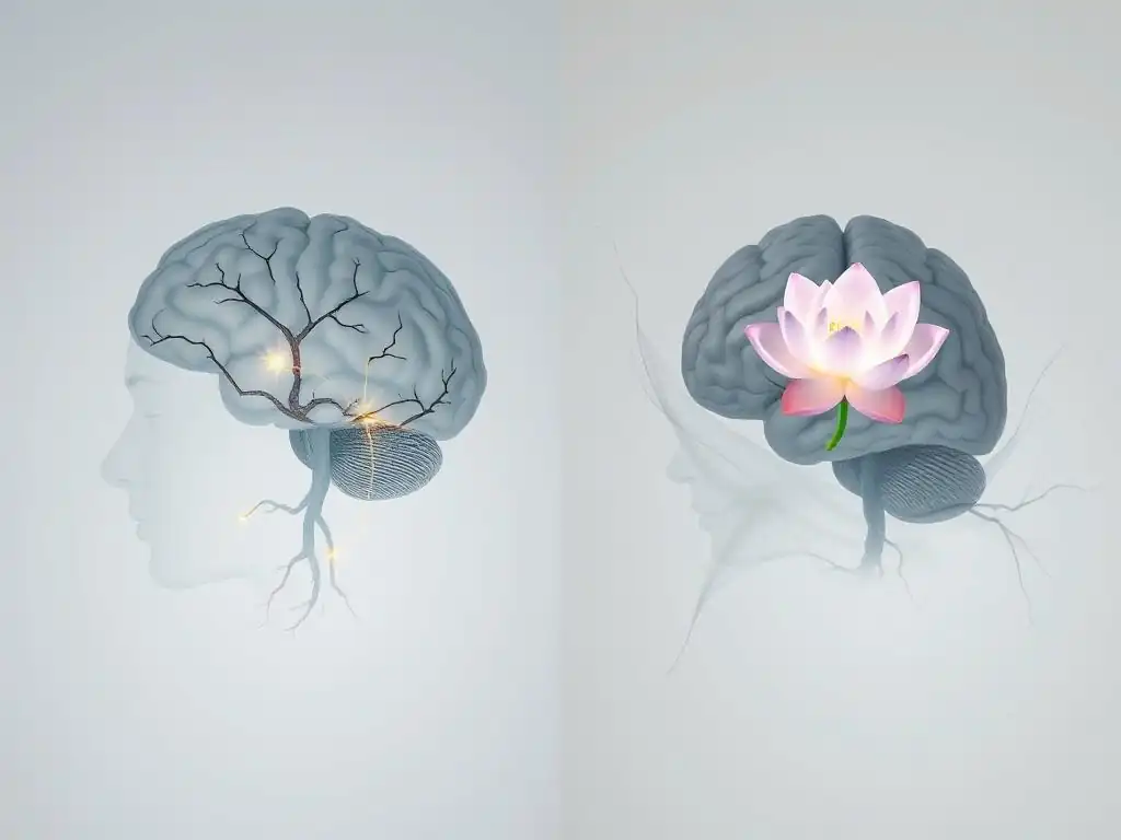
Cognitive Behavioral Therapy (CBT), particularly trauma-focused variations like Trauma-Focused CBT (TF-CBT) and Prolonged Exposure (PE), aims to help individuals identify and challenge maladaptive thought patterns and beliefs related to the trauma, and gradually confront trauma-related memories and situations in a safe environment. The underlying principle is that changing thoughts and behaviors can alter emotional responses and, consequently, brain function. MRI studies investigating the effects of trauma-focused CBT have provided compelling evidence of its neural impact.
Research, such as studies examining PE, has shown that successful treatment is associated with measurable changes in brain activity. For instance, one study involving veterans with PTSD treated with PE observed decreased activity in the amygdala when exposed to trauma-related stimuli post-treatment. Simultaneously, they found increased activation in areas of the prefrontal cortex, suggesting improved top-down regulation of the fear response. These findings indicate that CBT's focus on processing trauma memories and altering cognitive appraisals corresponds with a shift towards a more balanced and less hyper-reactive neural state, contributing to the observed trauma therapy brain changes.
Eye Movement Desensitization and Reprocessing (EMDR): Exploring Brain Activity Changes
Eye Movement Desensitization and Reprocessing (EMDR) therapy involves recalling distressing trauma memories while simultaneously engaging in bilateral stimulation, typically guided eye movements. The exact mechanisms are still debated, but theories suggest it may facilitate the processing of traumatic memories, allowing them to be stored in a less distressing way. MRI studies have sought to understand the neural correlates of EMDR and its contribution to trauma therapy brain changes.
Studies using fMRI have examined brain activity during and after EMDR sessions. Some research has shown that successful EMDR therapy is associated with decreased activation in the amygdala and hippocampus when processing trauma-related cues after treatment. For example, one study reported a significant reduction in amygdala reactivity in PTSD patients after EMDR compared to a control group. These findings support the idea that EMDR helps to "cool down" the brain's fear circuits and integrate traumatic memories more adaptively, leading to measurable shifts in neural activity patterns and contributing to recovery.
Other Effective Trauma Therapies and Their Neural Correlates: Somatic Experiencing, Narrative Exposure Therapy, Mindfulness-Based Interventions
Beyond CBT and EMDR, other therapeutic approaches also show promise in addressing trauma and inducing positive trauma therapy brain changes, although the neuroimaging research on them is still developing. Somatic Experiencing (SE), for example, focuses on the body's physiological responses to trauma, aiming to release trapped energy and regulate the nervous system. While direct MRI studies on SE are less common, its focus on the autonomic nervous system suggests potential impacts on brainstem and limbic system regulation, which could be explored with future imaging.
Narrative Exposure Therapy (NET) involves constructing a chronological narrative of one's life, focusing on traumatic events. This process aims to integrate fragmented trauma memories into a coherent life story. Conceptually, this could influence hippocampal function and connectivity, potentially improving the organization and recall of autobiographical memory. Mindfulness-Based Interventions (MBIs), such as Mindfulness-Based Stress Reduction (MBSR), teach techniques to increase present-moment awareness and non-judgmental observation. Research on MBIs in trauma populations suggests they can enhance prefrontal cortex activity and reduce amygdala reactivity, improving emotional regulation and stress response, adding to the growing evidence of diverse trauma therapy brain changes.
Specific MRI Findings: Mapping Brain Changes with Trauma Therapy
Increased Hippocampal Volume and Connectivity Following Trauma-Focused Therapy
One of the most consistent findings in neuroimaging research on trauma is the reduced volume of the hippocampus in individuals with chronic PTSD. This reduction is thought to reflect the neurotoxic effects of prolonged stress hormones like cortisol and disruptions in neurogenesis (the growth of new neurons). Encouragingly, studies on trauma therapy brain changes have provided evidence that effective treatment can mitigate or even reverse some of this volume loss.
Several studies using sMRI have reported increases in hippocampal volume following successful trauma-focused therapies like PE or TF-CBT. While the degree of increase varies between studies, some research suggests a measurable growth, particularly in specific subfields, after several months of treatment. Furthermore, fMRI studies indicate that connectivity between the hippocampus and other brain regions, such as the prefrontal cortex, may improve, suggesting better integration of memory and emotional regulation processes. These findings underscore the brain's capacity for recovery and structural repair in response to therapeutic intervention.
Reduced Amygdala Hyperactivity and Improved Emotional Regulation After Treatment
As previously mentioned, the amygdala often exhibits heightened activity in response to trauma-related cues in individuals with PTSD, contributing to symptoms like hyperarousal and exaggerated fear responses. A key aim of trauma therapy is to dampen this overactive fear response and improve emotional regulation. Neuroimaging studies consistently demonstrate that successful trauma therapy brain changes include a reduction in this amygdala hyperactivity.
Using fMRI, researchers have observed decreased amygdala activation when trauma survivors are shown trauma-relevant images or asked to recall traumatic memories after completing therapies like PE or EMDR. This reduction in reactivity is often correlated with a decrease in PTSD symptom severity. This suggests that therapy helps the brain differentiate between past danger and present safety, allowing the amygdala to return to a more normative level of responsiveness. The improved emotional regulation is further supported by observed increases in connectivity between the amygdala and regulatory regions in the prefrontal cortex.
Enhanced Prefrontal Cortex Function and Improved Cognitive Control
The prefrontal cortex (PFC) plays a critical role in executive functions, including attention, decision-making, and, importantly, the top-down regulation of emotions. In trauma survivors, decreased activity or impaired connectivity within the PFC is often observed, contributing to difficulties with emotional control and cognitive processing. Positive trauma therapy brain changes frequently involve improvements in PFC function.
Studies using fMRI have shown increased activity in specific areas of the PFC, such as the ventromedial PFC (vmPFC), after successful trauma therapy. The vmPFC is particularly important for inhibiting the amygdala and processing safety signals. Enhanced activity in this region indicates improved capacity to regulate fear responses. Furthermore, connectivity between the vmPFC and the amygdala often strengthens post-therapy, providing a neural basis for better emotional control. These findings highlight how therapy helps restore the brain's capacity for self-regulation and adaptive coping.
Case Studies: Real-World Examples of Brain Healing Through Therapy
Case Study 1: PTSD Reduction and Hippocampal Growth After Prolonged Exposure Therapy
While specific patient data is confidential, research provides composite pictures of typical outcomes. Consider a hypothetical case based on research findings: "Sarah," a 45-year-old veteran, presented with severe PTSD symptoms following combat exposure. An initial sMRI scan revealed reduced hippocampal volume compared to age-matched controls, consistent with chronic trauma exposure. She began a course of Prolonged Exposure (PE) therapy.
Over 12 weeks, Sarah engaged in imaginal exposure (revisiting the trauma memory) and in vivo exposure (gradually confronting feared situations related to the trauma). Her PTSD symptoms, measured by clinical scales, significantly decreased. A follow-up sMRI scan approximately six months after the start of therapy showed a measurable increase in the volume of her hippocampus, approaching the size seen in non-traumatized individuals in some studies. This illustrative case demonstrates how behavioral processing in therapy can correlate with structural changes in key memory regions, providing tangible evidence of trauma therapy brain changes.
Case Study 2: Improved Emotional Regulation and Amygdala Modulation Following EMDR
Another composite example, based on research, might be "David," a 30-year-old who experienced childhood abuse, leading to difficulties with emotional outbursts and hypervigilance. An initial fMRI scan, while David viewed images triggering his trauma response, showed significant hyperactivity in his amygdala. He opted for Eye Movement Desensitization and Reprocessing (EMDR) therapy.
David completed approximately 15 sessions of EMDR, processing several key traumatic memories. He reported a significant reduction in emotional reactivity and felt less constantly "on edge." A follow-up fMRI scan, conducted under similar conditions, revealed a noticeable decrease in the peak activity of his amygdala when presented with triggering stimuli. This reduction in amygdala response was accompanied by increased functional connectivity between his amygdala and areas of his prefrontal cortex. This example illustrates how EMDR's processing mechanisms can lead to shifts in emotional regulation pathways, impacting the brain's fear response center and demonstrating specific trauma therapy brain changes.
Long-Term Therapy Outcomes and Brain Changes: Sustaining the Benefits
Maintaining Brain Plasticity After Therapy: Strategies for Long-Term Well-being
The positive trauma therapy brain changes observed during and immediately after treatment highlight the brain's capacity for plasticity. However, maintaining these gains over the long term requires ongoing effort. The brain continues to be shaped by experiences, so sustaining a healing trajectory involves cultivating habits that support neural health and resilience.
Strategies for maintaining brain plasticity and well-being post-therapy include regular engagement in activities known to promote neurogenesis and healthy brain function:
- Consistent physical exercise: Aerobic exercise, in particular, has been shown to support hippocampal health.
- Mindfulness and meditation practices: These can continue to strengthen prefrontal regulation and reduce stress responses.
- Engaging in mentally stimulating activities: Learning new skills or challenging oneself cognitively helps maintain neural connections.
- Prioritizing healthy sleep: Sleep is crucial for memory consolidation and overall brain repair.
- Maintaining social connections: Positive social interactions support emotional regulation and reduce stress.
These lifestyle factors reinforce the positive neural pathways established during therapy.
The Role of Continued Therapy and Support in Preventing Relapse
While significant trauma therapy brain changes can occur during an initial course of treatment, recovery is often a non-linear process. Life stressors or unexpected triggers can sometimes lead to a resurgence of symptoms. For many individuals, ongoing support, including periodic check-ins with a therapist or participation in support groups, plays a vital role in consolidating gains and preventing relapse.
Continued therapeutic support can help individuals navigate new challenges, reinforce coping skills learned in therapy, and address any re-emerging symptoms before they escalate. This doesn't necessarily mean weekly therapy indefinitely, but rather having access to support as needed. This ongoing engagement acts as a form of maintenance for the positive neural architecture built during initial treatment, helping to sustain the beneficial trauma therapy brain changes and support long-term resilience.
Limitations and Future Directions in Trauma Therapy Brain Research
Challenges in Interpreting MRI Data and Establishing Causality
While MRI research provides valuable insights into trauma therapy brain changes, it's important to acknowledge its limitations. Interpreting complex brain imaging data is challenging; correlation does not equal causation. Observing a change in brain activity or structure after therapy doesn't definitively prove that the therapy caused the change, although well-designed studies with control groups strengthen this inference. Other factors, such as the passage of time, natural recovery processes, or engagement in other life changes, could also play a role.
Furthermore, the relationship between subjective symptom reduction and objective brain changes is not always perfectly aligned. Different individuals may show varying degrees of brain change for similar levels of symptom improvement. Methodological variations across studies, such as scanner differences, analysis techniques, and sample sizes, can also make it difficult to compare findings directly and draw universal conclusions about the neural effects of trauma treatment.
Future Research: Exploring Novel Therapies and Personalized Treatment Approaches
The field of trauma therapy brain research is continuously evolving. Future research directions aim to overcome current limitations and deepen our understanding of how healing occurs at the neural level. One key area is exploring the neural correlates of novel and emerging trauma therapies, such as psychedelic-assisted therapy or neurofeedback, to understand their specific impacts on brain function and structure.
Another critical direction is the development of personalized treatment approaches. By identifying specific patterns of brain dysfunction in individuals with trauma, researchers hope to predict who might respond best to particular types of therapy. This could involve using baseline MRI scans or other biomarkers to tailor treatment plans for maximum effectiveness. Understanding the nuances of trauma therapy brain changes in diverse populations and contexts will be crucial for optimizing recovery outcomes and developing more targeted interventions in the future.
Conclusion: The Power of Therapy to Heal the Traumatized Brain
Trauma Therapy Brain Changes: A Path to Recovery and Resilience
The accumulated evidence from neuroimaging studies provides a powerful and hopeful message: trauma therapy can lead to tangible, positive changes in the brain. The often-observed patterns of reduced amygdala hyperactivity, increased hippocampal volume and connectivity, and enhanced prefrontal cortex function following effective treatment underscore the brain's remarkable capacity for healing and adaptation. These trauma therapy brain changes are not just abstract scientific findings; they represent the biological foundation for symptom reduction, improved emotional regulation, and enhanced overall well-being experienced by individuals in recovery.
Understanding the neural mechanisms of trauma and therapy validates the experiences of survivors and highlights the profound impact of therapeutic work. It moves beyond viewing trauma solely as a psychological injury to recognizing its deep biological imprint, and crucially, demonstrating that this imprint can be altered through targeted intervention. The evidence of trauma therapy brain changes offers a scientific basis for hope, confirming that the brain is not permanently damaged by trauma but can be guided towards healing and resilience through therapeutic processes.
Empowering Individuals with Knowledge of Brain-Based Healing
Sharing the findings about trauma therapy brain changes is essential for empowering individuals affected by trauma. Knowing that therapy doesn't just teach coping skills but actively helps to repair and reorganize the brain can be incredibly validating and motivating. It provides a concrete understanding of why therapy works and reinforces the importance of committing to the therapeutic process.
By demystifying the connection between trauma, the brain, and therapy, we can reduce stigma and encourage more people to seek help. This knowledge underscores that seeking therapy for trauma is not a sign of weakness but a proactive step towards facilitating the brain's natural healing capabilities. As research continues to uncover the intricate details of how therapy changes the brain, it will further enhance our ability to provide effective, evidence-based care and support survivors on their path to recovery and a life of greater peace and resilience.
Frequently Asked Questions
What exactly does the article "Healing the Traumatized Brain: MRI Evidence of Therapy's Effects" reveal about trauma therapy brain changes?
The article showcases MRI evidence demonstrating that trauma therapy can lead to measurable changes in brain structure and function. These changes are often seen in regions associated with emotional regulation and memory processing.
How do MRIs help us understand the impact of trauma therapy on the brain?
MRIs provide detailed images of the brain's structure and activity. This allows researchers to observe how trauma therapy influences neural pathways and brain areas impacted by trauma, such as the amygdala and hippocampus.
What types of trauma therapy are likely to produce noticeable brain changes according to the article?
While the article might not specify particular therapies, effective trauma-focused therapies like EMDR (Eye Movement Desensitization and Reprocessing) and Cognitive Processing Therapy (CPT) are often associated with positive brain changes.
Can trauma therapy reverse some of the negative brain changes caused by traumatic experiences?
The article suggests that trauma therapy can indeed help to reverse some negative effects. It aims to promote neural plasticity, enabling the brain to adapt and reorganize, potentially mitigating the impact of the traumatic experience.
Mind Care Guide offers a range of resources, including articles, support groups, and directories of mental health professionals specializing in trauma therapy. Look for sections dedicated to mental well-being and trauma support.
Final Reflection: The Rewiring of Resilience
The groundbreaking research presented here demonstrates that trauma's impact on the brain isn't a life sentence; it's a condition that can be actively reshaped through targeted therapy. This isn't just about alleviating symptoms; it's about fundamentally altering the neural pathways that perpetuate suffering, offering a tangible path towards lasting healing and a brighter future for millions affected by trauma.
The implications of these MRI findings extend far beyond the clinical setting. We're witnessing a paradigm shift in our understanding of trauma, moving away from a purely psychological framework towards a more holistic, neurobiological perspective. This understanding is crucial for dismantling the stigma surrounding mental health and fostering a culture of empathy and support. As Bessel van der Kolk eloquently stated, The body keeps the score,
and these images provide powerful visual evidence of how therapy can help the body, and the brain, rewrite that score.
Consider the potential this research unlocks. Imagine a future where access to effective trauma-informed therapies is readily available, where early intervention prevents the long-term consequences of trauma, and where individuals are empowered to reclaim their lives and build resilience. This isn't just a hope; it's a call to action.
Take a moment to reflect on the power of healing, not just for yourself, but for the countless others who carry the weight of unresolved trauma. Seek support if you need it, and advocate for policies that prioritize mental health and access to evidence-based treatments. The journey to healing may be challenging, but the rewiring of resilience is possible, and the future is brighter than ever before.
Unleash Your Brain's Healing Power
Thanks for diving into the fascinating world of brain healing! You've learned how powerful therapy can be in reshaping the traumatized brain. Now it's time to take that knowledge and turn it into action.
- Schedule a consultation with a trauma-informed therapist: This is your first step towards healing and reclaiming your well-being. Don't delay the journey to a healthier you.
- Share this article with someone who might benefit: Let's spread awareness and support those navigating similar challenges. A simple share can make a world of difference.
- Explore mindfulness techniques: Even small daily practices can foster resilience and promote neurological healing. Discover what works for you.
What small step will you take today to nurture your brain's incredible healing capacity?