
From Chaos to Calm: Brain Scan View of Therapy for Bipolar Disorder

Welcome to Mind Care Guide, your compassionate companion on the journey to mental well-being. Ever wondered what really happens in the brain during bipolar disorder therapy? Our groundbreaking article, "From Chaos to Calm: Brain Scan View of Therapy for Bipolar Disorder," delves deep into the neuroscience, revealing fascinating insights gleaned from bipolar disorder therapy brain scans. Could these scans unlock new pathways to healing? Explore the potential and understand the science behind finding your calm.
From Chaos to Calm: Brain Scan View of Therapy for Bipolar Disorder
- Understanding Bipolar Disorder and the Brain
- The Role of Therapy in Managing Bipolar Disorder
- Brain Imaging of Bipolar Disorder Therapy: Revealing Neurological Changes
- Case Study: Impact of CBT on Brain Activity in a Patient with Bipolar I Disorder
- How Therapy Reshapes the Bipolar Brain: Specific Areas Affected
- Benefits of Monitoring Therapy with Brain Scans
- Limitations of Brain Scans in Bipolar Disorder Therapy
- Long-Term Effects of Therapy on the Brain in Bipolar Disorder
- Future Directions: Brain Scans and Bipolar Disorder Treatment
-
Frequently Asked Questions
- How can brain scans help in bipolar disorder therapy?
- What types of therapy are most effective for bipolar disorder according to brain scan research?
- Can brain scans predict the success of bipolar disorder therapy before it starts?
- Are there any risks associated with using brain scans for bipolar disorder therapy planning?
- How accessible are brain scans for individuals seeking bipolar disorder therapy?
- Final Reflection: The Illuminating Path to Bipolar Stability
- Unlock Your Inner Calm: Take Charge of Your Bipolar Journey
Understanding Bipolar Disorder and the Brain
The Neurological Impact of Bipolar Disorder
Bipolar disorder is a complex mood disorder characterized by significant shifts in mood, energy, activity levels, and the ability to carry out daily tasks. These shifts manifest as manic or hypomanic episodes alternating with major depressive episodes. Research utilizing advanced neuroimaging techniques, such as functional magnetic resonance imaging (fMRI), has revealed that these mood fluctuations are associated with distinct structural and functional abnormalities in the brain.
Key areas implicated include the prefrontal cortex, amygdala, hippocampus, and basal ganglia, which are crucial for regulating emotions, decision-making, memory, and reward processing. For instance, studies often show reduced gray matter volume in the prefrontal cortex and hippocampus in individuals with bipolar disorder compared to healthy controls, sometimes by as much as 5-10% in certain regions, suggesting a neurobiological basis for the disorder's symptoms.
The interplay between these brain regions is also often disrupted. For example, there is frequently observed dysregulation in the circuitry connecting the prefrontal cortex (involved in executive function and emotional regulation) and the amygdala (involved in processing emotions, particularly fear and threat). During manic episodes, the amygdala may show heightened activity, while the prefrontal control may be diminished, leading to impulsivity and elevated mood states.
Conversely, during depressive episodes, activity in prefrontal regions might be reduced, contributing to feelings of hopelessness and lack of motivation. Understanding these neurological underpinnings is vital for developing effective treatments, including therapy, and for exploring how bipolar disorder therapy brain scan studies can shed light on treatment mechanisms.
How Functional Magnetic Resonance Imaging (fMRI) Helps Understand Bipolar Disorder
Functional Magnetic Resonance Imaging (fMRI) is a non-invasive neuroimaging technique that measures brain activity by detecting changes in blood flow. When a brain region is more active, it requires more oxygenated blood, and fMRI captures these changes. This allows researchers to observe which parts of the brain are engaged during specific tasks or even at rest. In the context of bipolar disorder, fMRI has been instrumental in identifying the neural circuits involved in mood dysregulation. Studies using fMRI have shown altered connectivity patterns between various brain regions, such as the prefrontal cortex and limbic system, during different mood states.
For example, fMRI scans performed during emotional tasks often reveal exaggerated amygdala responses to negative stimuli in individuals with bipolar disorder, along with reduced activity in regulatory prefrontal areas. Resting-state fMRI, which examines brain activity when a person is not performing a specific task, has also identified abnormal functional connectivity within key networks, such as the default mode network (involved in self-referential thought) and the salience network (involved in detecting important stimuli).
These findings provide empirical support for the neurological basis of bipolar disorder symptoms and offer a potential avenue for tracking changes associated with interventions like bipolar disorder therapy using brain scan technology.
The Role of Therapy in Managing Bipolar Disorder
Cognitive Behavioral Therapy (CBT) and Dialectical Behavior Therapy (DBT) for Bipolar Disorder
While medication is often the cornerstone of bipolar disorder treatment, psychotherapy plays a crucial role in managing symptoms, preventing relapse, and improving overall quality of life. Cognitive Behavioral Therapy (CBT) is one of the most widely used therapeutic approaches. CBT for bipolar disorder helps individuals identify and challenge distorted thinking patterns (cognitive distortions) and unhelpful behaviors that contribute to mood swings.
Patients learn skills to recognize early warning signs of manic or depressive episodes, develop coping strategies, and improve problem-solving abilities. A typical course of CBT might involve 12-20 weekly sessions, focusing on mood monitoring, activity scheduling, and cognitive restructuring.
Dialectical Behavior Therapy (DBT), initially developed for borderline personality disorder, has also shown promise in treating bipolar disorder, particularly for individuals experiencing frequent mood swings, impulsivity, and difficulties with emotional regulation. DBT emphasizes four core skills: mindfulness, distress tolerance, emotion regulation, and interpersonal effectiveness.
Through individual therapy, group skills training, and phone coaching, patients learn to manage intense emotions, reduce impulsive behaviors, and build stable relationships. While research on DBT specifically for bipolar disorder is less extensive than for CBT, studies suggest it can help reduce hospitalization rates and improve overall functioning, offering another avenue for exploring the impact of bipolar disorder therapy via brain scan insights.
Beyond symptom reduction, therapy for bipolar disorder aims to help individuals achieve long-term mood stability and enhance their social and occupational functioning. By equipping patients with practical coping skills and strategies for managing stress and triggers, therapy empowers them to navigate the challenges of living with bipolar disorder more effectively. For instance, learning to recognize and respond to prodromal symptoms (early signs of an episode) can help prevent full-blown manic or depressive states, reducing the frequency and severity of mood swings.
Improved emotional regulation and interpersonal skills learned in therapy also significantly impact social functioning. Individuals with bipolar disorder often experience difficulties in relationships due to mood-related behaviors. Therapy helps them develop healthier communication patterns, set boundaries, and manage conflicts more constructively. Over time, consistent engagement in therapy can lead to greater stability in personal relationships, improved performance at work or school, and a higher sense of self-efficacy, contributing to a more fulfilling life despite the challenges of the disorder. The neurological changes underlying these improvements can potentially be observed through bipolar disorder therapy brain scan research.
Brain Imaging of Bipolar Disorder Therapy: Revealing Neurological Changes
Functional Magnetic Resonance Imaging (fMRI) and Positron Emission Tomography (PET) in Bipolar Disorder Research
Neuroimaging techniques like fMRI and Positron Emission Tomography (PET) have become invaluable tools in understanding the neurobiological underpinnings of mental disorders and the mechanisms by which treatments exert their effects. As discussed, fMRI measures brain activity by detecting blood flow changes, providing insights into which areas are active during tasks or at rest. PET, on the other hand, uses a radioactive tracer to measure metabolic activity, neurotransmitter binding, or blood flow in the brain. For instance, PET scans can be used to study dopamine or serotonin receptor availability, neurotransmitters often implicated in mood disorders.
In bipolar disorder research, both fMRI and PET are used to compare brain structure and function in individuals with the disorder versus healthy controls, and critically, to examine how these metrics change after therapeutic interventions. By performing scans before and after a course of therapy, researchers can identify specific brain regions or networks whose activity or connectivity patterns are altered, potentially correlating these changes with clinical improvements observed in patients.
This allows for a more objective, biological assessment of treatment efficacy, moving beyond purely subjective symptom reports and exploring the neural correlates of successful bipolar disorder therapy using brain scan technology.
Visualizing Changes in Brain Activity After Bipolar Disorder Therapy
The application of brain imaging, particularly fMRI, to study the effects of psychotherapy on the bipolar brain is a growing area of research. The hypothesis is that successful therapy leads to measurable changes in brain activity patterns that reflect improved mood regulation and cognitive function. Studies have begun to provide evidence supporting this idea. For example, research has shown that after receiving courses of therapies like CBT, individuals with bipolar disorder exhibit altered activity in brain regions known to be involved in emotional processing and cognitive control, such as the amygdala and prefrontal cortex.
These changes are often visualized by comparing brain activation maps from scans conducted before therapy to those conducted after therapy. Reductions in amygdala hyperactivity during emotional tasks, for instance, or increased activity in prefrontal regions during cognitive control tasks, can be observed. While results can vary depending on the specific study, patient population, and therapy type, the general trend suggests that therapy helps to normalize the dysregulated brain circuits associated with bipolar disorder, offering tangible evidence of its impact at a neural level through bipolar disorder therapy brain scan investigations.
Case Study: Impact of CBT on Brain Activity in a Patient with Bipolar I Disorder
Initial fMRI Findings Before CBT
Consider a hypothetical case study of a 45-year-old individual diagnosed with Bipolar I Disorder, experiencing recurrent depressive episodes and hypomanic phases. Before commencing Cognitive Behavioral Therapy (CBT), the individual undergoes an fMRI scan while performing an emotional face-matching task, a common paradigm used in affect research. The baseline fMRI results show heightened activity in the amygdala when presented with negative emotional stimuli (e.g., fearful faces) compared to neutral stimuli.
This increased amygdala response is consistent with findings in many individuals with bipolar disorder and is thought to reflect exaggerated processing of negative emotional information. Furthermore, connectivity analysis might reveal weaker functional coupling between the amygdala and the dorsolateral prefrontal cortex (DLPFC), a region involved in top-down emotional regulation. These initial findings provide a neural snapshot of the individual's emotional processing biases prior to therapeutic intervention, offering a baseline for future bipolar disorder therapy brain scan comparisons.
Additionally, resting-state fMRI before therapy might show altered connectivity within the default mode network (DMN), perhaps increased connectivity between DMN nodes implicated in self-referential rumination, which is common during depressive states. The salience network, involved in detecting and orienting to important stimuli, might also show dysregulation, potentially contributing to difficulties in prioritizing and focusing.
These initial fMRI findings not only help confirm the presence of neural patterns associated with bipolar disorder but also serve as crucial baseline data against which the effects of subsequent therapy can be evaluated. This pre-therapy scan is a critical component of any bipolar disorder therapy brain scan study aiming to track neurological changes.
Cognitive Behavioral Therapy Intervention
Following the baseline fMRI, the individual participates in a 16-session course of Cognitive Behavioral Therapy, specifically adapted for bipolar disorder. The therapy focuses on several key areas: psychoeducation about bipolar disorder and its symptoms; mood monitoring and identifying triggers; learning cognitive restructuring techniques to challenge negative or distorted thoughts related to depression and mania; developing behavioral strategies to promote stable routines (e.g., consistent sleep schedule, regular activity); and relapse prevention planning, including identifying early warning signs of mood episodes.
The therapist works collaboratively with the individual, using techniques such as Socratic questioning to help them examine the evidence for their thoughts and behavioral experiments to test the validity of their beliefs. Homework assignments are given between sessions to reinforce learned skills and encourage their application in daily life. The goal of this intervention is not just symptom reduction but also to equip the individual with lifelong tools for managing their mood, improving coping skills, and preventing future episodes, providing the clinical context for any subsequent bipolar disorder therapy brain scan analysis.
Follow-Up fMRI Results: Decreased Amygdala Activity
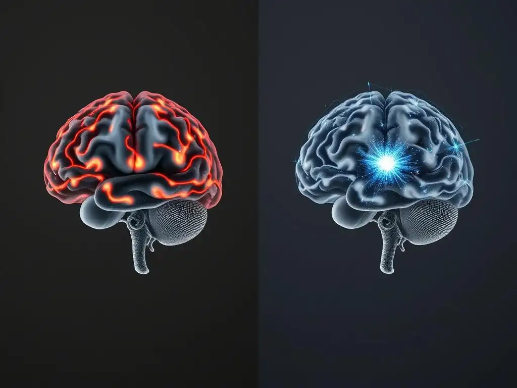
Approximately one month after completing the 16-session CBT course, the individual undergoes a follow-up fMRI scan using the same emotional face-matching task and resting-state protocols as the baseline scan. Clinically, the individual reports significant improvement in mood stability, reduced frequency and intensity of depressive symptoms, and better management of hypomanic tendencies. Crucially, the follow-up fMRI results reveal a notable change: decreased activity in the amygdala when presented with negative emotional stimuli compared to the baseline scan. The amygdala's response is now closer to that observed in healthy control groups.
Furthermore, analysis of functional connectivity shows increased coupling between the amygdala and the dorsolateral prefrontal cortex, suggesting enhanced top-down regulation of emotional responses by the prefrontal control regions. Resting-state fMRI might also show more normalized connectivity patterns within the DMN and salience network.
These findings provide compelling neural evidence that CBT has not only improved the individual's clinical symptoms but has also induced measurable changes in brain activity and connectivity patterns, supporting the efficacy of this bipolar disorder therapy through brain scan evidence. This case illustrates how neuroimaging can provide objective support for the effectiveness of psychotherapy at a biological level.
How Therapy Reshapes the Bipolar Brain: Specific Areas Affected
Dorsolateral Prefrontal Cortex: Enhanced Cognitive Control
The dorsolateral prefrontal cortex (DLPFC) is a key area involved in executive functions, including planning, decision-making, working memory, and cognitive control. It plays a crucial role in regulating thoughts and emotions. In individuals with bipolar disorder, the DLPFC often shows reduced activity or connectivity, particularly during mood episodes, which may contribute to difficulties with impulse control, emotional regulation, and cognitive flexibility. Therapy, especially CBT and related approaches, aims to enhance these executive functions by teaching patients strategies for challenging unhelpful thoughts, planning behaviors, and managing impulses.
Neuroimaging studies exploring the effects of bipolar disorder therapy using brain scan technology have provided evidence that successful therapy can lead to increased activation or connectivity in the DLPFC. For instance, research might show greater DLPFC engagement during tasks requiring emotional regulation or cognitive control after a course of therapy compared to before. This suggests that therapy helps strengthen the capacity of the DLPFC to exert top-down control over emotional responses originating in subcortical areas like the amygdala, contributing to greater mood stability and improved decision-making.
Amygdala: Modulation of Emotional Reactivity
The amygdala is a critical component of the brain's limbic system, heavily involved in processing emotions, particularly those related to fear, threat, and reward. In bipolar disorder, the amygdala is frequently found to be hyperactive, especially during manic or hypomanic states, and sometimes during depressive episodes as well. This heightened reactivity is thought to contribute to the intense emotional experiences and mood swings characteristic of the disorder. Therapeutic approaches like CBT and DBT specifically target emotional regulation skills.
Brain scan studies, particularly fMRI, have demonstrated that effective bipolar disorder therapy can lead to a modulation of amygdala activity. As seen in the case study example, a common finding is a reduction in amygdala hyperactivity in response to emotional stimuli after successful therapy. This suggests that therapy helps individuals process emotional information in a less reactive and more balanced way. By learning coping strategies and cognitive techniques, patients may be able to dampen the intensity of emotional responses originating in the amygdala, leading to greater emotional stability.
Ventral Striatum: Improved Reward Processing
The ventral striatum, including the nucleus accumbens, is a key region in the brain's reward pathway. It is involved in processing pleasure, motivation, and goal-directed behavior. Dysregulation in the ventral striatum is often implicated in bipolar disorder, particularly during manic episodes, which are characterized by increased goal-directed activity, pleasure-seeking, and risk-taking. During depressive episodes, activity in this region might be reduced, contributing to anhedonia (inability to experience pleasure) and lack of motivation.
While less extensively studied than the amygdala or prefrontal cortex, some research suggests that therapy can also influence activity in the ventral striatum in individuals with bipolar disorder. For example, behavioral activation techniques used in CBT for depression aim to increase engagement in rewarding activities, which could potentially normalize activity in this region.
Improved mood regulation through therapy might also indirectly impact the ventral striatum, leading to a more balanced response to rewards and a reduction in the extreme swings between excessive pleasure-seeking during mania and lack of motivation during depression. Further bipolar disorder therapy brain scan research is needed to fully understand therapy's impact on this region.
Benefits of Monitoring Therapy with Brain Scans
Quantifying Treatment Effectiveness Through Neural Correlates
One significant benefit of using brain scans in conjunction with bipolar disorder therapy is the potential to quantify treatment effectiveness at a biological level. While clinical assessments rely on subjective self-reports and observational data, brain imaging provides objective measures of neural changes. By observing shifts in regional brain activity, connectivity patterns, or gray matter volume before and after therapy, researchers and clinicians can gain a deeper understanding of whether and how the intervention is impacting the underlying neurobiology of the disorder.
For example, if a bipolar disorder therapy brain scan study consistently shows a reduction in amygdala hyperactivity correlating with reduced emotional reactivity reported by patients, it provides strong objective support for the therapy's mechanism of action. This can be particularly valuable in research settings for validating new therapeutic approaches or understanding why certain individuals respond better than others. While not yet standard clinical practice, this ability to find neural correlates offers a powerful tool for demonstrating the tangible impact of therapy on the brain.
Tailoring Treatment Strategies Based on Individual Brain Activity
Another potential future application of bipolar disorder therapy brain scan insights is the ability to tailor treatment strategies based on an individual's specific brain activity patterns. Just as genetic testing is beginning to inform medication choices, neuroimaging could potentially help personalize therapy. If a patient's baseline scan reveals particularly severe dysregulation in a specific brain circuit, such as weak prefrontal-amygdala connectivity, it might suggest that therapies emphasizing emotional regulation and cognitive control are particularly indicated.
Conversely, if scan data points to different patterns, it might guide clinicians towards other therapeutic modalities or combinations of treatments. For instance, individuals with significant reward pathway dysregulation might benefit more from therapies incorporating behavioral activation or motivational techniques. While this is still an area of active research, the hope is that by understanding the unique neural signature of a patient's bipolar disorder, clinicians could select or modify therapeutic interventions for optimal outcomes, moving towards a more precise, neurobiologically informed approach to therapy.
Identifying Individuals Unlikely to Respond to Standard Treatments
Brain scans might also potentially help identify individuals who are less likely to respond to standard psychotherapeutic approaches for bipolar disorder. By examining baseline brain activity or structural characteristics, researchers may be able to predict treatment response. For example, if certain severe structural abnormalities or highly atypical functional connectivity patterns are identified in a scan before therapy, it might suggest that the individual's condition is less amenable to change through standard talk therapy alone and may require more intensive or alternative interventions, possibly including different medication strategies or novel brain stimulation techniques.
This predictive capability, while still under investigation, could help avoid prolonged courses of ineffective treatment, saving time, resources, and reducing patient frustration. By identifying potential non-responders early, clinicians could pivot to alternative strategies sooner, improving the chances of finding an effective treatment plan. This highlights the potential of bipolar disorder therapy brain scan research not just for understanding treatment effects but also for guiding clinical decision-making and improving patient care pathways.
Limitations of Brain Scans in Bipolar Disorder Therapy
High Cost and Limited Availability of Advanced Imaging
Despite the promising insights offered by bipolar disorder therapy brain scan research, there are significant limitations that currently prevent it from becoming a routine clinical tool. One major barrier is the high cost of advanced neuroimaging techniques like fMRI and PET. Running an fMRI scan can cost hundreds or even thousands of dollars, making it prohibitively expensive for routine use in clinical practice, especially when repeated scans are needed to track changes over time.
Furthermore, the availability of the necessary equipment and trained personnel is often limited to specialized research centers and large hospitals. Many mental health clinics and private practices do not have access to these technologies. This lack of accessibility means that while brain scans are valuable research tools for understanding bipolar disorder and treatment mechanisms, they are not readily available for guiding the therapy of the average patient, creating a gap between research findings and clinical application.
Challenges in Interpreting Complex Brain Scan Data
Interpreting brain scan data, particularly fMRI and PET scans which capture dynamic activity, is a complex process that requires specialized expertise. The brain is a highly interconnected organ, and changes in one area can affect activity in many others. Isolating the specific effects of therapy from other factors (such as medication changes, life events, or spontaneous fluctuations in mood) is challenging.
Moreover, interpreting the meaning of observed changes in brain activity in terms of clinical outcomes is not always straightforward. A change in brain activity might be statistically significant but not necessarily translate into a clinically meaningful improvement for the patient.
There is also considerable variability in brain structure and function across individuals, even within the same diagnostic category. This heterogeneity makes it difficult to establish clear, universally applicable neural markers for treatment response. Researchers are still working to develop standardized protocols and analytical methods to make brain scan data more reliable and interpretable in the context of mental health treatment, including bipolar disorder therapy brain scan studies.
The Importance of Integrating Clinical Observations with Imaging Results
It is crucial to recognize that brain scan data, while informative, should not replace traditional clinical assessment and observation. A brain scan provides a snapshot of neural activity or structure, but it does not capture the full complexity of a person's lived experience, their subjective symptoms, their coping abilities, or their therapeutic relationship. Clinical observations, patient self-reports, and therapist assessments remain essential for understanding the individual's progress in therapy.
Effective clinical decision-making requires integrating information from multiple sources. Brain scan data can potentially supplement clinical judgment by providing objective biological insights, but it cannot substitute for the nuanced understanding gained through direct interaction with the patient. The most valuable approach involves combining the objective data from bipolar disorder therapy brain scan research with the rich, subjective information gathered through clinical interviews and assessments to create a comprehensive picture of the patient's condition and response to treatment.
Long-Term Effects of Therapy on the Brain in Bipolar Disorder
Sustaining Brain Health and Functional Connectivity
Long-term engagement in therapy for bipolar disorder, often alongside medication, is associated with better prognosis and sustained mood stability. While acute effects on brain activity might be observed shortly after a course of therapy, ongoing treatment and the consistent application of learned skills likely contribute to more lasting changes in brain structure and function. Regular therapy can help individuals maintain healthy routines, manage stress effectively, and prevent the disruptive effects of severe mood episodes, which themselves can be neurotoxic over time.
Preliminary research suggests that consistent therapeutic engagement may help sustain healthy functional connectivity patterns in the brain, particularly between regulatory prefrontal regions and limbic areas. By reinforcing adaptive cognitive and behavioral strategies, therapy may help "hardwire" more stable neural pathways. This sustained impact of bipolar disorder therapy on brain health, as potentially revealed by long-term brain scan studies, highlights the importance of therapy not just as an acute treatment but as a vital component of long-term management.
Preventing Relapse Through Maintenance Therapy
Relapse is a significant challenge in bipolar disorder. Even after achieving remission, individuals remain vulnerable to future mood episodes. Maintenance therapy, which involves ongoing therapeutic support after acute symptoms have subsided, plays a crucial role in preventing relapse. This might involve less frequent sessions focused on reinforcing coping skills, monitoring early warning signs, and addressing life stressors.
The mechanisms by which maintenance therapy prevents relapse likely involve the continued reinforcement of adaptive neural circuits. By regularly practicing skills like cognitive restructuring and emotional regulation, individuals keep the associated brain pathways active and strong. This sustained neural engagement, potentially observable through periodic bipolar disorder therapy brain scan evaluations, may make the brain more resilient to the internal and external triggers that can precipitate mood episodes. Therapy helps individuals build a robust set of psychological tools that have corresponding neural underpinnings, contributing to long-term stability.
Early Intervention and Consistent Treatment for Optimal Outcomes
Research consistently shows that early intervention and consistent, long-term treatment lead to the best outcomes in bipolar disorder. This includes both pharmacological treatment and psychotherapy. Starting therapy early in the course of the illness, perhaps after the first mood episode, may help prevent the cascading neurobiological changes that can occur with recurrent, untreated episodes. Each severe mood episode, particularly mania, is thought to potentially contribute to structural and functional changes in the brain over time.
Consistent engagement in therapy helps mitigate the impact of the disorder on the brain by reducing the frequency and severity of episodes. By providing ongoing support and skills, therapy empowers individuals to manage their condition proactively. The cumulative effect of consistent bipolar disorder therapy on brain structure and function, as potentially tracked by longitudinal brain scan studies, is likely significant, contributing to improved long-term prognosis, reduced disability, and a higher quality of life.
Future Directions: Brain Scans and Bipolar Disorder Treatment
Advancements in Diffusion Tensor Imaging (DTI) and Resting-State fMRI
The field of neuroimaging is continually advancing, offering new tools to understand the brain's structure and function. Diffusion Tensor Imaging (DTI), for instance, measures the diffusion of water molecules to map the brain's white matter tracts, which are the communication highways connecting different brain regions. Alterations in these tracts have been observed in bipolar disorder, and future research could use DTI to investigate how therapy impacts the structural integrity or connectivity of these pathways.
Resting-state fMRI, which examines brain activity and connectivity while the individual is at rest, is also becoming increasingly sophisticated. Analyzing the intricate patterns of connectivity within and between different brain networks (like the default mode, salience, and executive control networks) offers a powerful way to understand the global impact of bipolar disorder and how therapy modulates these network dynamics. Future bipolar disorder therapy brain scan studies will likely leverage these advancements to gain a more comprehensive picture of treatment effects.
Developing Targeted Interventions Based on Neural Circuits
A key future direction for bipolar disorder therapy brain scan research is the development of more targeted interventions based on specific neural circuit abnormalities identified through imaging. If research consistently links certain symptom profiles or treatment responses to particular brain circuit dysfunctions, it might be possible to design or refine therapies to specifically address those circuits. For example, therapies could be tailored to strengthen connectivity in a weak circuit or dampen activity in a hyperactive one.
This could involve adapting existing psychotherapies to focus more intensely on skills that specifically engage the target neural circuits, or even developing novel interventions, potentially integrating behavioral techniques with technologies like neurofeedback or transcranial magnetic stimulation (TMS) guided by imaging data. The goal is to move towards a future where treatments are precisely matched to the individual patient's neurobiological profile, leading to more effective and efficient care for bipolar disorder.
Personalized Medicine in Bipolar Disorder
Ultimately, the integration of brain scan technology with clinical practice holds the potential to move towards personalized medicine in bipolar disorder treatment. By combining detailed clinical assessments, genetic information, and neuroimaging data, clinicians could potentially build a more complete picture of an individual's unique presentation of bipolar disorder and predict their likely response to different treatment options, including specific psychotherapies.
While significant research is still needed, the vision is that a bipolar disorder therapy brain scan could, in the future, be one piece of data used to inform the selection of the most appropriate therapy type, intensity, and duration for a given patient. This personalized approach, guided by objective neurobiological data, could revolutionize the treatment of bipolar disorder, leading to better outcomes, reduced trial-and-error in treatment selection, and a more efficient use of healthcare resources.
Frequently Asked Questions
How can brain scans help in bipolar disorder therapy?
Brain scans, such as fMRI, can provide objective data about brain activity during different emotional states. This information can help therapists understand how the brain responds to treatment and tailor therapy accordingly.
What types of therapy are most effective for bipolar disorder according to brain scan research?
Brain scan studies suggest that therapies like Cognitive Behavioral Therapy (CBT) and Dialectical Behavior Therapy (DBT) can induce changes in brain activity associated with mood regulation, indicating their effectiveness.
Can brain scans predict the success of bipolar disorder therapy before it starts?
Potentially, yes. Some research explores using pre-treatment brain activity patterns to predict how well a person will respond to specific therapies for bipolar disorder. This is still an area of ongoing research.
Are there any risks associated with using brain scans for bipolar disorder therapy planning?
Generally, brain scans like fMRI are considered safe and non-invasive. However, the interpretation of scan results requires expertise, and misinterpretations can be detrimental.
How accessible are brain scans for individuals seeking bipolar disorder therapy?
Brain scans are not typically part of standard bipolar disorder treatment. Their use is usually limited to research settings or cases where more detailed information about brain function is deemed necessary.
Final Reflection: The Illuminating Path to Bipolar Stability
The integration of brain imaging technology with therapeutic interventions for bipolar disorder marks a pivotal moment in mental health care. Understanding the neurological underpinnings of this complex condition, as visualized through brain scans, offers a powerful path towards more personalized and effective treatments, moving beyond trial-and-error approaches to a future of precision medicine.
Witnessing the transformative power of therapy, as reflected in the changing patterns of brain activity, has profoundly impacted my understanding of bipolar disorder. It underscores the dynamic nature of the brain and its remarkable capacity for healing and adaptation. The brain is the most complex organ in the body, and it is capable of amazing things. With the right support and treatment, people with bipolar disorder can live full and productive lives.
This quote, while simple, encapsulates the hope and potential revealed by the research discussed in this article. The scans aren't just images; they are windows into the ongoing process of recovery, offering tangible evidence of the positive impact of therapeutic interventions.
The journey towards managing bipolar disorder is unique for every individual. This article has hopefully illuminated the scientific basis for effective treatment, offering a glimpse into the intricate workings of the brain during recovery. Now, I invite you to reflect on your own experiences, or those of loved ones, and consider how this new understanding might inform your approach to mental health. Seek out resources, advocate for better access to care, and remember that hope, coupled with the right support, can lead to a calmer, more fulfilling life.
Unlock Your Inner Calm: Take Charge of Your Bipolar Journey
Thanks for taking the time to learn more about the fascinating connection between brain activity and bipolar disorder treatment. We hope this article provided valuable insights into the effectiveness of therapeutic approaches. Now, let's put that knowledge into action!
- Prioritize your mental wellbeing: Schedule a consultation with a mental health professional to discuss personalized therapy options and explore how brain scan insights can benefit your treatment plan. This is your first and most important step.
- Share this article with someone you care about who might also find it helpful. Helping others is a powerful act of self-care.
- Explore relaxation techniques like mindfulness meditation or deep breathing exercises – simple practices can make a big difference in managing mood fluctuations.
What small step can you take today to nurture your mental wellness and begin your journey toward greater stability?