
Structural Changes in the Depressed PFC: What It Means for Executive Function


Welcome to Mind Care Guide, a place dedicated to understanding and nurturing your mental well-being.
Ever wondered how depression can actually change your brain? We delve into the fascinating neuroscience behind mental health, starting with a look at the Depressed PFC executive function. Discover how structural changes in the prefrontal cortex impact your ability to plan, focus, and make decisions. Ready to unravel the connection and explore potential pathways to recovery? Let's begin...
Structural Changes in the Depressed Brain: Impact on Executive Function
- Understanding the Depressed Brain and Executive Function
- Key Structural Changes in the Depressed Brain
- Executive Function Deficits Associated with Depressed Brain
- Molecular and Cellular Mechanisms Underlying Brain Changes in Depression
- How Depressed Brain Changes Affect Daily Life
- Diagnosing Executive Dysfunction in Depression
- Treatment Strategies Targeting Brain Dysfunction in Depression
- The Future of Research on Depressed Brain and Executive Function
-
Frequently Asked Questions
- How does depression affect the structure of the Prefrontal Cortex (PFC)?
- What is executive function, and why is it important?
- What are the key symptoms of depressed PFC executive function?
- Can changes in PFC structure due to depression be reversed?
- Where can I find resources and support for dealing with depression and its impact on executive function?
- Final Reflection: Rebuilding the Bridge to Executive Function
- Unlock Your Brain's Potential: Take Charge of Your Executive Function
Understanding the Depressed Brain and Executive Function
The Role of the Prefrontal Cortex in Executive Function
The prefrontal cortex (PFC) is a critical brain region located at the front of the brain, playing a central role in executive functions. These higher-level cognitive processes include planning, decision-making, working memory, cognitive flexibility, and inhibitory control. Different subregions of the PFC, such as the dorsolateral PFC (dlPFC), ventromedial PFC (vmPFC), and orbitofrontal cortex (OFC), are involved in distinct aspects of executive function and emotional regulation. The dlPFC, for instance, is heavily implicated in working memory and planning, while the vmPFC and OFC are more involved in decision-making based on emotional valuation and reward processing.
This complex network within the PFC allows individuals to regulate their thoughts, emotions, and behaviors to achieve goals and adapt to changing circumstances. Its extensive connections to other brain areas, including the limbic system (involved in emotion) and parietal cortex (involved in attention), highlight its role as an executive control center, integrating information and guiding goal-directed actions. Understanding the healthy function of the PFC is essential for appreciating how its disruption, as seen in depression, can lead to significant cognitive and emotional challenges, profoundly impacting depressed PFC executive function.
Depression's Impact on Brain Structure and Function
Major depressive disorder (MDD) is increasingly understood not just as a mood disorder but as a condition associated with significant alterations in brain structure and function. Research using neuroimaging techniques like MRI has consistently shown differences in brain regions involved in emotion regulation, reward processing, and executive function in individuals with depression compared to healthy controls. These changes are not merely functional but include observable structural differences, such as changes in gray matter volume and white matter integrity.
These structural and functional changes contribute significantly to the complex symptom profile of depression, extending beyond persistent sadness to include cognitive deficits. Impairments in executive functions, often linked to alterations in the prefrontal cortex and its connections, are common in depression and can persist even during periods of remission. This indicates that understanding the underlying neurological changes is crucial for developing effective treatments that address both the emotional and cognitive aspects of the disorder, particularly concerning depressed PFC executive function.
Key Structural Changes in the Depressed Brain
Reduced Gray Matter Volume in Depressed Individuals
Numerous studies have reported a reduction in gray matter volume in various brain regions in individuals diagnosed with depression. Gray matter consists primarily of neuronal cell bodies, dendrites, and synapses, and its volume is often seen as an indicator of the number of neurons and synaptic connections in a given area.
Meta-analyses of structural MRI studies have suggested widespread reductions, though the most consistent findings point to areas like the prefrontal cortex, hippocampus, and anterior cingulate cortex. For example, some research indicates a gray matter volume reduction of approximately 5-10% in the hippocampus of individuals with recurrent or chronic depression compared to healthy controls.
This reduction in gray matter volume is thought to reflect processes such as neuronal atrophy, decreased neurogenesis (the birth of new neurons), or synaptic loss. While the exact mechanisms are still being investigated, these structural changes are strongly correlated with the severity and duration of depressive episodes. The impact on regions critical for emotion regulation, memory, and executive function likely contributes directly to the symptoms experienced by individuals with depression, including the observed difficulties with depressed PFC executive function.
Gray Matter Loss in Specific Brain Regions (e.g., Hippocampus, Amygdala)
Beyond the prefrontal cortex, significant gray matter volume reductions are frequently observed in the hippocampus and amygdala in depressed individuals. The hippocampus is crucial for memory formation and retrieval, as well as contextual processing of emotions, and its volume reduction has been consistently reported, particularly in chronic or treatment-resistant depression. For instance, one large-scale study found hippocampal volume to be significantly smaller in depressed patients compared to controls, with the reduction being more pronounced in those with earlier onset or longer duration of illness.
The amygdala, a key region for processing emotions, especially fear and threat, often shows altered volume in depression, although findings are more mixed, sometimes showing increased volume in early stages or decreased volume in chronic cases. These changes in limbic structures, combined with alterations in the prefrontal cortex, disrupt the circuits involved in emotional processing and regulation. The interconnectedness of these regions means that structural changes in one area, like the hippocampus, can influence the function and structure of others, including the PFC, thereby impacting depressed PFC executive function.
Altered White Matter Integrity in Depression
White matter tracts, composed of myelinated axons, are the brain's communication highways, connecting different gray matter regions. Alterations in the integrity of these tracts, often measured using diffusion tensor imaging (DTI), are commonly observed in individuals with depression. DTI metrics like fractional anisotropy (FA) can indicate the coherence and density of white matter fibers. Studies have shown reduced FA in various white matter tracts in depressed patients, suggesting disrupted connectivity.
These white matter changes are found in pathways connecting areas involved in emotional regulation and cognitive control, such as tracts within the corpus callosum, the cingulum bundle, and fronto-limbic pathways. For example, reduced FA in the anterior cingulum bundle, which connects the anterior cingulate cortex to other frontal and limbic areas, has been linked to rumination and emotional dysregulation in depression. These disruptions in connectivity mean that information flow between crucial brain regions is impaired, contributing to the cognitive and emotional symptoms of depression, including the challenges with depressed PFC executive function.
White Matter and Connectivity in Key Brain Regions
The integrity of white matter tracts is crucial for efficient communication between brain regions. In depression, disrupted white matter integrity in pathways connecting the prefrontal cortex with subcortical and limbic areas is particularly relevant. For example, studies using DTI have shown reduced integrity in the uncinate fasciculus, a tract connecting the anterior temporal lobe (including the amygdala) with the orbitofrontal and ventromedial prefrontal cortices. This pathway is vital for emotional regulation and decision-making.
Disrupted connectivity in these fronto-limbic circuits can lead to impaired top-down control from the PFC over emotional responses generated in the amygdala. Similarly, changes in tracts connecting the dlPFC to parietal regions can impact attentional networks and working memory capacity. These widespread white matter alterations underscore that depression is not just a problem within isolated brain regions but involves disruptions in the complex network of communication pathways, significantly affecting how the depressed PFC executive function and interacts with other brain areas.
Neural Circuitry Disruptions in Depressed Brain
Depression is characterized by widespread disruptions in several key neural circuits rather than abnormalities in a single brain region. Prominent among these are circuits involved in emotional processing (like the amygdala-PFC circuit), reward processing (involving the nucleus accumbens and ventral PFC), and cognitive control (involving the dlPFC and parietal cortex).
Functional MRI studies show altered activity and connectivity within and between these networks in depressed individuals. For instance, there is often increased activity or connectivity within the default mode network (DMN) during rest, a network associated with self-referential thought and rumination, while task-related activity in cognitive control networks (like the central executive network) is often reduced.
These disruptions mean that the brain struggles to flexibly switch between internal thought processes and external task demands. The imbalance between hyperactive emotional processing circuits and hypoactive cognitive control circuits contributes to core depressive symptoms like persistent negative mood, anhedonia, and cognitive deficits. The structural changes discussed earlier—reduced gray matter and altered white matter—likely contribute to these functional circuit disruptions, underpinning the difficulties observed in depressed PFC executive function.
Impact on Cognitive and Emotional Processing Networks
The disruption of neural circuitry in depression has a profound impact on both cognitive and emotional processing networks. The prefrontal cortex, as a central hub, is intricately connected to both. Altered connectivity between the PFC and limbic regions impairs the ability to regulate emotions, leading to heightened reactivity to negative stimuli and difficulty recovering from emotional distress. For example, reduced functional connectivity between the vmPFC and amygdala is often observed, which is thought to impair the top-down inhibition of amygdala activity.
Simultaneously, disruptions within and between PFC subregions and their connections to parietal and other cortical areas impair cognitive control networks. This manifests as difficulties in sustaining attention, managing multiple pieces of information (working memory), and adapting strategies when faced with new challenges (cognitive flexibility). The interconnected nature of these deficits highlights how structural alterations, particularly in the prefrontal cortex, can simultaneously undermine emotional resilience and cognitive performance, thereby defining the challenges associated with depressed PFC executive function.
Executive Function Deficits Associated with Depressed Brain
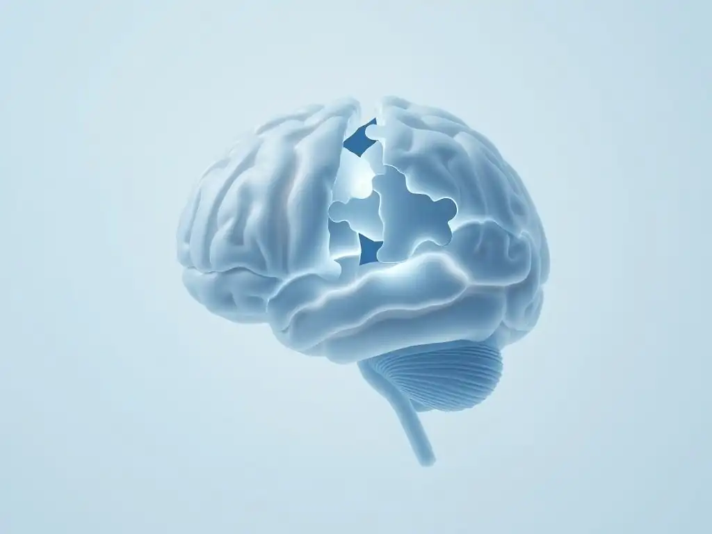
Working Memory Impairment in Depression
Working memory, the ability to temporarily hold and manipulate information, is a key executive function often impaired in depression. Individuals may report difficulty concentrating, remembering instructions, or keeping track of tasks. Neuroimaging studies link working memory deficits in depression to altered activity and structure in the dorsolateral prefrontal cortex (dlPFC) and parietal cortex, areas critical for this function. For instance, fMRI studies often show reduced activation in the dlPFC during working memory tasks in depressed patients compared to healthy controls.
This impairment isn't just a minor inconvenience; it can significantly impact daily functioning. Imagine trying to follow a complex recipe, remember a sequence of steps for a work task, or even hold a conversation without losing track of the topic – these all rely heavily on working memory. The compromised structural integrity and functional connectivity of the dlPFC, often observed as part of the changes in the depressed PFC executive function, directly contribute to these real-world difficulties.
Brain Regions' Role in Working Memory Capacity
Working memory is supported by a network of brain regions, with the dorsolateral prefrontal cortex (dlPFC) playing a central role in managing and manipulating information. The parietal cortex is involved in storing and attending to information, while the hippocampus contributes to binding information into coherent representations. Communication between these regions, facilitated by white matter tracts, is essential for efficient working memory.
In depression, structural changes like reduced gray matter volume in the dlPFC and parietal cortex, along with altered white matter integrity connecting these areas, can compromise this network. For example, studies show a correlation between reduced FA in tracts connecting frontal and parietal regions and poorer performance on working memory tests in depressed individuals. These structural underpinnings explain why working memory capacity and efficiency are often diminished, representing a core aspect of how the depressed PFC executive function is compromised.
Cognitive Flexibility and Set-Shifting Difficulties
Cognitive flexibility, the ability to switch between different mental sets, tasks, or strategies in response to changing demands, is another executive function frequently impaired in depression. This might manifest as difficulty adapting to unexpected changes, getting stuck on negative thoughts or problem-solving approaches, or struggling to see alternative perspectives. Tasks like the Wisconsin Card Sorting Test, which requires participants to shift their sorting strategy based on feedback, often reveal deficits in depressed individuals.
These difficulties are linked to dysfunction in the ventrolateral prefrontal cortex (vlPFC) and anterior cingulate cortex (ACC), regions involved in detecting conflicts, monitoring performance, and inhibiting prepotent responses to switch to a new rule. Structural changes, such as altered gray matter volume or connectivity in these areas, contribute to the observed inflexibility. This aspect of depressed PFC executive function impairment can make it challenging for individuals to cope with daily stressors or engage in effective problem-solving.
Structural Changes Affecting Cognitive Flexibility
The structural integrity of brain regions and pathways supporting cognitive flexibility is compromised in depression. Reductions in gray matter volume in the anterior cingulate cortex (ACC) and ventrolateral prefrontal cortex (vlPFC) are associated with difficulties in set-shifting and conflict monitoring. For example, research indicates that smaller ACC volume correlates with increased perseveration errors on cognitive flexibility tasks in depressed patients.
Furthermore, disruptions in white matter tracts connecting these frontal regions to other parts of the brain, such as the parietal cortex, also play a role. These structural alterations impair the efficiency of the neural circuits responsible for detecting when a current strategy is no longer working and implementing a new one. Thus, changes in the physical structure of these key brain areas, particularly within the broader context of the depressed PFC executive function network, directly undermine the capacity for flexible thinking.
Impaired Decision-Making and Planning
Decision-making and planning are complex executive functions that involve evaluating options, anticipating consequences, and organizing steps to achieve a goal. Individuals with depression often exhibit impairments in these areas, sometimes making poor choices, procrastinating, or struggling to initiate and complete tasks. This can range from difficulty making simple daily decisions to struggling with significant life choices.
These deficits are linked to alterations in the ventromedial prefrontal cortex (vmPFC) and orbitofrontal cortex (OFC), regions crucial for evaluating the value of potential outcomes and guiding behavior based on reward and punishment. Functional imaging studies often show reduced activity in these areas when depressed individuals are engaged in reward-based decision-making tasks. Structural changes, such as altered gray matter or connectivity in the vmPFC and OFC, contribute to this impaired ability to weigh options and plan effectively, reflecting a core deficit in depressed PFC executive function.
The Brain's Influence on Goal-Directed Behavior
Goal-directed behavior relies on the integration of information from various brain regions, orchestrated by the prefrontal cortex. The vmPFC and OFC are particularly important for assigning subjective value to potential outcomes and influencing choices. The dlPFC is involved in planning the steps required to achieve the desired outcome, while connections to the striatum (part of the basal ganglia) are crucial for motivation and action initiation.
In depression, structural and functional abnormalities in these interconnected regions disrupt this system. Reduced gray matter volume or altered connectivity in the vmPFC and OFC can lead to a reduced ability to experience pleasure or anticipate reward (anhedonia), making it difficult to assign value to goals. Dysfunction in the dlPFC impairs the planning component, and altered striatal connectivity can reduce motivation. These combined deficits in the brain's reward and planning circuitry fundamentally undermine goal-directed behavior, a critical aspect of depressed PFC executive function.
Attention and Focus Deficits
Difficulties with attention and concentration are common and distressing symptoms of depression. Individuals may find it hard to focus during conversations, while reading, or on tasks at work or school. This can manifest as distractibility, reduced sustained attention, or problems shifting attention appropriately.
These attentional deficits are associated with altered activity and structure in brain networks involving the prefrontal cortex (particularly the dlPFC and ACC) and parietal cortex, which are key components of the brain's attentional system. For example, studies using neuroimaging show reduced activation in the dlPFC and parietal regions during tasks requiring sustained attention in depressed individuals. Structural changes in these areas or the white matter tracts connecting them likely contribute to the observed difficulties with focus, reflecting another facet of impaired depressed PFC executive function.
Brain Activity and Sustained Attention
Sustained attention, the ability to maintain focus on a task over time, relies on the coordinated activity of the dorsal attention network, which includes the dorsolateral prefrontal cortex (dlPFC) and parts of the parietal cortex. The anterior cingulate cortex (ACC) is also involved in monitoring performance and detecting errors that might indicate a lapse in attention.
In depression, functional and structural alterations disrupt this network. Reduced functional connectivity within the dorsal attention network and between it and other networks (like the DMN) are often observed. Structurally, reduced gray matter volume in the dlPFC or ACC, or compromised white matter integrity in connections between these areas, can impair the efficiency of this network. These changes make it harder for the brain to maintain a consistent state of focused attention, contributing to the concentration problems characteristic of depression and impacting overall depressed PFC executive function.
Molecular and Cellular Mechanisms Underlying Brain Changes in Depression
Neurotransmitter Imbalances (Serotonin, Dopamine, Norepinephrine)
The monoamine hypothesis, while an oversimplification, highlights the role of neurotransmitters like serotonin, dopamine, and norepinephrine in depression. These chemicals are crucial for regulating mood, motivation, reward, and cognitive function. Imbalances in their levels or signaling are thought to contribute to the development of depressive symptoms. For instance, reduced serotonin activity is linked to mood regulation problems, while dopamine and norepinephrine are important for motivation, energy, and attention.
These neurotransmitters also influence brain structure and function more broadly. Serotonin, for example, plays a role in neurogenesis and synaptic plasticity. Dopamine is critical for the reward pathways and influences the activity of the prefrontal cortex. Norepinephrine affects arousal and attention. Alterations in these neurotransmitter systems can thus contribute to the structural changes observed in the depressed brain, indirectly impacting depressed PFC executive function by influencing neuronal health, connectivity, and activity levels.
Neurotransmitter Effects on Brain Structure
Neurotransmitters like serotonin, dopamine, and norepinephrine exert influence beyond just modulating immediate neural signaling; they also play a role in the long-term health and structure of neurons and their connections. For example, serotonin can influence the expression of genes involved in neurogenesis (the creation of new neurons) and synaptogenesis (the formation of new synapses), particularly in regions like the hippocampus and prefrontal cortex. Chronic stress, often a trigger for depression, can deplete these neurotransmitters and reduce levels of neurotrophic factors like Brain-Derived Neurotrophic Factor (BDNF), which supports neuronal survival and growth.
Reduced levels of BDNF, often observed in depression, are associated with decreased neurogenesis in the hippocampus and potential atrophy in the prefrontal cortex. Similarly, dopamine and norepinephrine are involved in regulating synaptic plasticity and the function of neuronal circuits, including those critical for executive functions in the PFC. Thus, imbalances in these neurotransmitter systems can contribute to the observed gray matter volume reductions, altered white matter integrity, and synaptic dysfunction that characterize the depressed brain, directly influencing depressed PFC executive function.
Neuroinflammation and Brain Dysfunction
Growing evidence points to the role of neuroinflammation, an inflammatory response within the brain, in the pathophysiology of depression. Studies have shown elevated levels of inflammatory markers, such as cytokines (e.g., IL-6, TNF-alpha, CRP), in the blood and cerebrospinal fluid of depressed individuals. These inflammatory molecules can cross the blood-brain barrier and affect brain function.
Neuroinflammation can impact brain structure and function through various mechanisms. It can impair neurotransmitter synthesis and reuptake, particularly affecting monoamines. It can also disrupt the integrity of the blood-brain barrier, leading to further influx of inflammatory cells. Furthermore, chronic inflammation can impair synaptic plasticity and neurogenesis, and potentially contribute to glial cell dysfunction (astrocytes and microglia), which are crucial for supporting neuronal health. This inflammatory state contributes to neuronal dysfunction and loss, impacting areas like the prefrontal cortex and hippocampus, thereby influencing depressed PFC executive function.
Inflammation's Role in Brain Changes
Inflammation within the brain can directly contribute to the structural changes seen in depression. Chronic exposure to inflammatory cytokines can lead to oxidative stress and excitotoxicity, damaging neurons and potentially leading to neuronal atrophy or death. For example, studies suggest that elevated levels of inflammatory markers are associated with reduced gray matter volume in the prefrontal cortex and hippocampus.
Moreover, neuroinflammation can impair the function of glial cells, which are essential for maintaining the health and function of neurons and synapses. Activated microglia, the brain's immune cells, can release neurotoxic substances when chronically stimulated by inflammation. This inflammatory environment hinders neurogenesis in the hippocampus and impairs synaptic plasticity, the brain's ability to form and strengthen connections. These processes collectively contribute to the observed structural deficits and functional impairments in the depressed brain, directly impacting the neural basis of depressed PFC executive function.
Reduced Neurogenesis and Synaptic Plasticity
Neurogenesis, the birth of new neurons, primarily occurs in the hippocampus in the adult brain, and synaptic plasticity, the ability of synapses to strengthen or weaken over time, is fundamental to learning and memory and occurs throughout the brain, including the prefrontal cortex. Both processes are crucial for brain health and function, and both are impaired in depression. Animal models of depression and post-mortem studies in humans suggest reduced rates of hippocampal neurogenesis in depressed states.
Similarly, synaptic plasticity, which is essential for the flexible functioning of neural circuits, appears to be compromised in depression. This can involve reduced expression of proteins involved in synaptic function and structure. For example, studies have shown reduced levels of synaptic markers in the prefrontal cortex of individuals with depression. Impaired neurogenesis and synaptic plasticity weaken the structural and functional foundation of brain circuits involved in mood, memory, and executive function, contributing significantly to the challenges associated with depressed PFC executive function.
Structural Impacts of Reduced Neurogenesis
Reduced neurogenesis, particularly in the hippocampus, has direct structural consequences. A lower rate of new neuron formation contributes to the observed reduction in hippocampal volume in depressed individuals. These newly born neurons are thought to play a role in integrating emotional and contextual information, and their depletion can impair the hippocampus's ability to modulate activity in other brain regions, including the prefrontal cortex.
Furthermore, chronic stress and inflammation, factors implicated in depression, can suppress neurogenesis and impair the survival and integration of new neurons. While the direct impact of reduced neurogenesis on PFC gray matter volume is less established than in the hippocampus, the interconnectedness of these regions means that hippocampal dysfunction can indirectly affect PFC function and potentially structure over time. Impaired synaptic plasticity in the PFC itself, a process influenced by factors that also affect neurogenesis, directly contributes to the altered connectivity and functional deficits underlying depressed PFC executive function.
How Depressed Brain Changes Affect Daily Life
Impact on Work and Academic Performance
The executive function deficits stemming from structural changes in the depressed brain, particularly affecting the depressed PFC executive function, can significantly impair performance in work and academic settings. Difficulties with concentration, working memory, planning, and decision-making make it challenging to complete tasks efficiently, manage deadlines, organize work, and learn new information. For example, a study might show that individuals with depression score significantly lower on tests of sustained attention and task switching, skills critical for academic and professional success.
These cognitive struggles, combined with symptoms like fatigue and loss of motivation, can lead to decreased productivity, increased errors, difficulty retaining employment, or challenges succeeding in school. The effort required to perform tasks that were previously routine can become overwhelming, leading to feelings of frustration and inadequacy, further exacerbating depressive symptoms.
Executive function deficits, particularly those related to cognitive flexibility, emotional regulation, and social cognition (which relies heavily on the vmPFC and OFC), can also complicate social interactions. Difficulty interpreting social cues, regulating emotional responses during conversations, or flexibly adapting communication style can strain relationships. Impaired planning and motivation can make it hard to initiate social activities or maintain commitments.
For instance, impaired cognitive flexibility might make it difficult to shift perspective during a disagreement, leading to conflict. Difficulty with emotional regulation, linked to disrupted fronto-limbic circuits, can result in heightened irritability or withdrawal. These challenges, stemming from the impact on depressed PFC executive function and its connections, can lead to social isolation, misunderstanding, and further feelings of loneliness and disconnection, perpetuating the cycle of depression.
Difficulties in Self-Care and Daily Routines
Maintaining daily routines and engaging in self-care activities often requires planning, motivation, and the ability to initiate and complete tasks – all executive functions. The impaired planning, decision-making, and initiation abilities associated with structural changes in the depressed brain make these basic activities difficult. Simple tasks like getting out of bed, showering, preparing meals, or managing finances can feel insurmountable.
This difficulty with self-care is a direct consequence of the impact on depressed PFC executive function. The reduced motivation linked to reward circuit dysfunction (involving the OFC and striatum) makes initiating tasks hard, while impaired planning (dlPFC) means organizing the steps feels overwhelming. This leads to neglect of personal needs, contributing to a decline in physical health and reinforcing feelings of helplessness and hopelessness.
Diagnosing Executive Dysfunction in Depression

Neuropsychological Testing for Executive Function
Neuropsychological testing is a standard method used to objectively assess executive function deficits in individuals with depression. These tests evaluate specific abilities such as working memory (e.g., digit span, N-back task), cognitive flexibility (e.g., Wisconsin Card Sorting Test, Trail Making Test Part B), planning and problem-solving (e.g., Tower of London), and attention (e.g., Continuous Performance Test).
Administering a battery of these tests provides a profile of an individual's cognitive strengths and weaknesses, helping to identify specific areas of executive dysfunction. For example, a person might show significant impairment on tasks requiring set-shifting but perform relatively normally on working memory tasks. These tests provide concrete evidence of how structural and functional changes in the brain, particularly impacting depressed PFC executive function, translate into measurable cognitive difficulties.
Clinical Interviews and Behavioral Observations
In addition to formal testing, clinical interviews and observations of an individual's behavior provide crucial information about executive function difficulties in daily life. Clinicians ask about challenges with concentration, memory, organization, decision-making, and task completion. They might inquire about specific examples of difficulties at work, school, or home.
Observing how a person approaches tasks during the interview, their level of organization, ability to stay on topic, and responses to questions requiring planning or problem-solving can also reveal executive function challenges. While subjective, this information complements neuropsychological test results by providing context on how depressed PFC executive function deficits manifest in real-world behavior and impact daily functioning.
Treatment Strategies Targeting Brain Dysfunction in Depression
Antidepressant Medications and Brain Activity
Antidepressant medications, particularly selective serotonin reuptake inhibitors (SSRIs) and serotonin-norepinephrine reuptake inhibitors (SNRIs), primarily target neurotransmitter systems. While their primary effect is on mood symptoms, research suggests they can also influence brain structure and function over time. For example, some studies indicate that successful antidepressant treatment can be associated with increases in gray matter volume in areas like the hippocampus and prefrontal cortex, potentially related to enhanced neurogenesis or reduced neuronal atrophy.
Antidepressants can also modulate activity and connectivity within neural circuits implicated in depression, including those involving the prefrontal cortex. By restoring more balanced neurotransmitter levels, they can improve the efficiency of communication between brain regions. While the effects on reversing structural changes are often gradual and partial, improvements in brain activity can contribute to improved executive function, offering hope for individuals struggling with depressed PFC executive function.
Cognitive Behavioral Therapy (CBT) and Executive Function
Cognitive Behavioral Therapy (CBT) is a psychotherapy that helps individuals identify and change negative thought patterns and behaviors. While not directly targeting brain structure, CBT can lead to functional and potentially structural changes in the brain over time, particularly in regions involved in cognitive control and emotional regulation, such as the prefrontal cortex and amygdala.
CBT helps individuals develop skills related to executive functions, such as problem-solving, planning, and cognitive restructuring (which involves cognitive flexibility to challenge and change negative thoughts). By practicing these skills, individuals can strengthen the neural pathways associated with healthier cognitive processing. Studies using fMRI have shown changes in PFC activity patterns after successful CBT, suggesting that this therapy can help improve depressed PFC executive function by teaching individuals more effective ways to use their cognitive resources.
Brain Stimulation Techniques (TMS, tDCS)
Brain stimulation techniques offer more direct ways to modulate brain activity and potentially influence structure. Transcranial Magnetic Stimulation (TMS) uses magnetic pulses to stimulate specific brain regions, typically the dorsolateral prefrontal cortex (dlPFC), which is often underactive in depression and crucial for executive function. Repeated TMS sessions can normalize activity in the dlPFC and improve connectivity within related circuits.
Transcranial Direct Current Stimulation (tDCS) uses low-level electrical currents to modulate neuronal excitability in targeted areas. Both TMS and tDCS applied to the PFC have shown promise in alleviating depressive symptoms and improving cognitive function, including aspects of executive function. These techniques represent approaches that directly attempt to restore healthier brain activity patterns and connectivity, offering potential benefits for depressed PFC executive function by directly engaging the affected brain regions.
Lifestyle Modifications and Brain Health
Lifestyle factors play a significant role in brain health and can influence neurogenesis, synaptic plasticity, and inflammation. Regular physical exercise, for instance, has been shown to increase BDNF levels, promote neurogenesis in the hippocampus, and improve executive function in both healthy individuals and those with depression. A healthy diet, particularly one rich in omega-3 fatty acids and antioxidants, can reduce inflammation and support neuronal health.
Adequate sleep is crucial for cognitive function and synaptic plasticity, and chronic sleep deprivation can negatively impact the PFC. Stress reduction techniques like mindfulness and meditation have been shown to alter brain structure and function, potentially increasing gray matter in the PFC and reducing amygdala volume. Engaging in mentally stimulating activities also supports cognitive function and may promote synaptic health. These lifestyle modifications offer accessible ways to support overall brain health and potentially mitigate some of the structural and functional changes associated with depression, thereby supporting depressed PFC executive function.
The Future of Research on Depressed Brain and Executive Function
Advancements in Neuroimaging Techniques
Future research into the depressed brain and executive function will heavily rely on advancements in neuroimaging techniques. Higher-resolution MRI and fMRI can provide more detailed insights into subtle structural changes and dynamic functional connectivity within specific subregions of the prefrontal cortex and their circuits. Techniques like diffusion spectrum imaging (DSI) or HARDI can offer a more detailed picture of complex white matter tract architecture.
Combining different imaging modalities, such as simultaneously measuring brain activity (fMRI) and neurotransmitter levels (PET), will allow researchers to better understand the interplay between molecular changes, structural alterations, and functional deficits. These advanced techniques will enable a more precise mapping of the neural correlates of specific executive function impairments in depression, leading to a deeper understanding of depressed PFC executive function at a granular level.
Personalized Treatment Approaches Based on Brain Biomarkers
A key goal of future research is to develop personalized treatment approaches based on individual brain characteristics or biomarkers. Identifying specific structural or functional patterns in a patient's brain (e.g., the severity of gray matter reduction in the dlPFC, specific white matter tract abnormalities) could help predict their response to different treatments, whether medication, therapy, or stimulation techniques.
For example, individuals with pronounced structural deficits in the dlPFC might respond better to TMS targeting that region, while those with more widespread connectivity issues might benefit from treatments that enhance overall brain network efficiency. Developing reliable brain biomarkers for different subtypes of depression characterized by specific patterns of depressed PFC executive function impairment could revolutionize treatment selection, moving towards more effective and targeted interventions.
Novel Therapeutic Targets for Brain Repair
Understanding the molecular and cellular mechanisms underlying structural brain changes in depression, such as neuroinflammation, reduced neurogenesis, and synaptic dysfunction, is opening doors for novel therapeutic targets aimed at brain repair or restoration. Future treatments might focus on developing compounds that promote neurogenesis in the hippocampus, enhance synaptic plasticity in the prefrontal cortex, or specifically target neuroinflammatory pathways.
Research is exploring agents that boost BDNF levels, modulate glial cell activity, or directly protect neurons from stress-induced damage. While still largely experimental, these approaches aim to address the root biological causes of structural and functional deficits in the depressed brain, offering the potential to not only alleviate mood symptoms but also restore cognitive function and improve depressed PFC executive function.
Frequently Asked Questions
How does depression affect the structure of the Prefrontal Cortex (PFC)?
Depression can lead to structural changes in the PFC, including a reduction in gray matter volume. This reduction primarily affects regions crucial for executive function.
What is executive function, and why is it important?
Executive function refers to a set of cognitive processes that control and regulate other cognitive abilities. It is crucial for planning, problem-solving, and decision-making.
What are the key symptoms of depressed PFC executive function?
Symptoms include difficulty concentrating, impaired decision-making, and a reduced ability to plan and organize. This significantly impacts daily life.
Can changes in PFC structure due to depression be reversed?
While challenging, treatment options like therapy and medication can help improve PFC function and potentially reverse some structural changes over time.
Where can I find resources and support for dealing with depression and its impact on executive function?
The Mind Care Guide offers information, support, and resources for various mental health challenges, including depression and its cognitive effects. You can also consult with a mental health professional.
Final Reflection: Rebuilding the Bridge to Executive Function
The compromised prefrontal cortex (PFC) in depression isn't just a neurological curiosity; it's a critical factor hindering millions in their daily lives. Understanding the structural changes within the PFC, as detailed in this article, is paramount to developing effective treatments and fostering a more compassionate understanding of the challenges faced by individuals struggling with depression. The implications extend far beyond the clinical setting, impacting relationships, careers, and overall well-being.
The ongoing struggle to bridge the gap between neurological understanding and practical application is a deeply personal one for me. Witnessing the debilitating effects of depression on executive function, from impaired decision-making to difficulty with planning and emotional regulation, underscores the urgency of further research and improved therapeutic interventions. The mind is not a vessel to be filled, but a fire to be kindled.
– Plutarch. This quote resonates deeply, as it highlights the potential for renewal and growth even in the face of significant neurological challenges. We must continue to seek ways to rekindle the "fire" of executive function in those affected by depression.
Consider the impact of these structural changes on your own life, and the lives of those around you. Perhaps you've experienced the frustration of struggling with tasks, or witnessed a loved one grappling with the complexities of depression. This article is not just an academic exploration; it's a call to action.
Let's advocate for increased research funding, improved access to mental healthcare, and a more empathetic societal understanding of the invisible struggles faced by individuals with depression. By fostering a culture of support and understanding, we can help build the bridges necessary to restore executive function and empower individuals to live fulfilling lives.
Unlock Your Brain's Potential: Take Charge of Your Executive Function
Thanks for exploring the fascinating world of prefrontal cortex function and its connection to depression with us! We hope you gained valuable insights into how structural changes impact your daily life. Ready to put this knowledge into action?
- Prioritize one small, manageable task today that challenges your executive function. This could be anything from planning your day meticulously to tackling a long-postponed project – even for just 15 minutes. Small wins build confidence!
- Share this article with a friend or family member who might also benefit from understanding the PFC's role in mental wellbeing. Starting a conversation can be incredibly powerful.
- Consider journaling about your experience with executive function challenges, noticing patterns and identifying areas for improvement. This mindful reflection can be surprisingly revealing.
What small step will you take today to strengthen your executive function?