
Seeing the Unseen: How Neuroimaging Reveals Inflammation in Mental Illness

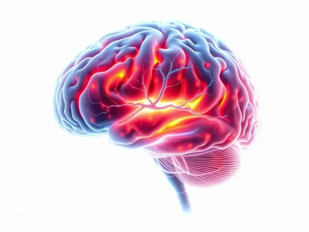
Welcome to Mind Care Guide, a space dedicated to understanding and nurturing your mental well-being. Are you curious about the hidden connections between your physical and mental health? Ever wondered if something unseen could be impacting your mood and cognition?
Our article, "Seeing the Unseen: How Neuroimaging Reveals Inflammation in Mental Illness," delves into cutting-edge research using neuroimaging to uncover the role of inflammation in mental health. Join us as we explore this fascinating intersection and discover how these insights could pave the way for new approaches to care. Read on to unlock a deeper understanding of the brain-body connection and its impact on mental well-being.
Seeing the Unseen: How Neuroimaging Reveals Inflammation in Mental Illness
- Introduction: Understanding the Link Between Inflammation and Mental Health
- Neuroimaging Techniques for Detecting Inflammation in Mental Illness
- The Significance of Neuroimaging Findings in Inflammation and Mental Illness
- Challenges and Future Directions in Neuroimaging and Inflammation Research
- Case Studies: Neuroimaging Illuminating Inflammation in Specific Mental Illnesses
- The Future of Neuroimaging in Understanding and Treating Mental Illness
- Conclusion: Neuroimaging as a Window into Inflammation and Mental Wellbeing
-
Frequently Asked Questions
- How does neuroimaging help us understand the link between inflammation and mental illness?
- What types of mental illnesses are most commonly associated with inflammation?
- What specific inflammatory markers are typically investigated using neuroimaging in relation to mental health?
- Can neuroimaging be used to predict the effectiveness of treatments targeting inflammation in mental illness?
- Are there any risks associated with using neuroimaging to study inflammation and mental illness?
- Final Reflection: Unveiling the Hidden Landscape of Mental Illness
- Unlock Your Brain's Potential: Discover the Power of Understanding Inflammation
Introduction: Understanding the Link Between Inflammation and Mental Health
For decades, mental illnesses were primarily understood through psychological and neurochemical lenses, focusing on neurotransmitter imbalances or cognitive patterns. However, a growing body of research is highlighting another critical player: inflammation. This biological process, typically associated with the body's response to injury or infection, is increasingly implicated in the onset and progression of various mental health conditions, including depression, schizophrenia, and anxiety disorders. The brain, once thought to be immune-privileged, is now known to have its own complex immune system, and when this system becomes dysregulated, it can contribute to psychiatric symptoms.
The connection between inflammation and mental health is complex and bidirectional. Chronic stress, poor diet, infections, and genetic factors can trigger inflammatory responses that impact brain function. Conversely, mental illnesses themselves can sometimes perpetuate inflammatory states. Understanding this intricate relationship is crucial for developing more effective diagnostic tools and treatments. This is where advanced techniques like neuroimaging play a transformative role, allowing researchers and clinicians to peer into the living brain and observe the subtle, and sometimes not so subtle, signs of inflammation.
The Emerging Role of Neuroinflammation in Mental Disorders
Neuroinflammation, the inflammatory response within the brain or spinal cord, is now recognized as a significant factor in the pathophysiology of numerous psychiatric conditions. Unlike acute inflammation which is a protective response, chronic or dysregulated neuroinflammation can lead to neuronal damage, synaptic dysfunction, and alterations in neural circuits critical for mood, cognition, and behavior.
Research indicates elevated inflammatory markers, such as cytokines like IL-6, TNF-alpha, and CRP, are often found in the blood and cerebrospinal fluid of individuals with major depressive disorder, bipolar disorder, and schizophrenia, compared to healthy controls. For instance, studies have shown that patients experiencing a depressive episode can have CRP levels up to 50% higher than non-depressed individuals.
Furthermore, post-mortem brain studies have revealed increased numbers and activation states of microglia and astrocytes – the brain's primary immune cells – in specific brain regions of individuals with severe mental illness. These findings underscore that inflammation is not merely a peripheral symptom but an active process occurring within the brain, directly impacting mental health.
Why Neuroimaging is Crucial for Studying Inflammation in the Brain
While blood and CSF markers provide valuable systemic information, they offer an indirect view of what is happening within the brain itself. Neuroimaging techniques provide a non-invasive way to visualize brain structure, function, and even molecular processes in living individuals. This capability is paramount for studying neuroinflammation in real-time.
Neuroimaging allows researchers to pinpoint where in the brain inflammation is occurring, how severe it is, and how it correlates with specific symptoms or cognitive impairments. Techniques like Positron Emission Tomography (PET) can use specific radiotracers that bind to markers of activated immune cells, providing quantitative data on inflammatory activity in different brain regions. Magnetic Resonance Imaging (MRI) can detect structural changes associated with inflammation, such as white matter alterations or changes in brain volume.
By combining these methods, scientists can build a more comprehensive picture of the relationship between inflammation mental illness neuroimaging findings reveal. This direct observation is vital for moving beyond correlational studies and establishing a clearer understanding of causal pathways and potential therapeutic targets.
Neuroimaging Techniques for Detecting Inflammation in Mental Illness
MRI and Inflammation: Visualizing Brain Structure and Activity
Magnetic Resonance Imaging (MRI) is a versatile neuroimaging technique that uses strong magnetic fields and radio waves to create detailed images of brain structures. While not directly measuring inflammation, MRI can reveal structural and functional changes that are often consequences or correlates of neuroinflammatory processes.
Different MRI sequences can highlight specific tissue properties. For instance, Diffusion Tensor Imaging (DTI), a type of MRI, measures the diffusion of water molecules, providing insights into the integrity of white matter tracts. Inflammation can disrupt myelin sheaths and axons, altering water diffusion patterns that are detectable with DTI. Functional MRI (fMRI) measures brain activity by detecting changes in blood flow; neuroinflammation can alter neurovascular coupling, impacting fMRI signals and indicating dysfunctional brain circuits. Structural MRI can also detect subtle volume changes in specific brain regions, which may be linked to chronic inflammatory damage or glial cell proliferation.
How MRI Detects Changes Related to Inflammation
MRI detects changes related to inflammation through various mechanisms. One key aspect is its sensitivity to changes in tissue water content and composition. Inflammation can lead to edema (swelling) or changes in cellular density, which alter the magnetic resonance signal. For example, T2-weighted MRI sequences are particularly sensitive to areas with increased water content, often seen in inflammatory lesions.
Furthermore, contrast agents containing gadolinium can be used in some MRI protocols. These agents typically do not cross the intact blood-brain barrier (BBB). However, inflammation can compromise the integrity of the BBB, allowing the contrast agent to leak into brain tissue, which is then visible on the MRI scan as areas of enhancement.
While primarily used for conditions like multiple sclerosis or tumors, this principle can sometimes be applied in research settings to assess BBB integrity in the context of neuroinflammation associated with mental illness. Advanced MRI techniques like quantitative MRI (qMRI) can also provide more specific measures of tissue properties, potentially revealing subtle microstructural changes linked to chronic inflammation that are not apparent on standard scans.
The Application of MRI in Major Depressive Disorder Research
MRI has been extensively used in research on Major Depressive Disorder (MDD), often revealing structural and functional abnormalities in brain regions known to be affected by inflammation, such as the prefrontal cortex, hippocampus, and amygdala. Studies using structural MRI have reported reduced hippocampal volume in patients with chronic depression, a finding potentially linked to the detrimental effects of chronic stress and inflammation on neurogenesis and neuronal survival.
Functional MRI studies in MDD have shown altered connectivity within neural networks involved in mood regulation, reward processing, and cognitive control. For instance, there's often hypoactivity in certain prefrontal areas and hyperactivity in the amygdala. While these findings are not solely attributable to inflammation, research integrating fMRI with peripheral inflammatory markers or other neuroimaging modalities suggests that inflammation likely contributes to these circuit dysfunctions.
DTI studies in MDD patients have also indicated impaired white matter integrity in tracts connecting these regions, which could be a consequence of inflammatory processes affecting glial cells and myelin. Collectively, MRI findings provide valuable structural and functional correlates that support the hypothesis of neuroinflammation playing a role in the pathogenesis of MDD.
PET Scans and Inflammation: Measuring Molecular Activity
Positron Emission Tomography (PET) is a powerful neuroimaging technique that allows for the quantitative measurement of specific molecular targets in the living brain. Unlike MRI, which primarily shows structure and blood flow, PET can directly visualize and quantify the distribution and binding of radioactive tracers (radioligands) that are designed to target specific molecules or receptors. This capability makes PET uniquely suited for studying neuroinflammation at a molecular level.
PET imaging enables researchers to measure parameters such as the density of activated immune cells, the expression of inflammatory enzymes, or the integrity of neurotransmitter systems that might be affected by inflammation. By injecting a small amount of a radioligand that binds to a marker of neuroinflammation, such as the Translocator Protein (TSPO), scientists can generate images that reflect the spatial distribution and intensity of inflammatory activity throughout the brain. The amount of radioactivity detected in a given brain region correlates with the concentration of the target molecule, providing a quantitative measure of neuroinflammation.
Radioligands: Targeting Inflammatory Markers with PET
The development of specific radioligands has been crucial for advancing PET imaging of neuroinflammation. The most widely used target for neuroinflammation PET is the Translocator Protein (TSPO), which is upregulated in activated microglia and astrocytes. Various TSPO-binding radioligands have been developed over the years, such as [11C]PK11195, [11C]PBR28, and [18F]FEPPA. These tracers bind to TSPO, and their uptake in brain regions reflects the level of glial cell activation, a key indicator of neuroinflammation.
However, TSPO radioligands have limitations, including genetic polymorphism effects on binding affinity for some tracers and the challenge of separating specific binding from non-specific binding. Researchers are also developing PET tracers targeting other inflammatory markers, such as cyclooxygenase-2 (COX-2), a key enzyme in the synthesis of prostaglandins involved in inflammation, or cytokine receptors. These novel radioligands hold promise for providing more specific and sensitive measures of different aspects of the inflammatory cascade in the brain, further enhancing the ability of PET to reveal inflammation mental illness neuroimaging research explores.
PET Imaging in Schizophrenia Studies
PET imaging using TSPO radioligands has provided compelling evidence of neuroinflammation in schizophrenia. Multiple studies have reported increased TSPO binding, indicative of microglial activation, in various brain regions of individuals with schizophrenia compared to healthy controls. These regions often include areas implicated in the pathophysiology of schizophrenia, such as the prefrontal cortex, temporal lobe, thalamus, and hippocampus.
For example, research using [11C]PBR28 has shown elevated TSPO binding in the frontal cortex and hippocampus of patients with chronic schizophrenia. Some studies suggest that the level of microglial activation correlates with symptom severity, particularly positive symptoms like hallucinations and delusions, or cognitive deficits.
While findings across studies are not entirely consistent, possibly due to variations in patient populations, illness stage, and methodology, the overall body of PET research supports the presence of neuroinflammatory processes in the schizophrenic brain. This provides crucial support for the hypothesis that inflammation plays a role in this complex disorder and highlights potential targets for novel anti-inflammatory therapies.
SPECT Scans: A Complementary Neuroimaging Approach
Single-Photon Emission Computed Tomography (SPECT) is another nuclear medicine neuroimaging technique that, like PET, uses radioactive tracers to visualize biological processes. While SPECT generally offers lower spatial resolution compared to PET, it is often more readily available and can utilize a wider range of radioisotopes with longer half-lives, making certain types of studies more feasible.
SPECT can be used to assess cerebral blood flow, which can be altered by neuroinflammation. Inflammatory processes can affect the microvasculature and disrupt blood flow patterns. Additionally, similar to PET, SPECT can utilize radioligands that bind to specific targets related to inflammation or immune function, although the development of SPECT tracers specifically for neuroinflammation markers like TSPO is less advanced than for PET. However, SPECT has been used to investigate aspects of brain function and receptor availability that may be indirectly influenced by inflammation.
SPECT Imaging of Neuroinflammation and Immune Response
SPECT imaging can provide insights into the brain's immune response and neuroinflammation, albeit often indirectly or through markers that are also accessible via PET. For instance, SPECT tracers targeting dopamine or serotonin transporters/receptors have been used to study alterations in these neurotransmitter systems, which are known to be affected by inflammatory cytokines. Changes in transporter or receptor availability observed with SPECT could potentially reflect the downstream consequences of neuroinflammatory processes on neuronal function.
While direct SPECT imaging of microglial activation using TSPO tracers is less common than with PET due to tracer limitations, some research explores this avenue. More broadly, SPECT studies assessing regional cerebral blood flow can reveal areas of hypo- or hyperperfusion, which might be indicative of metabolic or vascular changes associated with inflammation.
For example, reduced blood flow in certain brain regions could suggest impaired neuronal activity or vascular damage potentially linked to chronic inflammation. Thus, SPECT serves as a complementary tool, providing functional and receptor-level information that can be integrated with findings from other modalities to build a fuller picture of inflammation mental illness neuroimaging studies uncover.
Using SPECT to Investigate Bipolar Disorder
SPECT imaging has been employed in research on Bipolar Disorder (BD) to investigate functional abnormalities and potential links to neuroinflammation. Studies using SPECT to measure regional cerebral blood flow (rCBF) have reported altered perfusion patterns in individuals with BD, often showing changes in areas like the prefrontal cortex, amygdala, and basal ganglia. These findings vary depending on the mood state (manic, depressive, euthymic) of the patient.
While SPECT does not directly image inflammation markers as readily as PET, the observed rCBF abnormalities in BD could be influenced by neuroinflammatory processes affecting neurovascular coupling or neuronal activity. Some research suggests that inflammatory cytokines can impact neurotransmitter systems, particularly dopamine and serotonin, which are targets for SPECT radioligands.
Therefore, SPECT studies examining dopamine or serotonin transporter/receptor availability in BD patients could indirectly reflect the impact of inflammation on these systems. Although direct inflammation neuroimaging in BD is more commonly done with PET, SPECT contributes valuable functional data that complements the growing understanding of inflammation's role in this complex mood disorder.
The Significance of Neuroimaging Findings in Inflammation and Mental Illness
Identifying Biomarkers of Inflammation in Mental Disorders
Neuroimaging techniques, particularly PET with specific radioligands, are instrumental in identifying in vivo biomarkers of neuroinflammation in mental disorders. Unlike peripheral blood markers, which reflect systemic inflammation, neuroimaging allows researchers to visualize inflammatory processes occurring specifically within the brain.
For example, elevated TSPO binding measured by PET in certain brain regions can serve as a potential biomarker for neuroinflammation in conditions like depression or schizophrenia. The pattern and intensity of this binding could correlate with disease severity, symptom profiles, or treatment response. Identifying such imaging biomarkers is critical for several reasons: it helps validate the role of neuroinflammation in specific disorders, provides objective measures that can be used in clinical trials, and offers potential targets for diagnostic tests.
While still largely a research tool, the hope is that these neuroimaging markers could eventually be integrated into clinical practice to aid diagnosis and prognosis. Research is actively working to standardize these measures and determine their clinical utility.
Neuroimaging as a Tool for Early Diagnosis and Intervention
The ability of neuroimaging to detect subtle signs of neuroinflammation before overt structural damage occurs or severe symptoms manifest holds significant promise for early diagnosis and intervention in mental illness. If neuroinflammation is a contributing factor to the development of a disorder, detecting it early through neuroimaging could allow for preventative or early-stage treatments targeting inflammation.
For instance, identifying elevated microglial activation via PET in young individuals at high risk for developing schizophrenia could flag them for early intervention strategies, potentially including anti-inflammatory agents, alongside standard care. Similarly, detecting neuroinflammation in individuals experiencing their first episode of depression might indicate a specific inflammatory subtype of the disorder that could respond better to certain treatments. While still in the research phase, the potential for neuroimaging to serve as a predictive tool or an aid in early diagnosis, enabling timely and targeted interventions, is a major area of focus in inflammation mental illness neuroimaging research.
Personalized Treatment Approaches Based on Neuroimaging Results
Perhaps one of the most exciting implications of neuroimaging findings on inflammation in mental illness is the potential for personalized medicine. Mental disorders are highly heterogeneous, and individuals with the same diagnosis can have different underlying biological causes and symptom profiles. Neuroimaging could help stratify patients based on the presence and pattern of neuroinflammation.
If neuroimaging reveals significant neuroinflammation in a patient with depression, for example, they might be more likely to benefit from anti-inflammatory medications or lifestyle interventions aimed at reducing inflammation, compared to a patient with depression but no detectable neuroinflammation. This approach moves away from a one-size-fits-all model towards tailoring treatments to the individual patient's specific neurobiological profile. Clinical trials are beginning to explore whether using neuroimaging to identify inflammatory subtypes of mental illness can improve treatment outcomes, paving the way for more effective and personalized care.
Challenges and Future Directions in Neuroimaging and Inflammation Research
Limitations of Current Neuroimaging Techniques
Despite significant advancements, current neuroimaging techniques for studying inflammation in mental illness face several limitations. For PET imaging of TSPO, the genetic polymorphism of the TSPO gene affects tracer binding affinity for some radioligands, requiring genotyping of participants or the use of tracers less affected by this polymorphism. The signal-to-noise ratio for TSPO tracers can also be relatively low in certain brain regions.
MRI is sensitive to structural and functional changes but does not directly measure inflammatory molecules. Interpreting MRI findings in the context of inflammation often requires correlation with other markers or techniques. Furthermore, obtaining neuroimaging scans, especially PET, can be costly and requires specialized equipment and expertise, limiting their widespread use in routine clinical practice. The interpretation of complex neuroimaging data also requires sophisticated analysis techniques, and standardization across different research sites remains a challenge.
Developing More Sensitive and Specific Imaging Agents
A key future direction in inflammation mental illness neuroimaging research is the development of more sensitive and specific imaging agents. While TSPO tracers have been valuable, they reflect general glial activation and do not distinguish between the specific roles of microglia and astrocytes or different activation states (e.g., pro-inflammatory vs. anti-inflammatory).
Researchers are actively developing novel PET and potentially SPECT radioligands that target other aspects of the inflammatory cascade. This includes tracers for specific cytokine receptors, enzymes like COX-2, or markers of specific immune cell subtypes or activation pathways. The goal is to create tracers that can provide a more nuanced understanding of the specific inflammatory processes involved in different mental illnesses. More specific tracers could improve the accuracy of neuroimaging as a diagnostic tool and help identify precise molecular targets for therapeutic interventions.
Integrating Neuroimaging with Other Biological Markers
The future of understanding the complex interplay between inflammation and mental illness lies in integrating neuroimaging findings with other biological markers. Neuroimaging provides spatial and temporal information about brain activity and pathology, but combining it with data from genomics, proteomics, metabolomics, and peripheral inflammatory markers can provide a more complete picture.
For example, combining PET imaging of neuroinflammation with genetic data (e.g., identifying risk genes associated with inflammatory pathways) could help pinpoint individuals at higher risk or reveal specific genetic influences on brain inflammation. Integrating neuroimaging with blood or CSF cytokine levels can help determine the relationship between systemic and neuroinflammation in specific patient populations. This multi-modal approach, combining insights from inflammation mental illness neuroimaging studies with other 'omics' data, is essential for unraveling the complex biological networks underlying mental disorders and developing truly comprehensive and personalized treatment strategies.
Case Studies: Neuroimaging Illuminating Inflammation in Specific Mental Illnesses

Case Study 1: Neuroinflammation in Major Depressive Disorder (MDD)
Major Depressive Disorder (MDD) is a debilitating condition, and research increasingly points to neuroinflammation as a contributing factor, particularly in treatment-resistant depression or depression with atypical features. Neuroimaging has been crucial in providing in vivo evidence for this link.
Neuroimaging Evidence of Increased Inflammation in MDD
Multiple PET studies using TSPO radioligands have reported evidence of increased microglial activation in specific brain regions of individuals with MDD compared to healthy controls. These areas commonly include the anterior cingulate cortex, prefrontal cortex, insula, and amygdala – regions involved in mood regulation, reward processing, and cognitive control. For instance, a meta-analysis of TSPO PET studies in MDD found significantly elevated binding in the anterior cingulate cortex.
Some studies also show correlations between the degree of TSPO binding and the severity of depressive symptoms or the presence of anxiety. While findings can vary based on the specific radioligand used and patient characteristics, the cumulative neuroimaging evidence strongly supports the presence of neuroinflammation in subsets of patients with MDD.
These findings are complemented by MRI studies showing structural changes like reduced hippocampal volume or altered white matter integrity in MDD patients, changes that could be consequences of chronic neuroinflammation. Functional MRI studies showing altered connectivity in mood circuits also align with the idea that inflammation disrupts normal brain function. Together, these neuroimaging modalities provide converging evidence that neuroinflammatory processes are active in the brains of many individuals experiencing depression.
Implications for Treatment Strategies in MDD
The neuroimaging evidence of neuroinflammation in MDD has significant implications for treatment strategies. It suggests that for a subset of patients, targeting inflammation could be a viable therapeutic approach. Clinical trials are currently investigating the efficacy of anti-inflammatory agents, such as non-steroidal anti-inflammatory drugs (NSAIDs), cytokine inhibitors, or minocycline (an antibiotic with anti-inflammatory properties), as adjuncts to standard antidepressant treatment, particularly in patients with evidence of elevated inflammation (e.g., high CRP levels in blood).
Neuroimaging could potentially help identify which patients are most likely to respond to these anti-inflammatory treatments. For example, a patient with MDD exhibiting high TSPO binding in mood-related brain regions on a PET scan might be a better candidate for anti-inflammatory intervention than a patient without such evidence of neuroinflammation. This moves towards a precision medicine approach, tailoring treatment based on the individual's specific neurobiological profile revealed by techniques like inflammation mental illness neuroimaging.
Case Study 2: Neuroinflammation in Schizophrenia
Schizophrenia is a severe psychiatric disorder characterized by hallucinations, delusions, disorganized thinking, and cognitive deficits. While neurotransmitter dysfunction (particularly dopamine) has been a primary focus, research increasingly points to neurodevelopmental factors and immune system involvement, including neuroinflammation.
Neuroimaging Findings Related to Inflammation and Schizophrenia Symptoms
PET studies using TSPO radioligands have consistently reported evidence of increased microglial activation in the brains of individuals with schizophrenia, both in first-episode psychosis and chronic stages of the illness. Elevated TSPO binding has been observed in widespread brain regions, including the prefrontal cortex, temporal lobes, hippocampus, thalamus, and basal ganglia. For instance, research has shown significantly higher TSPO binding in the hippocampus and temporal cortex of patients with schizophrenia compared to controls.
Some studies suggest a correlation between the degree of microglial activation and symptom severity, particularly positive symptoms or cognitive impairment. Longitudinal studies are beginning to explore whether neuroinflammation is present early in the course of the illness and how it changes over time.
MRI studies in schizophrenia often reveal structural abnormalities like reduced gray matter volume in specific areas and white matter disruptions, which could be partially mediated by chronic inflammatory processes. Together, these neuroimaging findings strongly support the hypothesis that neuroinflammation is a key component in the pathophysiology of schizophrenia, potentially contributing to both structural changes and symptom manifestation.
Potential Therapeutic Targets for Schizophrenia Based on Neuroimaging
The robust neuroimaging evidence of neuroinflammation in schizophrenia highlights immune and inflammatory pathways as potential therapeutic targets. Beyond standard antipsychotic medications, novel treatment strategies could involve targeting activated microglia or specific inflammatory mediators.
For example, interventions aimed at modulating microglial activity, reducing pro-inflammatory cytokine production, or enhancing anti-inflammatory processes could potentially alleviate symptoms or slow disease progression in patients with schizophrenia who exhibit significant neuroinflammation. Clinical trials are exploring the use of anti-inflammatory agents, such as aspirin, NSAIDs, or minocycline, as adjunctive treatments in schizophrenia.
Neuroimaging, specifically PET imaging of neuroinflammation, could potentially help identify which patients are most likely to benefit from these immune-modulating therapies, moving towards a more stratified approach to treating this complex disorder. The goal is to translate the insights gained from inflammation mental illness neuroimaging research into tangible clinical benefits.
Case Study 3: Neuroinflammation in Alzheimer's Disease
While primarily considered a neurodegenerative disorder, Alzheimer's Disease (AD) has a significant neuroinflammatory component that is increasingly recognized as playing a crucial role in its pathogenesis and progression. Neuroimaging has been vital in understanding the interplay between amyloid plaques, tau tangles, and neuroinflammation in AD.
Neuroimaging Studies Linking Inflammation to Alzheimer's Disease
PET imaging has provided compelling evidence of neuroinflammation in AD. TSPO PET studies have shown elevated microglial activation in brain regions affected by AD pathology, such as the temporal lobe, parietal lobe, and cingulate cortex, in individuals with AD dementia and even in those with mild cognitive impairment (MCI) or preclinical AD (with amyloid but no symptoms). For instance, studies using [11C]PK11195 or newer TSPO tracers consistently show increased binding in areas correlating with amyloid plaque deposition and neurodegeneration.
Crucially, multimodal neuroimaging studies combining amyloid PET, tau PET, and TSPO PET have shown that neuroinflammation is present early in the disease process, often coinciding with or even preceding significant tau pathology and cognitive decline. Some research suggests that the level of microglial activation correlates with the rate of cognitive decline. MRI studies in AD show characteristic patterns of brain atrophy, which are likely the result of prolonged neurodegenerative processes potentially exacerbated by chronic inflammation.
Understanding the Role of Inflammation in Alzheimer's Disease Progression
Neuroimaging findings strongly support the view that neuroinflammation is not merely a passive response to amyloid and tau pathology but an active contributor to neuronal dysfunction and loss in AD. Activated microglia, while initially attempting to clear amyloid, can become chronically activated and dysfunctional, releasing pro-inflammatory mediators that damage surrounding neurons and synapses.
PET imaging allows researchers to track these processes in vivo, observing how microglial activation changes over time and its relationship to amyloid accumulation, tau spread, and cognitive decline. This understanding, significantly advanced by inflammation mental illness neuroimaging techniques, is opening new avenues for therapeutic intervention in AD. Strategies aimed at modulating the inflammatory response, such as targeting specific inflammatory pathways or shifting microglia from a pro-inflammatory to a protective state, are being actively investigated as potential disease-modifying treatments for AD.
The Future of Neuroimaging in Understanding and Treating Mental Illness
Advances in Neuroimaging Technology and Analysis
The field of neuroimaging is continuously evolving, with ongoing advancements promising even greater insights into the role of inflammation in mental illness. New MRI techniques with higher field strengths offer increased spatial resolution and sensitivity to subtle tissue changes. Developments in PET scanner technology, such as total-body PET, allow for higher sensitivity and lower radiation doses, enabling longer scan times or the use of tracers with lower binding potential.
Crucially, the development of novel and more specific radioligands for PET and potentially SPECT remains a high priority. Tracers targeting specific subsets of glial cells, different inflammatory receptors or enzymes, or even downstream effects of inflammation on neurotransmitter systems will provide a more granular view of the complex neuroinflammatory landscape. Furthermore, advancements in data analysis techniques, including machine learning and artificial intelligence, are enabling researchers to extract more meaningful patterns from complex neuroimaging datasets and integrate them with other biological information.
Translating Research Findings into Clinical Practice
A major challenge and future direction is translating the wealth of research findings from inflammation mental illness neuroimaging studies into routine clinical practice. Currently, neuroimaging of neuroinflammation using techniques like TSPO PET is primarily a research tool. For it to become clinically useful, standardized protocols, validated biomarkers, and clear clinical guidelines are needed.
The high cost and limited availability of PET scanners are also practical barriers. However, as technology advances and costs potentially decrease, and as the clinical utility of identifying inflammatory subtypes of mental illness becomes clearer, neuroimaging might play a larger role in diagnosis, prognosis, and treatment selection. The integration of neuroimaging findings into clinical decision-making pathways requires collaboration between researchers, clinicians, and industry partners.
The Promise of Neuroimaging for Improved Mental Health Outcomes
The integration of neuroimaging insights into the understanding and treatment of mental illness holds immense promise for improving patient outcomes. By providing a window into the biological processes occurring in the brain, neuroimaging can help refine diagnostic categories, identify individuals at risk, and guide the selection of personalized treatments.
For conditions where neuroinflammation plays a significant role, neuroimaging can help identify the specific inflammatory pathways involved in a given patient, allowing for targeted anti-inflammatory or immune-modulating therapies. Furthermore, neuroimaging can be used to monitor treatment response, assessing whether an intervention is effectively reducing neuroinflammation or improving brain function. This ability to visualize and quantify underlying biological processes offers a path towards more effective, personalized, and ultimately, more successful interventions for mental illness, moving beyond symptom management to addressing core disease mechanisms.
Conclusion: Neuroimaging as a Window into Inflammation and Mental Wellbeing
Neuroimaging has revolutionized our understanding of the brain, and its application to the study of inflammation in mental illness is revealing a previously unseen landscape of biological activity. Techniques like MRI, PET, and SPECT provide powerful tools to visualize and quantify neuroinflammatory processes in the living brain, offering crucial insights into the complex relationship between the immune system and psychiatric disorders.
From identifying neuroinflammation biomarkers in depression and schizophrenia to understanding its role in neurodegenerative conditions like Alzheimer's, inflammation mental illness neuroimaging is fundamentally changing how we conceptualize these conditions. While challenges remain in developing more specific tracers and translating research findings into widespread clinical use, the potential for neuroimaging to enhance early diagnosis, guide personalized treatment, and ultimately improve mental health outcomes is undeniable.
As technology and research continue to advance, neuroimaging will undoubtedly play an increasingly vital role in unraveling the mysteries of mental illness and paving the way for more effective interventions that target the root causes of these debilitating conditions.
Frequently Asked Questions
How does neuroimaging help us understand the link between inflammation and mental illness?
Neuroimaging techniques like MRI and PET scans allow researchers to visualize brain activity and identify markers of inflammation. This helps establish a direct correlation between inflammatory processes and specific mental health conditions.
What types of mental illnesses are most commonly associated with inflammation?
Research suggests a strong link between inflammation and conditions such as depression, anxiety disorders, and schizophrenia. Further studies are exploring the role of inflammation in other mood and psychotic disorders.
What specific inflammatory markers are typically investigated using neuroimaging in relation to mental health?
Neuroimaging can indirectly detect inflammation by observing changes in brain metabolism and blood flow, as well as the presence of activated immune cells. These changes can indicate underlying inflammatory processes.
Can neuroimaging be used to predict the effectiveness of treatments targeting inflammation in mental illness?
Potentially, yes. By using neuroimaging to monitor changes in brain inflammation during treatment, clinicians may be able to predict treatment response. This personalized approach could improve outcomes.
Are there any risks associated with using neuroimaging to study inflammation and mental illness?
Neuroimaging techniques like MRI are generally considered safe, but PET scans involve exposure to small amounts of radiation. Also, the interpretation of neuroimaging data can be complex and requires specialized expertise.
The intricate relationship between inflammation and mental illness is no longer a theoretical concept; it's a rapidly evolving field with profound implications for diagnosis and treatment. Neuroimaging techniques are providing unprecedented insights into this complex interplay, offering the potential to revolutionize how we understand and address mental health challenges. This means moving beyond solely focusing on neurotransmitter imbalances and embracing a more holistic view of the brain and body's interconnectedness.
The journey of understanding the role of inflammation in mental illness has been a long and winding one, but the progress made, particularly through advancements in neuroimaging, is truly inspiring. It underscores the power of scientific inquiry to unravel the mysteries of the human mind. The ability to visualize inflammation in the living brain, previously unimaginable, is a testament to human ingenuity and perseverance. The greatest triumphs of science are often born from the most persistent questions.
This quote perfectly encapsulates the spirit of research in this field, driving us to seek answers where previously only speculation existed.
The implications of this research extend far beyond the laboratory. Consider the potential for earlier and more accurate diagnoses, leading to more effective and personalized treatments. This is not just about scientific advancement; it's about improving the lives of millions struggling with mental illness.
You, the reader, are now equipped with a deeper understanding of this crucial connection. I urge you to consider how this knowledge might inform your own perspectives on mental health, whether personally or professionally. Engage in conversations, advocate for increased research funding, and remember that every step forward in understanding inflammation's role brings us closer to a future where mental illness is better understood, treated, and ultimately, prevented.
Unlock Your Brain's Potential: Discover the Power of Understanding Inflammation
Thanks for taking the time to explore the fascinating connection between inflammation and mental health! We hope you gained valuable insights into neuroimaging's role in uncovering this hidden link. Now, let's take action:
- Dive deeper: Research local mental health professionals specializing in inflammation-related treatments. Many are now incorporating this knowledge into their practices!
- Share your thoughts: Join the conversation! Leave a comment below and share your experiences or questions about how this information resonates with you. Let's learn from each other.
- Explore further: Search MindCareGuide.org for related articles on holistic mental wellness strategies that consider inflammation.
What steps will you take today to understand and potentially manage inflammation's impact on your mental well-being?