
Understanding the Biology of PTSD in the Brain: A StressInduced Condition

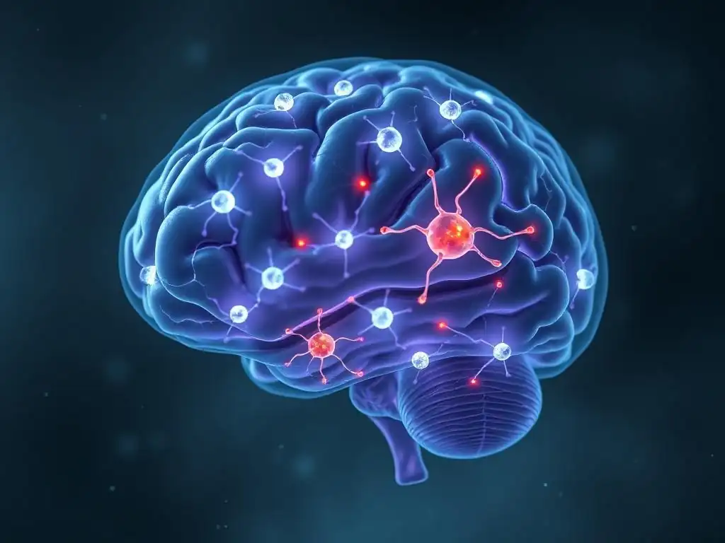
Welcome to Mind Care Guide, a safe space where understanding begins.
Feeling lost in the complexities of PTSD? You're not alone. We delve into the often-overlooked world of neuroscience to bring you clarity and hope. This article, " Understanding the Biology of PTSD in the Brain: A StressInduced Condition," offers an empathetic and insightful exploration of what happens within the brain during and after trauma. Discover how stress impacts brain structure and function, and what this means for recovery. Ready to unlock the secrets of your brain and begin your healing journey?
Understanding the Biology of PTSD in the Brain: A Stress-Induced Condition
- Introduction: PTSD Brain Biology Explained for Mind Care Guide Readers
- The Brain's Response to Trauma: A Foundation for Understanding PTSD
- Biological Mechanisms Underlying PTSD Brain Changes
- Structural and Functional Brain Alterations in PTSD
- The Role of Inflammation in PTSD Brain Biology
- The Impact of Chronic Stress on the PTSD Brain
- Diagnosing PTSD: Biological Markers and Clinical Assessments
- Treatment Strategies Targeting PTSD Brain Biology
- The Future of PTSD Research: Advancements in Understanding Brain Biology
- Conclusion: Reclaiming Mental Well-being Through Understanding PTSD Brain Biology
- Frequently Asked Questions
- Final Reflection: The Enduring Impact of Trauma on the Brain
- Unlock Your Brain's Resilience: Take Charge of Your Well-being
Introduction: PTSD Brain Biology Explained for Mind Care Guide Readers
Post-Traumatic Stress Disorder (PTSD) is a psychiatric condition that can develop in individuals who have experienced or witnessed a traumatic event, such as military combat, a natural disaster, a serious accident, or physical or sexual assault. Unlike the typical stress response, which is acute and resolves once the threat passes, PTSD involves a persistent, dysregulated reaction to trauma.
This disorder is characterized by a constellation of symptoms grouped into four main clusters: re-experiencing the trauma (e.g., flashbacks, nightmares), avoidance of trauma-related stimuli, negative alterations in cognition and mood (e.g., difficulty remembering aspects of the trauma, feeling detached), and alterations in arousal and reactivity (e.g., hypervigilance, exaggerated startle response). Understanding the core symptoms provides the foundation for exploring the underlying biological changes.
The diagnostic criteria for PTSD, as outlined in the DSM-5 (Diagnostic and Statistical Manual of Mental Disorders, Fifth Edition), require symptoms to persist for more than one month and cause clinically significant distress or impairment in social, occupational, or other important areas of functioning. It's estimated that approximately 6-8% of the U.S. population will experience PTSD at some point in their lives, though prevalence varies significantly based on trauma exposure.
For instance, combat veterans and survivors of sexual assault have notably higher rates. The chronic nature and severity of symptoms underscore the need to understand how PTSD impacts the brain and body at a biological level.
The Neuroscience of Mental Health: PTSD and the Brain
The field of neuroscience has made significant strides in revealing how mental health conditions, including PTSD, are rooted in specific changes within the brain. Far from being purely psychological phenomena, conditions like PTSD involve measurable alterations in brain structure, function, and neurochemistry. This biological perspective is crucial for demystifying the disorder and reducing stigma, highlighting that symptoms arise from real changes in brain circuits responsible for processing fear, memory, and stress. Exploring the neuroscience of mental health provides a framework for understanding why individuals with PTSD experience such profound and persistent symptoms.
Research utilizing advanced neuroimaging techniques has consistently shown differences in brain activity and structure in individuals with PTSD compared to healthy individuals. Areas like the amygdala, hippocampus, and prefrontal cortex, which are critical for processing emotions, forming memories, and regulating behavior, show distinct patterns of dysfunction. This focus on the PTSD brain biology explained through neuroscience offers valuable insights into the mechanisms driving re-experiencing, avoidance, and hyperarousal symptoms. By examining these neurological underpinnings, we can better appreciate the complexity of PTSD and the biological challenges individuals face in recovery.
Scope of this Guide: Exploring the Biological Impact of PTSD
This guide aims to provide a comprehensive overview of the biological changes associated with Post-Traumatic Stress Disorder. We will delve into the specific brain regions most affected by trauma and how their altered function contributes to PTSD symptoms. The discussion will cover the intricate biological mechanisms at play, including the dysregulation of the stress response system and imbalances in key neurotransmitters. Furthermore, we will explore the observable structural and functional alterations in the brains of individuals with PTSD, supported by findings from neuroimaging studies.
Beyond the immediate brain changes, this guide will also touch upon the role of inflammation, the long-term effects of chronic stress, and the interplay of genetic and environmental factors in shaping an individual's vulnerability to PTSD. We will then bridge the gap between biological understanding and clinical practice by discussing how biological markers and neuroimaging techniques are used in diagnosis and how current and emerging treatment strategies target these underlying biological processes.
The goal is to offer a detailed look at PTSD brain biology explained in a way that is both informative for Mind Care Guide readers and empathetic to the challenges faced by those living with this condition.
The Brain's Response to Trauma: A Foundation for Understanding PTSD
The Amygdala: Processing Fear and Emotional Memories in PTSD
The amygdala, a small almond-shaped structure deep within the temporal lobe, plays a central role in processing emotions, particularly fear. It acts as the brain's alarm system, quickly detecting potential threats and triggering the body's fight-or-flight response.
In response to a traumatic event, the amygdala becomes highly activated, encoding the emotional intensity of the experience and linking it to sensory cues present during the trauma. This heightened activity is adaptive in the moment, preparing the individual to respond to danger, but in PTSD, this system becomes dysregulated, leading to persistent hyperarousal and exaggerated fear responses to non-threatening stimuli that were associated with the trauma.
Numerous studies using fMRI (functional Magnetic Resonance Imaging) have shown that individuals with PTSD exhibit increased amygdala activity when exposed to trauma-related cues compared to healthy controls or individuals exposed to trauma who did not develop PTSD. For example, research on combat veterans often shows heightened amygdala responses to images or sounds reminiscent of combat situations.
This overactivity contributes directly to symptoms like exaggerated startle responses, hypervigilance, and intrusive thoughts or flashbacks, reflecting a brain stuck in a state of perceived threat, even when the actual danger has passed. This hyperresponsiveness is a key component of the PTSD brain biology explained by its impact on fear processing.
The hippocampus, another structure in the temporal lobe, is critical for forming and consolidating new memories, particularly episodic memories (memories of specific events) and contextual memories (memories of the time and place where an event occurred). It works closely with the amygdala to integrate the emotional aspects of an experience with its factual and contextual details.
In the face of overwhelming trauma, the hippocampus's ability to properly process and contextualize the event can be impaired. Instead of filing the traumatic memory away as a past event, it may remain fragmented or poorly integrated, leading to the feeling that the trauma is happening now, characteristic of flashbacks.
One of the most consistent findings in PTSD brain biology explained through neuroimaging is reduced hippocampal volume in individuals with chronic PTSD. While the exact cause-and-effect relationship is still debated (did the smaller hippocampus precede the trauma, or did the trauma cause the reduction? ), it is clear that hippocampal dysfunction contributes significantly to memory problems seen in PTSD.
This includes difficulty recalling details of the traumatic event itself (dissociative amnesia) but also problems with general memory and learning. Furthermore, impaired contextual processing by the hippocampus may explain why individuals with PTSD struggle to distinguish between safe situations and genuinely dangerous ones, leading to avoidance behaviors and hypervigilance even in benign environments.
The Prefrontal Cortex: Executive Function and Emotional Regulation in PTSD
The prefrontal cortex (PFC), the most evolved part of the brain located behind the forehead, is responsible for higher-level cognitive functions, including decision-making, planning, working memory, and crucially, emotional regulation. It acts as the "executive control center," modulating the activity of subcortical structures like the amygdala to inhibit inappropriate fear responses and help an individual respond flexibly to changing circumstances. In a healthy stress response, the PFC helps the individual appraise the situation, determine if the threat is real or past, and regulate the emotional reaction.
In individuals with PTSD, studies often reveal decreased activity in certain areas of the prefrontal cortex, particularly the medial prefrontal cortex (mPFC) and the ventromedial prefrontal cortex (vmPFC). This reduced top-down control from the PFC means that the overactive amygdala is less effectively regulated. This impaired communication contributes to difficulty controlling fear and anxiety, problems with emotional regulation, and challenges with executive functions like concentration and decision-making. The weakened ability of the PFC to dampen the fear response is a critical aspect of the PTSD brain biology explained as a disorder of impaired emotional processing and regulation.
Biological Mechanisms Underlying PTSD Brain Changes
HPA Axis Dysregulation in PTSD: The Stress Response System
The Hypothalamic-Pituitary-Adrenal (HPA) axis is the body's main stress response system. When a threat is perceived, the hypothalamus releases corticotropin-releasing hormone (CRH), which signals the pituitary gland to release adrenocorticotropic hormone (ACTH). ACTH then signals the adrenal glands to release cortisol, the primary stress hormone. Cortisol prepares the body for action (increasing heart rate, blood sugar, etc.) and, in a healthy system, also signals back to the brain (particularly the hippocampus and PFC) to shut down the stress response once the threat is gone, a process called negative feedback.
In many individuals with PTSD, the HPA axis becomes dysregulated, though the pattern can be complex and varies between individuals. While some studies initially suggested elevated cortisol, a more consistent finding, particularly in chronic PTSD, is lower baseline cortisol levels compared to healthy individuals, coupled with an exaggerated response to subsequent stress.
This paradoxical pattern, often involving increased sensitivity of glucocorticoid receptors (the receptors for cortisol), may contribute to the persistent hyperarousal and exaggerated stress reactivity seen in the disorder. This altered HPA axis function is a core component of the PTSD brain biology explained as a chronic stress-induced condition.
Neurotransmitter Imbalances in PTSD: Serotonin, Norepinephrine, and Dopamine
Neurotransmitters are chemical messengers that transmit signals between neurons in the brain. Several key neurotransmitters are implicated in the biological changes associated with PTSD, including serotonin, norepinephrine, and dopamine. Serotonin is involved in regulating mood, sleep, appetite, and anxiety. Research suggests altered serotonin function in PTSD, which is why Selective Serotonin Reuptake Inhibitors (SSRIs), medications that increase serotonin availability, are often effective treatments.
Norepinephrine is crucial for the body's fight-or-flight response, increasing alertness, heart rate, and blood pressure. In PTSD, there is often evidence of increased norepinephrine activity, contributing to symptoms like hypervigilance, exaggerated startle, and nightmares. Medications targeting norepinephrine, like SNRIs (Serotonin-Norepinephrine Reuptake Inhibitors), are also used in treatment.
Dopamine, involved in reward, motivation, and emotional processing, may also be altered, potentially contributing to symptoms like anhedonia (loss of pleasure) and emotional numbing. Understanding these neurotransmitter imbalances is vital for comprehending the neurobiological underpinnings of PTSD symptoms and guiding pharmacological interventions aimed at correcting the PTSD brain biology explained by these chemical messengers.
Genetic Predisposition and Environmental Factors in PTSD Development
While trauma is the necessary trigger for PTSD, not everyone exposed to trauma develops the disorder. This variability points to the influence of individual vulnerability factors, including genetics and pre-existing environmental exposures. Certain genetic variations have been associated with an increased risk of developing PTSD, though the impact of individual genes is typically small. For example, variations in genes related to the serotonin transporter, the HPA axis components (like the glucocorticoid receptor gene), and brain-derived neurotrophic factor (BDNF) have been studied for their potential roles in modulating risk and shaping the PTSD brain biology explained by genetic factors.
Moreover, early life experiences, such as childhood maltreatment or chronic stress, can significantly impact brain development and increase vulnerability to PTSD later in life, even before exposure to the specific trauma that triggers the disorder. This highlights the concept of biological sensitivity to context, where early environmental factors can "program" the stress response system and brain circuits, making an individual more susceptible to the long-term effects of trauma. The interplay between genetic predisposition and environmental factors, particularly early adversity, creates a complex vulnerability landscape that influences how an individual's brain responds to and recovers from traumatic stress.
Structural and Functional Brain Alterations in PTSD
Reduced Hippocampal Volume: Impact on Memory and Contextualization
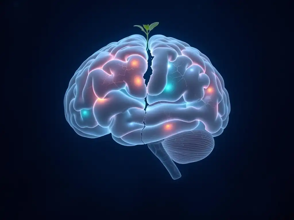
As mentioned earlier, one of the most frequently reported structural changes in the brains of individuals with PTSD is a reduction in the volume of the hippocampus. Meta-analyses of neuroimaging studies have consistently shown this effect, with estimated volume reductions ranging from 5% to 10% on average compared to trauma-exposed individuals without PTSD or healthy controls. This structural difference is thought to impair the hippocampus's ability to process and contextualize traumatic memories effectively, leaving them fragmented and prone to intrusive re-experiencing.
The functional consequence of reduced hippocampal volume and activity in PTSD is a diminished capacity for contextual fear extinction – the ability to learn that a previously threatening stimulus is no longer dangerous in a safe context. For example, a veteran might experience intense anxiety triggered by a car backfiring (a sound associated with combat) even when they are safely at home. A healthy hippocampus would help the brain differentiate the current safe context from the past dangerous one.
In PTSD, this contextual discrimination is impaired, leading to generalized fear responses and contributing to avoidance behaviors as the individual tries to avoid any situation that might trigger a reminder of the trauma. This structural change is a key aspect of PTSD brain biology explained by its impact on memory and context processing.
Increased Amygdala Activity: Heightened Fear and Anxiety Responses
Functional neuroimaging studies, particularly fMRI, have provided compelling evidence of heightened amygdala reactivity in individuals with PTSD when exposed to trauma-related stimuli or even general emotional cues. This increased activity reflects an exaggerated fear response and contributes significantly to the hyperarousal and anxiety symptoms characteristic of the disorder. The amygdala's persistent state of alert means that the individual is constantly scanning for threats, leading to symptoms like hypervigilance, an exaggerated startle response, and difficulty relaxing.
This amygdala hyperactivation is often observed in conjunction with decreased activity in regulatory brain regions like the prefrontal cortex, suggesting an imbalance where the fear-processing center is overactive and the control center is underactive. For example, studies might show a stronger amygdala response to fearful faces in individuals with PTSD compared to controls, alongside weaker connections or less activity in PFC regions involved in emotional regulation. This persistent state of heightened fear reactivity is a central feature of the PTSD brain biology explained as a disorder of a dysregulated fear circuit.
Decreased Prefrontal Cortex Activation: Impaired Emotional Regulation
Functional neuroimaging studies consistently show reduced activation in specific areas of the prefrontal cortex (PFC), particularly the medial PFC (mPFC) and ventral mPFC, when individuals with PTSD are engaged in tasks requiring emotional regulation or fear extinction. These areas are crucial for inhibiting the amygdala's fear response and integrating emotional information with cognitive processes. The decreased activity in these regions suggests a weakened capacity for top-down control over emotional reactions.
This impaired prefrontal function contributes to several core PTSD symptoms. It makes it difficult for individuals to modulate their emotional responses to reminders of the trauma, leading to intense anxiety or distress. It also impacts executive functions necessary for daily life, such as concentration, decision-making, and planning, which require the PFC. The weakened regulatory capacity of the PFC in the face of an overactive amygdala is a critical imbalance in the PTSD brain biology explained as a failure of the brain's emotional control system.
The Role of Inflammation in PTSD Brain Biology
Inflammation and Neurodegeneration in PTSD
Emerging research suggests that chronic low-grade inflammation in the brain and body may play a significant role in the development and maintenance of PTSD, contributing to neurobiological changes and symptoms. Trauma and chronic stress can activate the immune system, leading to the release of pro-inflammatory molecules called cytokines. While acute inflammation is part of the body's healing process, chronic inflammation can be detrimental to brain health, potentially contributing to neuronal dysfunction and even neurodegeneration over time.
Studies have found elevated levels of inflammatory markers, such as C-reactive protein (CRP) and various cytokines, in the blood and cerebrospinal fluid of individuals with PTSD compared to healthy controls. This chronic inflammatory state is hypothesized to impact brain regions involved in PTSD, such as the hippocampus and prefrontal cortex, potentially contributing to structural changes like reduced hippocampal volume and functional impairments. The link between inflammation and alterations in the PTSD brain biology explained is an active area of research, suggesting that the disorder might involve an inflammatory component alongside traditional neurological changes.
Cytokine Involvement in PTSD Pathophysiology
Cytokines are small proteins secreted by immune cells that act as signaling molecules. Pro-inflammatory cytokines, such as Interleukin-1 beta (IL-1β), Interleukin-6 (IL-6), and Tumor Necrosis Factor-alpha (TNF-α), are elevated in many individuals with PTSD. These molecules can cross the blood-brain barrier and directly influence brain function. In the brain, they can affect neurotransmitter systems, impair neurogenesis (the creation of new neurons, particularly in the hippocampus), increase neuronal excitability, and contribute to synaptic dysfunction.
For example, elevated levels of IL-6 have been correlated with the severity of PTSD symptoms, including re-experiencing and hyperarousal. These cytokines can disrupt the delicate balance of neurochemical signaling and neuronal plasticity, potentially exacerbating the dysregulation of the fear circuit and stress response system characteristic of PTSD. Understanding the specific roles of different cytokines is crucial for unraveling the complex PTSD brain biology explained by neuroimmune interactions.
Potential Therapeutic Targets: Anti-Inflammatory Approaches for PTSD
Given the evidence linking inflammation to PTSD pathophysiology, researchers are exploring whether targeting inflammatory pathways could offer novel therapeutic strategies. This could involve using existing anti-inflammatory medications or developing new compounds specifically designed to modulate neuroinflammation. While still largely experimental, this area of research holds promise for individuals who may not respond adequately to traditional treatments.
Approaches being investigated include targeting specific cytokines or their receptors, using omega-3 fatty acids known for their anti-inflammatory properties, or exploring other compounds that can dampen chronic immune activation in the brain. For instance, some studies are looking at whether medications like minocycline, an antibiotic with anti-inflammatory effects, could improve outcomes when used alongside standard PTSD treatments. Modulating inflammation is a promising avenue for intervening in the PTSD brain biology explained by immune system dysregulation, potentially offering new hope for recovery.
The Impact of Chronic Stress on the PTSD Brain
Long-Term Effects of Trauma Exposure
Exposure to a single, overwhelming traumatic event is the defining feature of PTSD. However, the chronicity of the stress response that follows is what drives many of the observed long-term biological changes. Unlike acute stress, which the brain and body are designed to handle temporarily, chronic stress associated with PTSD symptoms (like persistent hypervigilance, disrupted sleep, and intrusive thoughts) keeps the stress response system activated. This prolonged activation floods the brain with stress hormones like cortisol and neurotransmitters like norepinephrine, leading to cumulative damage and maladaptive changes.
The long-term effects include the persistent structural alterations observed in the brain, such as reduced hippocampal volume and changes in prefrontal cortex connectivity. Chronically elevated stress hormones can impair neurogenesis, particularly in the hippocampus, and potentially contribute to neuronal atrophy in vulnerable areas. Furthermore, chronic stress can exacerbate neuroinflammation, creating a vicious cycle that perpetuates brain dysfunction. This chronic stress exposure is central to understanding the lasting impact on the PTSD brain biology explained as a consequence of prolonged physiological and psychological strain.
The Stress-Diathesis Model of PTSD
The stress-diathesis model provides a framework for understanding why some individuals develop PTSD after trauma while others do not. It posits that PTSD results from an interaction between a pre-existing vulnerability (diathesis) and the stressor (trauma). The diathesis can include genetic predispositions, prior trauma exposure (especially in childhood), pre-existing mental health conditions, and personality traits. These vulnerabilities influence an individual's biological and psychological reactivity to stress.
According to this model, individuals with a higher diathesis may be more susceptible to the neurobiological changes associated with PTSD following trauma. Their HPA axis might be more reactive, their fear circuitry (amygdala-PFC balance) might be less resilient, or their inflammatory response might be more pronounced. The model helps explain the heterogeneity seen in PTSD development and severity, emphasizing that the disorder isn't solely about the trauma itself, but also about the individual's biological and psychological landscape before the trauma. This interaction highlights the complexity of the PTSD brain biology explained by individual differences in vulnerability.
Resilience Factors and Protective Mechanisms
Conversely, certain factors can promote resilience and protect individuals from developing PTSD despite trauma exposure. These can include genetic factors (e.g., variations associated with better stress regulation), biological factors (e.g., a more robust and flexible HPA axis response), psychological factors (e.g., effective coping strategies, a sense of self-efficacy), and social factors (e.g., strong social support networks). Resilience is not simply the absence of vulnerability but involves active biological and psychological processes that facilitate adaptation and recovery after stress.
Biologically, resilience might be linked to a more efficient negative feedback loop in the HPA axis, preventing chronic stress hormone elevation. It could also involve stronger functional connectivity between the prefrontal cortex and the amygdala, allowing for better regulation of fear responses. Furthermore, factors promoting neurogenesis and synaptic plasticity might enhance the brain's ability to adapt and recover from trauma's impact. Understanding these protective mechanisms and resilience factors offers insights into potential targets for prevention and intervention strategies aimed at buffering the PTSD brain biology explained by trauma.
Diagnosing PTSD: Biological Markers and Clinical Assessments
Neuroimaging Techniques for PTSD Diagnosis (MRI, fMRI, PET)
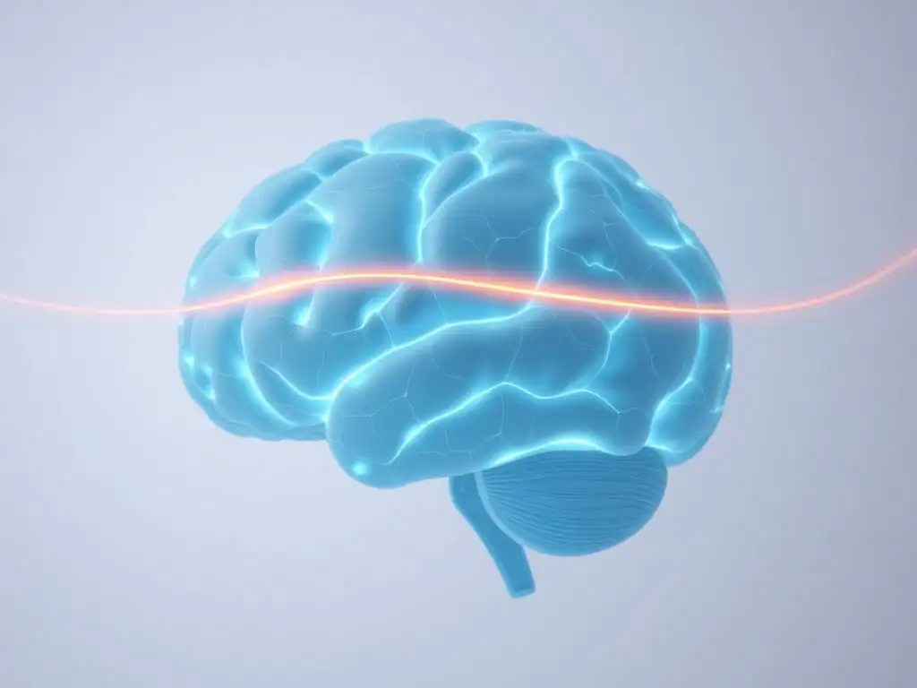
While PTSD is currently diagnosed based on clinical interviews and symptom criteria, research is actively exploring the potential of neuroimaging techniques as objective diagnostic aids or tools for understanding treatment response. Structural MRI (Magnetic Resonance Imaging) can measure brain volume and identify structural differences, such as the aforementioned reduced hippocampal volume. Although not consistently present in every individual with PTSD and also seen in other conditions, this finding is a significant marker in group studies.
Functional MRI (fMRI) measures brain activity by detecting changes in blood flow. fMRI studies in PTSD often show patterns like increased amygdala activity and decreased prefrontal cortex activity in response to trauma cues, which could potentially serve as functional biomarkers.
PET (Positron Emission Tomography) scans can measure metabolic activity or the distribution of specific neurotransmitter receptors in the brain, offering insights into neurochemical changes. While these techniques are primarily research tools today, advancements in neuroimaging hold promise for providing objective support for a PTSD diagnosis or predicting treatment outcomes by revealing aspects of the PTSD brain biology explained through visual brain activity patterns.
Biomarkers in PTSD: Potential for Early Detection
Beyond neuroimaging, researchers are investigating various biological markers (biomarkers) in blood, saliva, or cerebrospinal fluid that could indicate the presence or severity of PTSD, or even predict vulnerability. These include markers related to HPA axis function (e.g., cortisol levels, CRH levels), inflammation (e.g., cytokines, CRP), and genetic variations. For instance, specific patterns of cortisol reactivity in response to a stress test have been explored as potential indicators of PTSD risk or diagnosis.
The goal of identifying reliable biomarkers is to improve early detection, stratify patients based on their specific biological profile, and potentially predict which treatments might be most effective. For example, if a specific inflammatory profile is linked to a subtype of PTSD, anti-inflammatory approaches might be particularly beneficial for those individuals. While no single biomarker is currently used for routine clinical diagnosis, the ongoing search for reliable biological indicators is crucial for advancing our understanding and management of the condition by objectively assessing the PTSD brain biology explained by physiological measures.
Integrating Biological Findings with Clinical Criteria
Current clinical diagnosis of PTSD relies on the detailed symptom criteria outlined in diagnostic manuals like the DSM-5, gathered through interviews and questionnaires. This approach captures the subjective experience and observable behaviors associated with the disorder. While biological findings from research have provided invaluable insights into the mechanisms underlying these symptoms, they are not yet precise or consistent enough across individuals to replace clinical assessment for diagnosis.
However, the integration of biological findings with clinical criteria represents the future of precision psychiatry for PTSD. Understanding an individual's specific biological profile – perhaps through a combination of genetic testing, biomarker analysis, and potentially advanced neuroimaging in the future – could complement clinical assessment.
This integrated approach could lead to more accurate diagnoses, better prediction of who is at risk after trauma, and ultimately, more personalized treatment plans tailored to the individual's unique PTSD brain biology explained by their specific biological profile. This integration is essential for moving beyond a purely symptom-based approach to a more biologically informed understanding and treatment of PTSD.
Treatment Strategies Targeting PTSD Brain Biology
Psychotherapy and Brain Plasticity: Rewiring Neural Circuits
Psychotherapy, particularly trauma-focused therapies like Trauma-Focused Cognitive Behavioral Therapy (TF-CBT) and Eye Movement Desensitization and Reprocessing (EMDR), is considered a first-line treatment for PTSD. These therapies work by helping individuals process traumatic memories and change maladaptive thoughts and behaviors related to the trauma. Crucially, effective psychotherapy is believed to exert its effects by promoting neuroplasticity – the brain's ability to reorganize itself by forming new neural connections throughout life.
Successful trauma-focused therapy can lead to measurable changes in brain activity and structure, essentially "rewiring" the dysfunctional circuits involved in PTSD. Studies using neuroimaging have shown that effective treatment can lead to decreased amygdala reactivity, increased activity and connectivity in the prefrontal cortex (restoring top-down control), and potentially even increases in hippocampal volume or function. These therapies help the brain to properly process the traumatic memory, extinguish inappropriate fear responses, and regain regulatory control over emotions, demonstrating how psychological interventions can directly impact the PTSD brain biology explained by altered neural pathways.
Pharmacological Interventions: SSRIs, SNRIs, and Other Medications
Medications, primarily Selective Serotonin Reuptake Inhibitors (SSRIs) and Serotonin-Norepinephrine Reuptake Inhibitors (SNRIs), are also commonly used to treat PTSD symptoms. These medications work by modulating the levels of neurotransmitters like serotonin and norepinephrine in the brain, aiming to correct the imbalances discussed earlier. SSRIs like sertraline (Zoloft) and paroxetine (Paxil) are FDA-approved for PTSD and can help reduce core symptoms, particularly hyperarousal and avoidance.
Other medications may be used off-label to target specific symptoms. For example, prazosin, an alpha-1 adrenergic receptor antagonist, is sometimes used to reduce trauma-related nightmares, potentially by blocking the effects of norepinephrine in certain brain circuits. Beta-blockers have been explored for use shortly after trauma to potentially dampen the consolidation of fear memories, although their efficacy in preventing chronic PTSD is debated. These pharmacological approaches directly intervene in the neurochemical aspects of the PTSD brain biology explained by neurotransmitter dysfunction, aiming to restore a more balanced state.
Novel Therapies: TMS, Neurofeedback, and Emerging Approaches
Beyond traditional psychotherapy and medication, novel therapeutic approaches are being investigated for their potential to treat PTSD by directly targeting brain function. Transcranial Magnetic Stimulation (TMS) is a non-invasive procedure that uses magnetic pulses to stimulate specific areas of the brain, often the prefrontal cortex, to modulate neuronal activity. TMS is FDA-approved for depression and OCD and is being explored for its ability to improve emotional regulation and reduce symptoms in PTSD, potentially by enhancing PFC control over the amygdala.
Neurofeedback is another technique that allows individuals to learn to regulate their own brain activity using real-time feedback from EEG or fMRI. Individuals can be trained to increase activity in areas like the PFC or decrease activity in the amygdala, aiming to restore a healthier balance in the fear circuit. Emerging research is also exploring interventions like MDMA-assisted therapy, which is thought to work by enhancing empathy and reducing fear responses, potentially facilitating trauma processing. These novel therapies represent exciting frontiers in treating PTSD by directly addressing the PTSD brain biology explained through dysfunctional brain circuits and activity patterns.
The Future of PTSD Research: Advancements in Understanding Brain Biology
Personalized Medicine for PTSD: Tailoring Treatments to Individual Brain Profiles
One of the major goals for the future of PTSD research is the development of personalized medicine approaches. Given the heterogeneity of PTSD symptoms and responses to treatment, understanding an individual's unique biological profile – including genetic factors, biomarkers, and specific patterns of brain activity or structure – could allow for more tailored and effective interventions. Instead of a one-size-fits-all approach, treatments could be matched to the individual's specific PTSD brain biology explained by their unique vulnerabilities and mechanisms.
For instance, future research might identify specific biological markers that predict responsiveness to SSRIs versus trauma-focused therapy, or indicate which individuals might benefit most from novel treatments like TMS. Genomic sequencing could reveal genetic predispositions that inform risk assessment or guide treatment choices. Advances in computational neuroscience and machine learning could analyze complex biological data to identify subtypes of PTSD with distinct biological underpinnings, leading to subtype-specific treatments. This move towards personalized medicine holds immense promise for improving outcomes for individuals with PTSD.
Gene-Environment Interactions in PTSD: A Complex Interplay
Further research into gene-environment interactions is crucial for a complete understanding of PTSD. It's not just about having a particular gene or experiencing a specific environment; it's about how the two interact to shape vulnerability and resilience. For example, a genetic variation might only increase PTSD risk in the context of specific types of trauma or if combined with early life adversity. Conversely, protective genetic factors might buffer the impact of severe trauma.
Advanced genetic studies and longitudinal research tracking individuals exposed to trauma are needed to unravel these complex interactions. Understanding precisely how genes and environments conspire to influence the PTSD brain biology explained by these interactions can inform targeted prevention strategies. For instance, identifying individuals with high genetic risk and specific environmental exposures could allow for early intervention or enhanced support to mitigate the likelihood of developing chronic PTSD.
Prevention Strategies: Mitigating the Biological Impact of Trauma
Ultimately, a deeper understanding of the PTSD brain biology explained by trauma's impact offers opportunities for developing more effective prevention strategies. If we can identify biological vulnerabilities or early biological responses to trauma that predict chronic PTSD, interventions could be implemented in the immediate aftermath of a traumatic event to interrupt the pathological process.
This could include pharmacological interventions aimed at modulating the stress response or fear memory consolidation in high-risk individuals, or early psychological interventions designed to promote healthy brain plasticity and fear extinction. Research into factors that promote resilience is also key, as identifying biological and psychological markers of resilience could inform strategies to enhance these protective mechanisms in vulnerable populations. The future of PTSD prevention lies in leveraging our growing knowledge of its biological underpinnings to intervene early and effectively.
Conclusion: Reclaiming Mental Well-being Through Understanding PTSD Brain Biology
Key Takeaways: PTSD and the Brain
Understanding the PTSD brain biology explained by research offers crucial insights into this complex stress-related condition. Key takeaways include recognizing that PTSD is associated with measurable alterations in brain structure and function, particularly affecting the amygdala (leading to heightened fear), the hippocampus (impairing memory and context), and the prefrontal cortex (disrupting emotional regulation). The body's stress response system, the HPA axis, is often dysregulated, and imbalances in key neurotransmitters like serotonin and norepinephrine contribute to symptoms. Furthermore, chronic inflammation and the interplay of genetic and environmental factors play significant roles in vulnerability and symptom persistence.
These biological changes are not static; they are dynamic and can be influenced by effective treatments. Psychotherapy works by promoting neuroplasticity and helping to "rewire" dysfunctional circuits, while medications target neurochemical imbalances. Emerging therapies aim to directly modulate brain activity. Recognizing the biological basis of PTSD is vital for reducing stigma, appreciating the challenges faced by individuals living with the disorder, and guiding the development of more effective, perhaps personalized, interventions.
Resources for Further Learning and Support
For individuals seeking more information about PTSD or needing support, numerous resources are available. Learning about the PTSD brain biology explained in articles like this can be empowering, but professional help is often necessary for recovery.
- Mental health professionals specializing in trauma can provide accurate diagnosis and evidence-based treatments like TF-CBT and EMDR.
- Support groups offer connection with others who understand the challenges of living with PTSD.
- Organizations such as the National Center for PTSD (part of the U.S. Department of Veterans Affairs, though resources are for everyone) and the Anxiety & Depression Association of America (ADAA) provide extensive information, find-a-therapist tools, and support resources.
- Crisis hotlines are available for immediate support during times of distress.
Seeking help is a sign of strength, and effective treatments are available that target the underlying biological and psychological aspects of PTSD.
Mind Care Guide: Your Partner in Mental Health
Mind Care Guide is dedicated to providing comprehensive information, support, and resources for individuals navigating mental health challenges, including understanding conditions like PTSD. Our goal is to empower you with knowledge about the PTSD brain biology explained and its impact, connecting you with reliable information and pathways to support.
We believe that understanding the biological underpinnings of mental health conditions is a vital step towards recovery and well-being. By offering accessible information on topics like the neuroscience of mental health and the effects of stress on the brain, we aim to demystify these conditions and highlight the importance of seeking evidence-based care. Mind Care Guide is here to be your partner on your journey towards reclaiming mental well-being.
Frequently Asked Questions
How does PTSD affect the amygdala in the brain?
PTSD often leads to increased activity in the amygdala, the brain's fear center. This heightened activity can result in exaggerated fear responses and anxiety.
What role does the hippocampus play in PTSD?
The hippocampus, crucial for memory formation, is often smaller in individuals with PTSD. This can contribute to difficulties in distinguishing safe from unsafe memories.
How are stress hormones like cortisol involved in the biology of PTSD?
Chronic stress associated with trauma leads to dysregulation of the hypothalamic-pituitary-adrenal (HPA) axis, affecting cortisol levels. This hormonal imbalance can impair stress response mechanisms.
Does PTSD alter neural pathways in the brain?
Yes, PTSD can lead to alterations in neural pathways involved in fear, memory, and emotional regulation. These changes can contribute to heightened reactivity and difficulty processing trauma.
Can understanding the brain biology of PTSD help with treatment?
Absolutely. Understanding the biological mechanisms underlying PTSD allows for more targeted and effective treatments. This includes therapies and medications aimed at regulating brain activity and hormonal balance.
Final Reflection: The Enduring Impact of Trauma on the Brain
Understanding the biology of PTSD is not merely an academic exercise; it's a crucial step towards effective treatment and a more compassionate understanding of those who suffer. The intricate interplay of the amygdala, hippocampus, and prefrontal cortex, as detailed in this article, reveals the profound and lasting impact of trauma on the brain's structure and function. This knowledge is not just theoretical; it directly informs the development of targeted therapies and offers hope for healing.
The ongoing research into PTSD's neurological underpinnings is transforming our approach to mental health. It challenges the outdated stigma surrounding trauma and mental illness, fostering a more empathetic and informed societal response. The greatest barrier to recovery is often the silence surrounding trauma.
This quote highlights the importance of open dialogue and the crucial role of education in breaking down societal barriers and fostering support for those affected. The more we understand the biological mechanisms at play, the better equipped we are to provide effective support and advocate for improved mental healthcare access.
Consider the implications of this research on your own life. Perhaps you know someone struggling with PTSD, or maybe you've experienced trauma yourself. Take a moment to reflect on the power of understanding. This knowledge can empower you to seek help, offer support to others, or simply foster greater empathy and compassion for those navigating the complex landscape of trauma and recovery. Let the insights shared in this article serve as a catalyst for positive change, both within yourself and within your community.
Unlock Your Brain's Resilience: Take Charge of Your Well-being
Thanks for taking the time to delve into the complexities of PTSD and its impact on the brain. Understanding the biology behind this condition is a powerful first step towards healing and resilience. Now it's time to turn that knowledge into action.
Here's how you can move forward:
- Prioritize self-care strategies: Identify at least one new self-care practice—whether it's mindfulness meditation, regular exercise, or spending time in nature—and commit to incorporating it into your daily routine this week.
- Connect with a support network: Reach out to a trusted friend, family member, or therapist to share your insights and seek further support. Consider joining a relevant online support group for added connection.
- Share this article: Help spread awareness about PTSD and its biological underpinnings by sharing this article with someone who might benefit from the information.
What small step towards better brain health will you take today?