
How Stress Affects Brain Volume: A Biological Look at Specific Regions

Welcome to Mind Care Guide, a safe space for understanding your mental well-being.
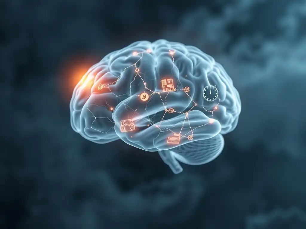
Ever wondered if stress is actually shrinking your brain? In this article, we delve into the neuroscience behind "Stress brain volume reduction," exploring how chronic stress can impact specific regions and what you can do to protect your cognitive health. Discover the biological mechanisms at play and empower yourself with knowledge to navigate stress more effectively. Let's explore this crucial topic together.
- Stress-Induced Brain Volume Reduction: A Biological Perspective on Vulnerable Regions
- Neuroscience of Stress: Deciphering the Biological Mechanisms
- Specific Brain Regions Affected by Stress-Related Volume Loss
- Measuring Brain Volume Changes: Imaging Techniques
- Mitigating Stress and Protecting Brain Volume: Strategies for Prevention
- Conclusion: Preserving Brain Health in the Face of Stress
-
Frequently Asked Questions
- What is the main connection between stress and brain volume reduction?
- Which brain regions are most vulnerable to volume reduction due to chronic stress?
- How does cortisol contribute to stress-related brain volume loss?
- Is the brain volume reduction caused by stress permanent?
- What are some practical strategies to protect brain volume from the negative impacts of stress?
- Final Reflection: The Silent Shrinkage of the Stressed Mind
- Unleash Your Brain's Potential: Take Control of Your Stress
Stress-Induced Brain Volume Reduction: A Biological Perspective on Vulnerable Regions
Introduction: The Detrimental Impact of Chronic Stress on Brain Health
In contemporary society, the escalating prevalence of stress has become a significant concern, impacting millions globally. From demanding workloads to personal challenges, chronic stress is no longer an occasional inconvenience but a persistent fixture in many lives. This sustained exposure to stressors activates the body's physiological response mechanisms, which, while adaptive in short bursts, can become detrimental over prolonged periods. Understanding the profound effects of this persistent stress on our physical and mental health is crucial for well-being.
Beyond the commonly known psychological symptoms like anxiety and depression, chronic stress exerts tangible physical changes within the body, particularly within the brain. Research in neuroscience has increasingly pointed to a phenomenon known as stress-induced brain volume reduction, where specific areas of the brain actually decrease in size.
This structural alteration is not merely theoretical; it's observable through advanced imaging techniques and is associated with significant impairments in cognitive function, emotional regulation, and overall mental resilience. This article will delve into the biological mechanisms underlying this stress brain volume reduction and highlight the specific brain regions most vulnerable to its impact.
Neuroscience of Stress: Deciphering the Biological Mechanisms
The Hypothalamic-Pituitary-Adrenal (HPA) Axis and Cortisol's Role
The body's primary system for responding to stress is the Hypothalamic-Pituitary-Adrenal (HPA) axis. This complex network involves the hypothalamus, which signals the pituitary gland, which in turn signals the adrenal glands to release stress hormones, primarily cortisol. In response to a perceived threat, the HPA axis orchestrates the "fight or flight" response, increasing heart rate, blood pressure, and making energy readily available. Once the threat passes, the HPA axis is designed to return to a baseline state. However, chronic stress keeps this axis constantly engaged, leading to persistently high levels of cortisol circulating throughout the body and brain.
Elevated cortisol levels, a hallmark of chronic stress, have a profound and often damaging impact on various brain structures. While cortisol is essential for regulating many bodily functions, excessive exposure acts like a toxin to certain neurons, particularly in areas rich in cortisol receptors. Studies have shown that sustained high cortisol can impair neurogenesis (the creation of new neurons), reduce synaptic plasticity (the ability of connections between neurons to change), and even cause neuronal death. This cascade of effects contributes directly to the observed stress brain volume reduction in vulnerable areas, fundamentally altering brain architecture and function.
Chronic stress doesn't just involve hormonal changes; it also triggers a persistent low-grade inflammatory response in the brain, known as neuroinflammation. When the body is under prolonged stress, immune cells within the brain, like microglia, become overactive. These cells release inflammatory molecules called cytokines (such as TNF-alpha, IL-1 beta, and IL-6) that are meant to protect the brain from injury or infection but become harmful when chronically elevated. These cytokines can interfere with neurotransmitter systems, disrupt synaptic function, and impair the survival of neurons.
The continuous presence of inflammatory cytokines in the brain under chronic stress is a significant contributor to brain atrophy and stress brain volume reduction. Neuroinflammation can impair the function and structure of vital brain cells, including neurons and glial cells, leading to a loss of gray matter density. This inflammatory process exacerbates the damage caused by excessive cortisol and creates a hostile environment for brain health, undermining the brain's ability to repair itself and maintain optimal structure and function.
Hippocampus: Memory Processing and Stress Vulnerability
The hippocampus, a seahorse-shaped structure deep within the temporal lobe, is critical for forming new memories, learning, and spatial navigation. It is also one of the brain regions with the highest concentration of cortisol receptors, making it particularly vulnerable to the effects of chronic stress. The intricate neural networks within the hippocampus are highly sensitive to the disruptions caused by elevated cortisol and neuroinflammation.
Numerous studies, including research on individuals with conditions like post-traumatic stress disorder (PTSD) or major depressive disorder which are often linked to chronic stress, have consistently shown a significant reduction in hippocampal volume. For instance, meta-analyses of MRI studies have reported hippocampal volume reductions of up to 10-15% in some populations exposed to severe or prolonged stress compared to healthy controls. This stress brain volume reduction in the hippocampus is directly associated with impairments in cognitive functions, including declarative memory (memory for facts and events), learning new information, and navigating spatial environments.
Prefrontal Cortex: Executive Function and Emotional Regulation
The prefrontal cortex (PFC), located at the front of the brain, is the command center for executive functions. These include complex cognitive processes like decision-making, planning, working memory, impulse control, and regulating emotions. The PFC allows us to think critically, anticipate consequences, and manage our responses to challenging situations. It plays a crucial role in our ability to cope effectively with stress.
However, chronic stress can lead to structural and functional changes in the PFC, including a reduction in gray matter volume. Studies using MRI have observed thinner cortical layers or reduced volume in specific subregions of the PFC in individuals experiencing prolonged stress. This stress brain volume reduction in the PFC impairs its ability to exert top-down control over more primitive brain regions involved in fear and emotion, such as the amygdala. The consequence is often difficulty with planning, reduced working memory capacity, impaired decision-making, and challenges in regulating emotions, contributing to increased anxiety and poor stress coping.
Amygdala: Fear and Anxiety Processing
The amygdala, a small almond-shaped structure, is a key player in processing emotions, particularly fear and anxiety. It acts as the brain's alarm system, quickly responding to perceived threats and triggering the stress response. While acute stress activates the amygdala appropriately, chronic stress can lead to maladaptive changes in this region.
Unlike the hippocampus and prefrontal cortex, the amygdala often shows increased activity or even enlargement in response to chronic stress. This hyper-responsivity means the amygdala becomes overly sensitive, triggering fear and anxiety responses even in the absence of real danger.
This altered state of the amygdala, coupled with impaired regulation from a compromised PFC, contributes significantly to the development and maintenance of anxiety disorders and heightened fear responses observed in individuals under chronic stress. Although the focus is often on stress brain volume reduction, changes like amygdala enlargement highlight the complex, region-specific structural adaptations the brain undergoes in response to persistent stress.
Other Brain Regions Potentially Affected by Stress
While the hippocampus, prefrontal cortex, and amygdala are the most extensively studied regions regarding stress-induced changes, other areas of the brain are also potentially affected. The anterior cingulate cortex (ACC), involved in conflict monitoring, pain processing, and emotional regulation, has shown altered activity and sometimes structural changes under chronic stress. The insula, which processes internal bodily states and emotions, is another region implicated, potentially contributing to the physical sensations associated with stress and anxiety.
These additional regions, along with the core HPA axis circuit areas, demonstrate that the impact of chronic stress is widespread throughout the brain. While specific patterns of stress brain volume reduction are prominent in certain areas like the hippocampus and PFC, the overall network is affected, contributing to the complex array of cognitive and emotional difficulties experienced by individuals facing prolonged stress. Understanding these broader impacts is crucial for developing comprehensive strategies for mitigating stress and protecting brain health.
Measuring Brain Volume Changes: Imaging Techniques
MRI (Magnetic Resonance Imaging) for Assessing Brain Structure
Magnetic Resonance Imaging (MRI) is a cornerstone non-invasive technique used extensively in neuroscience research to visualize and assess the structure of the brain. By using strong magnetic fields and radio waves, MRI scanners can create detailed, cross-sectional images of brain tissue, differentiating between gray matter (containing neuronal cell bodies), white matter (containing nerve fibers), and cerebrospinal fluid. This allows researchers and clinicians to examine the overall size and shape of the brain, as well as the volume of specific brain regions.
In the context of studying stress-related brain changes, MRI is invaluable. It enables researchers to compare the brain structure of individuals exposed to chronic stress (e.g., those with stress-related disorders or high self-reported stress levels) with that of healthy control groups. While MRI provides excellent spatial resolution, allowing for detailed structural analysis, it captures a snapshot in time. Longitudinal studies, involving multiple MRI scans over months or years, are necessary to track progressive changes in brain volume, including stress brain volume reduction, as stress exposure continues or as interventions are implemented.
Volumetric Analysis: Quantifying Brain Atrophy
To move beyond qualitative observation and precisely quantify changes in brain size, researchers employ computational techniques known as volumetric analysis. This involves processing the detailed MRI scans using specialized software that can automatically segment the brain into different regions (like the hippocampus, PFC, or amygdala) based on anatomical atlases. Once segmented, the software calculates the precise volume of each identified region.
Volumetric analysis allows researchers to detect even subtle differences in brain volume between groups or changes within the same individual over time. By comparing the measured volumes of specific brain regions in stressed populations versus control groups, studies can statistically determine if chronic stress is associated with significant stress brain volume reduction in particular areas. This quantitative approach provides objective evidence for the structural impact of stress and helps researchers understand the magnitude of atrophy associated with conditions like PTSD, chronic anxiety, or depression, reinforcing the biological reality of stress brain volume reduction.
Mitigating Stress and Protecting Brain Volume: Strategies for Prevention
Stress Management Techniques: Mindfulness, Meditation, and Exercise
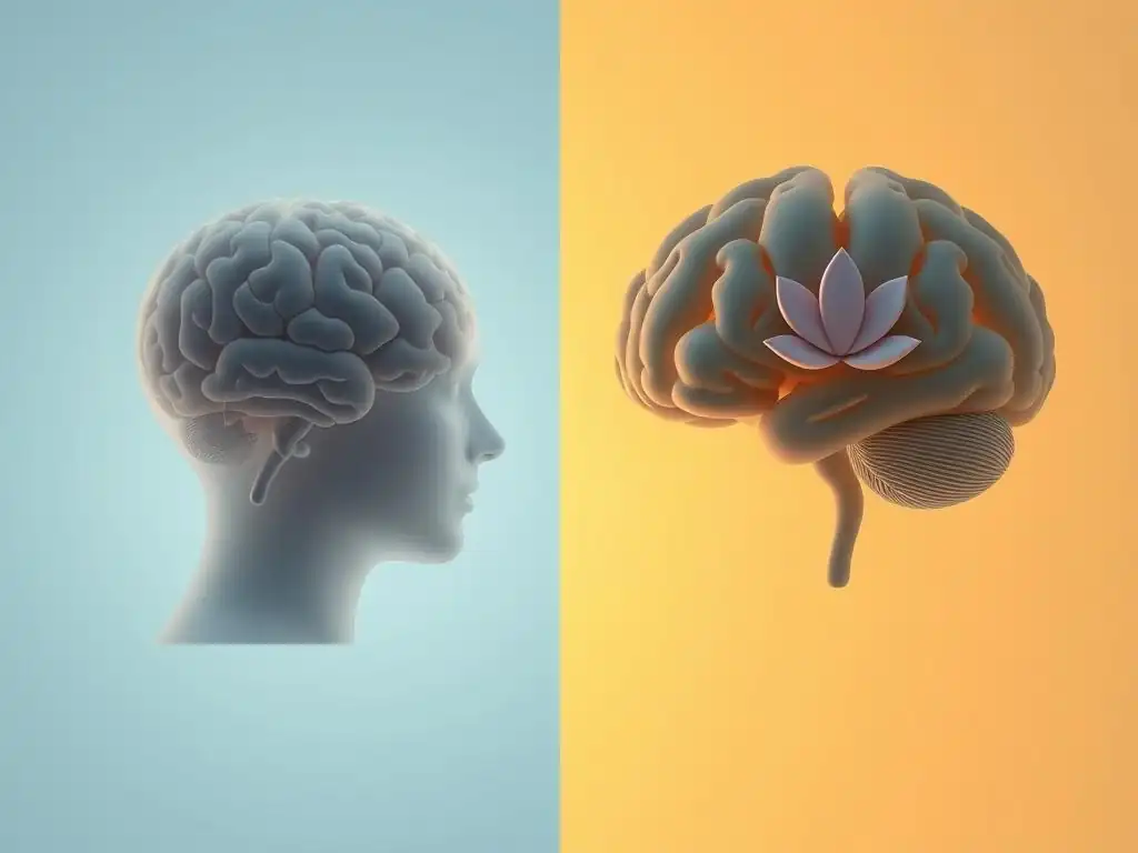
Implementing effective stress management techniques is a proactive way to buffer the brain against the damaging effects of chronic stress and potentially mitigate stress brain volume reduction. Practices such as mindfulness and meditation, which involve focusing on the present moment and cultivating non-judgmental awareness, have been shown in studies to reduce activity in stress-sensitive areas like the amygdala and potentially even increase gray matter density in regions like the hippocampus and PFC. Regular practice can help calm the nervous system and reduce overall cortisol levels.
Physical exercise is another powerful tool for stress reduction and brain health. Engaging in regular aerobic exercise, for example, not only helps to reduce stress hormones but also promotes the release of neurotrophic factors, such as Brain-Derived Neurotrophic Factor (BDNF). BDNF supports the survival, growth, and function of neurons and synapses, particularly in the hippocampus. Consistent physical activity can counteract some of the negative structural changes associated with stress, offering a protective effect against stress brain volume reduction and preserving cognitive function.
Beyond specific techniques, fundamental lifestyle modifications play a crucial role in building resilience to stress and supporting brain health. Adequate and restorative sleep is essential, as sleep deprivation exacerbates stress responses and impairs cognitive function. Aiming for 7-9 hours of quality sleep per night allows the brain to consolidate memories, clear metabolic waste products, and regulate hormone levels, including cortisol.
A healthy, balanced diet rich in antioxidants, omega-3 fatty acids, and complex carbohydrates can also protect the brain. Nutrients like omega-3s are vital components of brain cell membranes and have anti-inflammatory properties, helping to counteract the neuroinflammation driven by chronic stress. Conversely, diets high in processed foods and sugar can promote inflammation, potentially worsening stress-related brain damage.
Finally, strong social support networks provide emotional buffering against stress. Connecting with others releases oxytocin, a hormone that can counteract the effects of cortisol and promote feelings of calm and security, offering a vital protective factor against the physiological impacts of stress, including potential stress brain volume reduction.
Therapeutic Interventions: Cognitive Behavioral Therapy (CBT) and Other Approaches
For individuals struggling with chronic stress or stress-related disorders that are impacting brain health, therapeutic interventions offer structured support and coping strategies. Cognitive Behavioral Therapy (CBT) is a widely effective approach that helps individuals identify and challenge negative thought patterns and behaviors contributing to their stress response. By learning new ways to perceive and react to stressors, individuals can reduce the intensity and frequency of their stress activation, potentially mitigating the long-term biological consequences like stress brain volume reduction.
Other therapeutic modalities, such as Dialectical Behavior Therapy (DBT), Acceptance and Commitment Therapy (ACT), and trauma-focused therapies like Eye Movement Desensitization and Reprocessing (EMDR), may also be beneficial depending on the specific nature and severity of the stress-related difficulties. These therapies equip individuals with practical skills for emotional regulation, distress tolerance, and processing traumatic experiences, all of which can help dampen the chronic stress response and support the brain's capacity for resilience and recovery. In severe cases, pharmacological interventions might be considered in conjunction with therapy to manage symptoms like severe anxiety or depression under medical supervision.
Conclusion: Preserving Brain Health in the Face of Stress
Chronic stress poses a significant threat to brain health, leading to observable structural changes, most notably stress brain volume reduction in key areas like the hippocampus and prefrontal cortex, while potentially altering others like the amygdala. These biological alterations are not isolated phenomena but are directly linked to impairments in memory, executive function, emotional regulation, and an increased risk of developing mental health disorders. Understanding the mechanisms involving the HPA axis, cortisol, and neuroinflammation provides crucial insight into how stress impacts the brain at a biological level.
The good news is that the brain possesses remarkable plasticity, meaning it has the capacity to change and adapt. While chronic stress can cause damage, actively managing stress through proven techniques and lifestyle modifications offers a powerful pathway to protect and potentially restore brain volume and function.
By prioritizing stress management, engaging in regular exercise, ensuring adequate sleep, maintaining a healthy diet, nurturing social connections, and seeking professional help when needed, individuals can build resilience against the effects of stress and preserve their cognitive and emotional well-being, counteracting the potential for stress brain volume reduction and promoting a healthier brain throughout life.
Frequently Asked Questions
What is the main connection between stress and brain volume reduction?
Chronic stress triggers a cascade of hormonal and neurochemical changes that can lead to shrinkage in specific brain regions, particularly those involved in memory and emotional regulation. This is mainly seen in the hippocampus and prefrontal cortex.
Which brain regions are most vulnerable to volume reduction due to chronic stress?
The hippocampus, responsible for memory formation, and the prefrontal cortex, crucial for executive functions and emotional control, are particularly susceptible. Amygdala volume can sometimes increase in response to stress, leading to heightened anxiety.
Elevated levels of cortisol, a stress hormone, can interfere with neurogenesis (the creation of new brain cells) and promote neuronal atrophy (shrinkage) in vulnerable brain regions. This leads to decreased volume over time.
Is the brain volume reduction caused by stress permanent?
The effects of stress on brain volume aren't always permanent. With stress management techniques, lifestyle changes, and potentially therapy, some degree of recovery and neurogenesis is possible.
What are some practical strategies to protect brain volume from the negative impacts of stress?
Implementing stress reduction techniques like mindfulness, meditation, exercise, and ensuring adequate sleep can help. Engaging in social activities and seeking professional support when needed are also crucial.
Final Reflection: The Silent Shrinkage of the Stressed Mind
The pervasive nature of stress in modern life is undeniable, and its impact on brain volume is a stark reminder of its insidious power. We've explored the biological mechanisms by which chronic stress shrinks specific brain regions, impacting memory, emotional regulation, and cognitive function. This isn't just an abstract scientific concept; it's a tangible threat to our mental and physical well-being, impacting millions worldwide.
The ongoing research into stress-induced brain volume reduction underscores the urgent need for a societal shift in how we approach stress management. We must move beyond viewing stress as simply an inconvenience and acknowledge its profound, lasting effects on our brains. The brain is not a static organ; it is constantly changing and adapting in response to our experiences.
This adaptability, however, is not limitless, and chronic stress pushes it to its breaking point. This realization has deeply impacted my understanding of mental health and the importance of proactive self-care.
Consider the implications of this research on your own life. Are you prioritizing stress reduction techniques? Are you aware of the subtle ways stress might be impacting your cognitive abilities and emotional resilience? Take this opportunity to reflect on your relationship with stress and actively seek strategies to mitigate its effects. Whether it's through mindfulness practices, regular exercise, improved sleep hygiene, or seeking professional support, investing in your brain health is an investment in your overall well-being. The future of your mental clarity and emotional stability depends on it.
Unleash Your Brain's Potential: Take Control of Your Stress
Thank you for taking the time to explore the fascinating connection between stress and brain volume! We hope you gained valuable insights into how stress impacts specific brain regions. Now, let's put this knowledge into action.
Here's what you can do next:
- Prioritize stress-reducing activities: Identify one small, manageable change you can make today to reduce daily stress levels. This could be anything from taking a 10-minute walk to practicing mindful breathing. Make it a daily habit.
- Explore further: Research different stress management techniques that resonate with you – yoga, meditation, journaling – and try one out this week!
- Share your experience: Tell us (or a friend!) about the stress-reducing strategy you chose. What challenges did you face? What surprised you?
Ready to make a positive change in your brain health? What's your first step toward a less stressful tomorrow?