
The Synapse: Where Neurotransmitters Do Their Critical Work Between Neurons

Welcome to Mind Care Guide, your sanctuary for understanding and nurturing mental well-being.
Ever wondered how your brain orchestrates your thoughts, feelings, and behaviors? Our journey begins with the very foundation of this intricate process: neurotransmitter communication. Dive into our latest exploration of The Synapse: Where Neurotransmitters Do Their Critical Work Between Neurons and unlock the secrets of how these tiny messengers profoundly impact your mental health. Prepare to delve into the fascinating world of neuroscience and discover how understanding these mechanisms can empower you on your path to a healthier mind.
The Synapse: Central Hub for Neural Communication and Mental Health
- Synaptic Transmission: The Foundation of Brain Function and Mental Well-being
- Neurotransmitters and Mental Illness: Chemical Imbalances at the Synapse
- What is a Synapse? The Fundamental Unit of Neural Communication
- Neurotransmitter Release: Orchestrating Neural Communication
- Neurotransmitter Receptors: Binding and Signal Transduction
- Neurotransmitter Reuptake and Degradation: Regulating Synaptic Activity
- Synaptic Plasticity: Adapting and Changing Neural Connections
- The Synapse and Mental Disorders: A Direct Connection
-
Therapies Targeting the Synapse: Restoring Neurotransmitter Balance
- Antidepressants: Selective Serotonin Reuptake Inhibitors (SSRIs), SNRIs, and Tricyclic Antidepressants
- Antipsychotics: Dopamine Receptor Antagonists and Atypical Antipsychotics
- Anxiolytics: Benzodiazepines and Other GABAergic Enhancers
- Neuromodulation Techniques: TMS, DBS, and VNS Targeting Synaptic Function
- The Future of Synaptic Research: New Avenues for Mental Health Treatment
- Promoting Synaptic Health: Lifestyle and Environmental Factors
-
Frequently Asked Questions
- What is the role of a synapse in neurotransmitter communication and mental health?
- How does impaired neurotransmitter communication affect mental well-being?
- What factors can disrupt neurotransmitter communication at the synapse?
- What treatments are available to improve neurotransmitter communication for mental health?
- How can lifestyle changes impact neurotransmitter communication and mental well-being?
- Final Reflection: Bridging the Gap in Understanding Mental Health
- Unlock Your Brain's Potential: Dive Deeper into Neural Communication!
Synaptic Transmission: The Foundation of Brain Function and Mental Well-being
The Role of Synapses in Cognition, Emotion, and Behavior
Synapses are the fundamental points of connection between neurons, serving as the critical junctions where nerve impulses are transmitted from one cell to another. This intricate process, known as synaptic transmission, underpins every aspect of brain function, from the simplest reflex to the most complex thought.
Consider the act of learning a new skill, like riding a bike; this involves the strengthening and formation of specific synaptic connections within motor and memory areas of the brain. Similarly, the synapses in areas like the amygdala and prefrontal cortex are crucial for processing emotions, determining how we react to fear, joy, or sadness.
Beyond basic processing, the collective activity and modulation of synapses across vast neural networks are what allow for complex cognitive functions such as attention, decision-making, and problem-solving. The speed and efficiency of synaptic transmission, facilitated by neurotransmitter communication, directly influence how quickly we can process information and respond to our environment. Disruptions in these finely tuned synaptic processes can therefore have profound effects, manifesting as difficulties in concentration, memory issues, or altered emotional states, highlighting the indispensable role of healthy synapses in overall mental well-being.
Neurotransmitters and Mental Illness: Chemical Imbalances at the Synapse
How Neurotransmitter Communication Impacts Mental Health Disorders
Dysfunction in neurotransmitter communication at the synapse is strongly implicated in the development and manifestation of numerous mental health disorders. Conditions such as depression, anxiety, schizophrenia, and bipolar disorder are often associated with imbalances in the levels or activity of key neurotransmitters like serotonin, dopamine, norepinephrine, and GABA. For instance, research suggests that lower levels of serotonin in synaptic clefts within certain brain regions are linked to depressive symptoms, affecting mood, sleep, and appetite regulation.
The impact of altered neurotransmitter communication extends beyond simple deficits; it can involve issues with neurotransmitter synthesis, storage, release, receptor binding, or reuptake. For example, excessive dopamine activity in specific brain pathways is hypothesized to contribute to the positive symptoms of schizophrenia, such as hallucinations and delusions, while reduced dopamine in other areas might contribute to negative symptoms like lack of motivation. Understanding these complex interactions at the synapse is crucial for developing effective treatments that target these chemical imbalances, aiming to restore optimal neurotransmitter communication for improved mental health outcomes.
What is a Synapse? The Fundamental Unit of Neural Communication
Defining the Synapse: Structure and Function
A synapse is a specialized junction where one neuron (the presynaptic neuron) communicates with another neuron or target cell (the postsynaptic neuron or cell). This microscopic gap, typically measuring about 20 to 40 nanometers wide, is called the synaptic cleft. The synapse is not just a passive space but a highly organized structure containing specific proteins and molecules essential for signal transmission. On the presynaptic side, there's the terminal button containing synaptic vesicles filled with neurotransmitters. On the postsynaptic side, the membrane is studded with receptors designed to bind specific neurotransmitters.
The primary function of the synapse is to convert an electrical signal (the action potential) from the presynaptic neuron into a chemical signal (neurotransmitter release) that crosses the synaptic cleft and is then converted back into an electrical or biochemical signal in the postsynaptic neuron. This conversion allows for amplification, modulation, and integration of neural signals, enabling complex information processing throughout the brain. The efficiency and reliability of this conversion process are paramount for healthy brain function and robust neurotransmitter communication essential for mental health.
The Presynaptic Neuron: Neurotransmitter Synthesis, Storage, and Release
The presynaptic neuron is the sending neuron in synaptic communication. Within its cell body or axon terminal, it synthesizes neurotransmitters from precursor molecules, often obtained from the diet or produced metabolically. These neurotransmitters are then packaged into tiny sacs called synaptic vesicles, which are stored in the axon terminal, typically clustered near the presynaptic membrane. This storage protects the neurotransmitters from degradation and makes them readily available for rapid release.
When an electrical signal, known as an action potential, arrives at the presynaptic terminal, it triggers a cascade of events leading to the release of neurotransmitters. This involves the opening of voltage-gated calcium channels; the influx of calcium ions into the terminal is the key signal that prompts the synaptic vesicles to move towards and fuse with the presynaptic membrane. This fusion process, exocytosis, releases the neurotransmitter molecules into the synaptic cleft, ready to interact with the postsynaptic neuron.
The Postsynaptic Neuron: Receptor Binding and Signal Transduction
The postsynaptic neuron is the receiving cell in synaptic communication. Its membrane facing the synaptic cleft is rich in specialized proteins called neurotransmitter receptors. These receptors are like locks, and the neurotransmitters released from the presynaptic neuron are the keys. Each receptor is specific for a particular type of neurotransmitter (or a small group of similar neurotransmitters). When a neurotransmitter molecule binds to its corresponding receptor on the postsynaptic membrane, it initiates a response in the postsynaptic neuron.
The binding of neurotransmitter to receptor triggers a change in the postsynaptic neuron's activity. This can be either excitatory, making the postsynaptic neuron more likely to fire its own action potential, or inhibitory, making it less likely to fire. The type of response depends on the specific neurotransmitter and the type of receptor it binds to. This signal transduction process, converting the chemical signal back into a cellular response, is critical for propagating neural information and underlies the sophisticated nature of neurotransmitter communication in mental health processes.
The Synaptic Cleft: Extracellular Space and Neurotransmitter Diffusion
The synaptic cleft is the narrow, fluid-filled space separating the presynaptic and postsynaptic membranes. While small (around 20-40 nanometers), this gap is crucial as it prevents the electrical signal from jumping directly from one neuron to the next. Instead, the signal must be conveyed chemically via the neurotransmitter molecules released into this space. Once released from the presynaptic vesicles, neurotransmitters rapidly diffuse across the synaptic cleft.
The diffusion process is very fast due to the short distance. As neurotransmitter molecules diffuse, they encounter the postsynaptic membrane and bind to their specific receptors. The concentration of neurotransmitters in the cleft rises and falls rapidly, ensuring that the signal is brief and precise. Mechanisms for clearing neurotransmitters from the cleft, such as reuptake and enzymatic degradation, are also located within or near this space, playing a vital role in terminating the signal and preparing the synapse for the next round of communication, thereby regulating neurotransmitter communication and its impact on mental health.
Neurotransmitter Release: Orchestrating Neural Communication
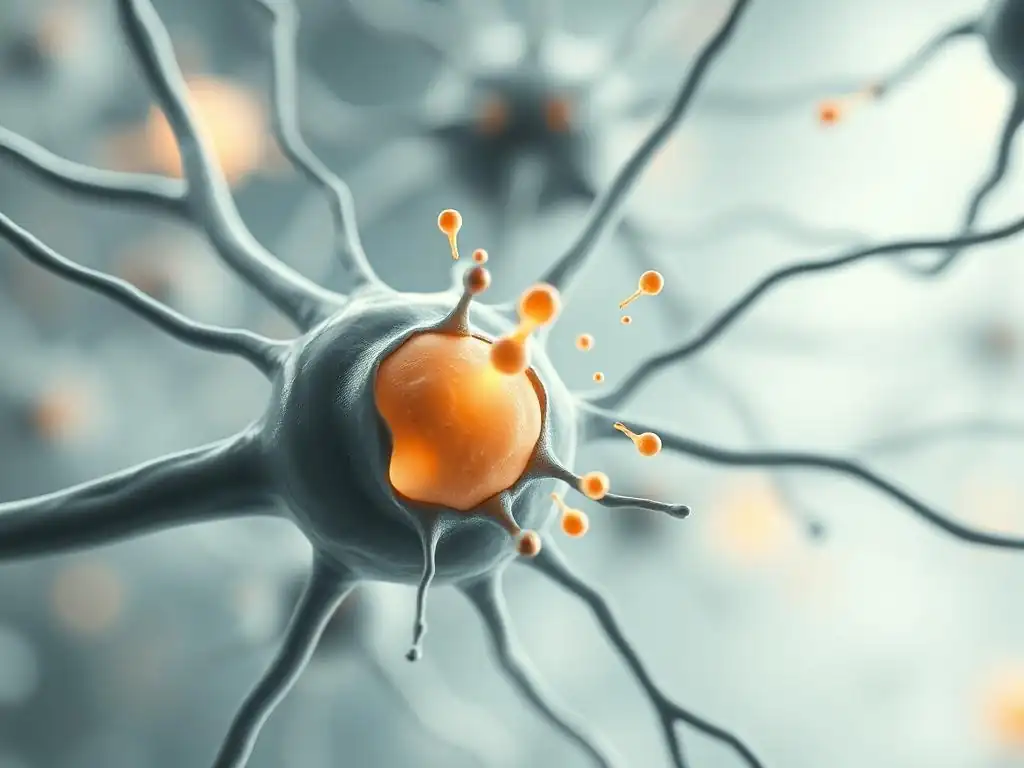
Action Potential Propagation and Neurotransmitter Release
The journey of a neural signal begins with an action potential, an electrical impulse that travels down the axon of the presynaptic neuron. When this action potential reaches the axon terminal, it causes a depolarization of the presynaptic membrane. This depolarization is the critical trigger that opens voltage-gated calcium channels embedded in the membrane. Calcium ions (Ca²⁺), which are present in higher concentration outside the neuron, then flow into the presynaptic terminal.
The influx of calcium ions is the essential signal that initiates the process of neurotransmitter release. Calcium binds to specific proteins associated with synaptic vesicles, such as synaptotagmin. This binding changes the conformation of these proteins, enabling the vesicles to dock with and fuse to the inner surface of the presynaptic membrane. Without sufficient calcium influx, the vesicles would not release their contents, demonstrating the direct link between the electrical action potential and the chemical release of neurotransmitters into the synaptic cleft.
Vesicle Fusion and Exocytosis: Delivering Neurotransmitters into the Synaptic Cleft
Vesicle fusion with the presynaptic membrane is a complex process orchestrated by a set of proteins collectively known as the SNARE complex. These proteins, including synaptobrevin on the vesicle membrane and syntaxin and SNAP-25 on the presynaptic membrane, act together like a molecular machine to pull the vesicle and the presynaptic membrane very close to each other. The calcium influx mentioned previously facilitates the final steps of this process.
Once the vesicle membrane fuses with the presynaptic membrane, a pore opens, and the neurotransmitter molecules stored inside the vesicle are rapidly released into the synaptic cleft. This process, called exocytosis, is incredibly fast, often occurring within a fraction of a millisecond after the action potential arrives. This rapid delivery ensures that the chemical signal is transmitted quickly and efficiently across the synapse, allowing for the high speed of information processing in the brain and regulating neurotransmitter communication crucial for mental health.
Key Neurotransmitters Involved in Mental Health: Glutamate, GABA, Dopamine, Serotonin, Norepinephrine
-
Glutamate: This is the primary excitatory neurotransmitter in the central nervous system. It plays a crucial role in learning, memory, and synaptic plasticity. Approximately 90% of excitatory synapses in the human brain use glutamate. Dysregulation of glutamatergic signaling has been implicated in conditions like schizophrenia, where altered glutamate receptor function is observed, and in anxiety disorders, where excessive glutamate activity can contribute to heightened neural excitability.
Glutamate acts through various receptor types, including NMDA, AMPA, and kainate receptors (all ionotropic), and metabotropic glutamate receptors (mGluRs). The balance between glutamate and GABA activity is critical for maintaining neural stability. Imbalances in glutamate signaling are also being investigated in neurodegenerative diseases and mood disorders, highlighting its pervasive influence on brain function and neurotransmitter communication underlying mental health.
-
GABA (gamma-aminobutyric acid): As the principal inhibitory neurotransmitter, GABA counteracts the excitatory effects of glutamate, helping to regulate neural activity and prevent over-excitation. It's estimated that around 20-40% of synapses in the mammalian brain are GABAergic. GABA's main function is to reduce neuronal excitability, often by increasing chloride ion influx into the postsynaptic neuron, making it less likely to fire an action potential.
Disruptions in GABAergic signaling are strongly associated with anxiety disorders, where reduced GABA activity can lead to increased neural firing and a state of heightened arousal. Benzodiazepine medications, commonly prescribed for anxiety, work by enhancing the effects of GABA at GABA-A receptors. Epilepsy and certain sleep disorders are also linked to GABA system dysfunction, underscoring its vital role in maintaining neural balance and healthy brain function related to mental well-being.
-
Dopamine: This neurotransmitter is involved in reward, motivation, pleasure, motor control, and executive functions. Dopamine pathways originating in the midbrain project to various brain regions, including the prefrontal cortex, nucleus accumbens, and striatum. Dysregulation of dopamine is centrally involved in several mental health conditions.
In schizophrenia, hyperactivity in certain dopamine pathways is linked to positive symptoms, while hypoactivity in other pathways may contribute to cognitive deficits. Dopamine dysfunction is also implicated in addiction due to its role in the brain's reward system. Furthermore, reduced dopamine activity in areas like the nucleus accumbens is associated with anhedonia (loss of pleasure), a common symptom of depression. Understanding dopamine's complex roles is key to understanding neurotransmitter communication and mental health challenges like substance use disorders and mood disorders.
-
Serotonin (5-HT): Serotonin is a monoamine neurotransmitter that profoundly influences mood, sleep, appetite, digestion, learning ability, and memory. Serotonergic neurons are primarily located in the brainstem's raphe nuclei, projecting widely throughout the brain. Serotonin is perhaps most famously linked to mood disorders like depression and anxiety.
Many antidepressant medications, such as Selective Serotonin Reuptake Inhibitors (SSRIs), work by increasing the amount of serotonin available in the synaptic cleft, thereby enhancing serotonergic signaling. While the exact mechanisms are still being researched, increasing serotonin levels is thought to help regulate mood and reduce symptoms of depression and anxiety. Serotonin also plays a role in conditions like Obsessive-Compulsive Disorder (OCD) and Panic Disorder, highlighting its broad impact on emotional and cognitive states.
-
Norepinephrine (Noradrenaline): Another monoamine neurotransmitter, norepinephrine, is involved in the body's "fight or flight" response, alertness, arousal, attention, and stress regulation. Neurons producing norepinephrine are mainly found in the locus coeruleus in the brainstem, projecting to many brain areas.
Norepinephrine dysfunction is implicated in mood disorders and anxiety. Reduced levels of norepinephrine are sometimes observed in depression, contributing to symptoms like fatigue, difficulty concentrating, and lack of motivation. Conversely, excessive norepinephrine activity can contribute to the heightened arousal, vigilance, and physical symptoms experienced in anxiety and panic attacks. Some antidepressants, like SNRIs (Serotonin-Norepinephrine Reuptake Inhibitors), target both serotonin and norepinephrine systems to address these imbalances and improve neurotransmitter communication for mental health benefits.
Neurotransmitter Receptors: Binding and Signal Transduction
Ionotropic Receptors: Ligand-Gated Ion Channels and Fast Synaptic Transmission
Ionotropic receptors are a class of neurotransmitter receptors that are also ion channels. When a neurotransmitter binds to an ionotropic receptor, it causes a conformational change in the receptor protein, which directly opens or closes an ion channel pore located within the receptor itself. This opening or closing allows specific ions (such as sodium, potassium, calcium, or chloride) to flow across the postsynaptic membrane, leading to a rapid change in the membrane potential of the postsynaptic neuron.
Because the receptor binding directly and quickly affects ion flow, ionotropic receptors mediate fast synaptic transmission. For example, the binding of glutamate to AMPA receptors or GABA to GABA-A receptors causes almost instantaneous changes in membrane potential, leading to rapid excitation or inhibition of the postsynaptic neuron. This speed is essential for rapid processing of sensory information and motor control, demonstrating a key aspect of dynamic neurotransmitter communication.
Metabotropic Receptors: G Protein-Coupled Receptors and Slower, Modulatory Effects
Metabotropic receptors are another major class of neurotransmitter receptors, distinct from ionotropic receptors. Unlike ionotropic receptors, they are not ion channels themselves. Instead, when a neurotransmitter binds to a metabotropic receptor, it activates an intracellular signaling cascade, often involving G proteins and secondary messengers (like cyclic AMP or calcium ions). This cascade then indirectly affects ion channels or other cellular processes.
Because this signaling pathway involves multiple steps, the effects of metabotropic receptors are slower but typically longer-lasting and more diffuse than those of ionotropic receptors. They can modulate the excitability of the postsynaptic neuron, alter the strength of synaptic connections (synaptic plasticity), or influence gene expression and protein synthesis. Many neurotransmitters, including dopamine, serotonin, norepinephrine, and acetylcholine, primarily act through metabotropic receptors, allowing for complex modulation of neural circuits and influencing mood, motivation, and cognition – all critical aspects of neurotransmitter communication in mental health.
Receptor Subtypes: Diversity and Specificity in Neurotransmitter Signaling
For many neurotransmitters, there isn't just one type of receptor; there are multiple subtypes, often with different structures, locations, and signaling properties. For example, there are at least five main types of dopamine receptors (D1-D5), fourteen known serotonin receptor subtypes (5-HT1 to 5-HT7, with further subdivisions), and multiple subtypes for glutamate (e.g., NMDA, AMPA, mGluR1-8) and GABA (GABA-A, GABA-B). This diversity allows a single neurotransmitter to have varied effects depending on which receptor subtype it binds to and where that receptor is located in the brain.
The existence of receptor subtypes adds a layer of complexity and specificity to neurotransmitter communication. It means that targeting a specific receptor subtype with a drug can have more precise effects than simply altering the overall level of a neurotransmitter. For instance, some antipsychotic medications primarily block D2 dopamine receptors, which are heavily involved in the pathways implicated in schizophrenia, while having less effect on other dopamine receptor subtypes. This specificity is a key goal in developing more effective and targeted treatments for mental health conditions, aiming to fine-tune synaptic signaling.
Neurotransmitter Reuptake and Degradation: Regulating Synaptic Activity
Reuptake Transporters: Recycling Neurotransmitters from the Synaptic Cleft
After neurotransmitter molecules are released into the synaptic cleft and bind to postsynaptic receptors, their action must be terminated to allow the synapse to reset and respond to subsequent signals. One of the primary mechanisms for clearing neurotransmitters from the cleft is reuptake. This process involves specialized protein pumps, called reuptake transporters, located on the presynaptic membrane or sometimes on surrounding glial cells.
These transporters actively pump neurotransmitter molecules back into the presynaptic terminal, where they can be repackaged into vesicles for future release or broken down. For example, the serotonin transporter (SERT), dopamine transporter (DAT), and norepinephrine transporter (NET) are responsible for reuptake of their respective neurotransmitters. Many antidepressant and stimulant medications work by blocking these transporters, increasing the concentration and dwell time of the neurotransmitter in the synaptic cleft, thereby enhancing neurotransmitter communication.
Enzymatic Degradation: Breaking Down Neurotransmitters in the Synapse
Another mechanism for terminating neurotransmitter signaling is enzymatic degradation. Specific enzymes located in the synaptic cleft or within the postsynaptic neuron break down neurotransmitter molecules into inactive metabolites. This process is particularly important for certain neurotransmitters, such as acetylcholine, which is rapidly broken down by the enzyme acetylcholinesterase in the synaptic cleft.
Monoamine neurotransmitters like dopamine, norepinephrine, and serotonin are also subject to enzymatic degradation, though this often occurs after reuptake into the presynaptic terminal or glial cells. Enzymes like monoamine oxidase (MAO) and catechol-O-methyltransferase (COMT) play crucial roles in metabolizing these neurotransmitters. Some older antidepressant medications, like MAO inhibitors, work by blocking the activity of these enzymes, leading to increased levels of monoamines in the brain.
The Importance of Regulation in Maintaining Neurotransmitter Balance and Mental Health
The precise regulation of neurotransmitter levels in the synaptic cleft through mechanisms like reuptake and degradation is absolutely critical for maintaining healthy brain function and mental well-being. The timing and amplitude of the postsynaptic signal depend on how long the neurotransmitter remains in the cleft and at what concentration. If neurotransmitters are cleared too slowly, the postsynaptic neuron can become overstimulated or desensitized. If they are cleared too quickly, the signal may be too weak to be effective.
Imbalances in these regulatory processes are directly linked to various mental health conditions. For instance, a genetic variation affecting the serotonin transporter (SERT) function has been associated with an increased risk of anxiety and depression in some individuals, particularly when combined with environmental stress. The effectiveness of many psychotropic medications hinges on their ability to modulate reuptake or degradation, highlighting how maintaining appropriate neurotransmitter balance through regulated synaptic activity is fundamental to addressing issues in neurotransmitter communication and mental health.
Synaptic Plasticity: Adapting and Changing Neural Connections
Long-Term Potentiation (LTP): Strengthening Synaptic Connections
Synaptic plasticity refers to the ability of synapses to strengthen or weaken over time in response to changes in their activity level. Long-Term Potentiation (LTP) is a persistent strengthening of synaptic connections, resulting in a larger postsynaptic response to the same presynaptic stimulus. It is considered a primary cellular mechanism underlying learning and memory. LTP typically occurs at glutamatergic synapses, particularly those involving NMDA and AMPA receptors.
The induction of LTP often requires strong or repeated stimulation of the presynaptic neuron, leading to significant depolarization of the postsynaptic neuron. This depolarization is necessary to remove a magnesium block from NMDA receptors, allowing calcium ions to enter the postsynaptic neuron. This calcium influx triggers a cascade of intracellular events, including the insertion of more AMPA receptors into the postsynaptic membrane and structural changes that enlarge the synaptic contact area, making the synapse more efficient at transmitting signals in the future.
Long-Term Depression (LTD): Weakening Synaptic Connections
Conversely, Long-Term Depression (LTD) is a persistent weakening of synaptic connections. Like LTP, LTD is a form of synaptic plasticity and is also thought to be important for learning and memory, particularly in processes like forgetting or clearing old information to make space for new. LTD typically occurs when the presynaptic neuron is stimulated at a low frequency for an extended period, or when the presynaptic and postsynaptic neurons are active out of sync in a specific way.
Similar to LTP, LTD at glutamatergic synapses involves NMDA receptors and calcium influx, but often the pattern and amount of calcium influx are different, triggering a different set of intracellular signaling pathways. These pathways can lead to the removal of AMPA receptors from the postsynaptic membrane or other structural changes that decrease the strength of the synaptic connection. The balance between LTP and LTD is crucial for the dynamic remodeling of neural circuits that underlies adaptation and learning throughout life.
Synaptic Plasticity and its Role in Learning, Memory, and Adaptation in Mental Health
Synaptic plasticity, through mechanisms like LTP and LTD, is the biological basis for how our brains learn, form memories, and adapt to new experiences. Every time we learn something new or recall a memory, specific synapses are being strengthened or weakened. This constant remodeling of neural circuits allows for flexibility and adaptation in our behavior and responses.
In the context of mental health, disruptions in synaptic plasticity are thought to contribute to various disorders. For example, impaired LTP in areas like the hippocampus might underlie memory deficits seen in depression or PTSD. Altered synaptic plasticity in the prefrontal cortex could contribute to executive function deficits in schizophrenia. Furthermore, therapeutic interventions, including psychotherapy and some medications, may work, in part, by promoting beneficial synaptic plasticity, helping individuals to unlearn maladaptive patterns and build new, healthier neural connections, demonstrating the potential of targeting the synapse to improve neurotransmitter communication and mental health.
The Synapse and Mental Disorders: A Direct Connection
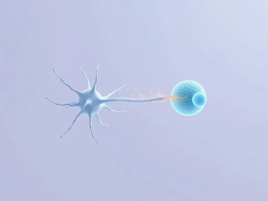
Schizophrenia: Dopaminergic and Glutamatergic Dysfunction
Schizophrenia is a complex mental disorder strongly linked to dysfunction at the synapse, particularly involving dopamine and glutamate systems. The "dopamine hypothesis" of schizophrenia suggests an overactivity of dopamine signaling in certain brain pathways (mesolimbic), contributing to positive symptoms like hallucinations and delusions. Conversely, reduced dopamine activity in other areas (mesocortical) may contribute to negative symptoms (e.g., apathy, social withdrawal) and cognitive deficits. Antipsychotic medications primarily target dopamine receptors, supporting this link.
However, research also points to significant abnormalities in glutamatergic neurotransmission in schizophrenia. NMDA receptor hypofunction is a leading hypothesis, suggesting that reduced activity at these crucial glutamate receptors contributes to both positive and negative symptoms, as well as the severe cognitive impairment characteristic of the disorder. Postmortem studies have shown reduced levels of NMDA receptor subunits in certain brain regions of individuals with schizophrenia. The interplay between dopamine and glutamate systems at the synapse is complex, and understanding their combined dysfunction is key to unraveling the neurobiology of this illness and improving neurotransmitter communication for mental health.
Depression: Serotonergic, Noradrenergic, and Dopaminergic Dysregulation
Major depressive disorder is associated with dysregulation across multiple monoamine neurotransmitter systems, including serotonin, norepinephrine, and dopamine, with synaptic dysfunction being a central feature. The "monoamine hypothesis" posits that a deficit in the activity of these neurotransmitters in certain brain circuits contributes to depressive symptoms. While overly simplistic, it led to the development of effective medications targeting these systems.
Studies have shown altered levels of these neurotransmitters or their metabolites in the cerebrospinal fluid or postmortem brains of depressed individuals, though these findings are not always consistent. More robust evidence comes from the effectiveness of antidepressants that increase the availability of serotonin (SSRIs), norepinephrine (SNRIs), or both in the synaptic cleft. Dopaminergic dysfunction, particularly in reward pathways, is also implicated, contributing to anhedonia and lack of motivation. This highlights how widespread synaptic imbalances in monoamine neurotransmitter communication can profoundly impact mental health and mood regulation.
Anxiety Disorders: GABAergic and Glutamatergic Imbalances
Anxiety disorders, including generalized anxiety disorder, panic disorder, and phobias, are often linked to an imbalance between excitatory and inhibitory neurotransmission, particularly involving GABA and glutamate at the synapse. Reduced activity of the inhibitory neurotransmitter GABA is strongly implicated. Lower levels of GABA or fewer GABA receptors could lead to excessive neural firing and heightened states of arousal and fear. Medications like benzodiazepines enhance GABA's effects at the GABA-A receptor, calming neural activity and reducing anxiety symptoms.
Conversely, excessive activity of the excitatory neurotransmitter glutamate may also contribute to anxiety by increasing neural excitability. The amygdala, a brain region critical for processing fear and anxiety, is rich in both GABAergic and glutamatergic synapses. An imbalance here, with too much excitation or too little inhibition, can lead to a hyperactive fear response. Research continues to explore the precise synaptic mechanisms in different anxiety subtypes, aiming to develop more targeted therapies that restore the balance of neurotransmitter communication and improve mental health outcomes.
The Impact of Synaptic Dysfunction on Cognitive and Emotional Processing in Mental Disorders
Synaptic dysfunction has a pervasive impact on the complex cognitive and emotional processing that is disrupted in mental disorders. For example, the working memory deficits seen in schizophrenia and depression are linked to altered synaptic function in the prefrontal cortex, a region critical for executive functions. Changes in synaptic plasticity, including impaired LTP and LTD, may underlie difficulties in learning and memory observed in these conditions.
Emotional processing is also heavily reliant on healthy synaptic function within circuits involving the amygdala, prefrontal cortex, hippocampus, and cingulate cortex. Dysregulation of neurotransmitter communication at synapses in these areas can lead to distorted emotional responses, such as excessive fear in anxiety disorders, pervasive sadness in depression, or inappropriate emotional reactions in schizophrenia. Understanding how specific synaptic alterations disrupt these neural circuits provides crucial insights into the lived experience of these disorders and guides the search for interventions that can restore healthy neurotransmitter communication and mental health.
Therapies Targeting the Synapse: Restoring Neurotransmitter Balance
Antidepressants: Selective Serotonin Reuptake Inhibitors (SSRIs), SNRIs, and Tricyclic Antidepressants
Many of the most common and effective treatments for depression and anxiety directly target synaptic function by altering neurotransmitter levels. Selective Serotonin Reuptake Inhibitors (SSRIs), such as fluoxetine (Prozac) and sertraline (Zoloft), work by blocking the serotonin transporter (SERT) on the presynaptic neuron. This prevents the reuptake of serotonin from the synaptic cleft, increasing its concentration and prolonging its binding to postsynaptic receptors, thereby enhancing serotonergic neurotransmitter communication.
Serotonin-Norepinephrine Reuptake Inhibitors (SNRIs), like venlafaxine (Effexor) and duloxetine (Cymbalta), block both the serotonin transporter (SERT) and the norepinephrine transporter (NET), increasing levels of both neurotransmitters in the cleft. Tricyclic Antidepressants (TCAs), an older class of drugs, also block the reuptake of serotonin and norepinephrine, though they tend to have more side effects due to actions on other receptors. These medications illustrate how modulating the reuptake process at the synapse can help restore neurotransmitter balance and improve mental health symptoms.
Antipsychotics: Dopamine Receptor Antagonists and Atypical Antipsychotics
Antipsychotic medications, used primarily to treat schizophrenia and bipolar disorder, largely exert their effects by targeting dopamine receptors at the synapse. First-generation or "typical" antipsychotics, such as haloperidol, are potent antagonists of dopamine D2 receptors. By blocking these receptors, they reduce excessive dopamine signaling, particularly in the mesolimbic pathway, which helps alleviate positive symptoms like hallucinations and delusions.
Second-generation or "atypical" antipsychotics, such as clozapine and risperidone, have a broader range of action. While they also block D2 receptors, they often have a weaker affinity for D2 receptors and also block serotonin 5-HT2A receptors.
This broader profile is thought to contribute to their effectiveness against both positive and negative symptoms, as well as potentially reducing the risk of certain side effects compared to typical antipsychotics. Their action at the synapse underscores the critical role of dopamine and serotonin systems in the pathophysiology and treatment of psychosis, highlighting how targeted synaptic intervention can improve neurotransmitter communication and mental health.
Anxiolytics: Benzodiazepines and Other GABAergic Enhancers
Anxiolytic medications, used to treat anxiety disorders, often work by enhancing the effects of the inhibitory neurotransmitter GABA at the synapse. Benzodiazepines, such as diazepam (Valium) and alprazolam (Xanax), bind to a specific site on the GABA-A receptor, distinct from the GABA binding site. When a benzodiazepine is bound, it increases the frequency of chloride channel opening when GABA binds to the receptor.
This increased chloride influx hyperpolarizes the postsynaptic neuron, making it less excitable and reducing overall neural activity. This calming effect helps alleviate symptoms of anxiety, panic attacks, and insomnia. Other drugs, like buspirone, work on different neurotransmitter systems (partially as a serotonin 5-HT1A receptor agonist), demonstrating that multiple synaptic targets can influence anxiety levels. These examples highlight the importance of GABAergic signaling and the synapse as a target for reducing excessive neural activity associated with anxiety, thereby improving neurotransmitter communication and mental health.
Neuromodulation Techniques: TMS, DBS, and VNS Targeting Synaptic Function
Beyond pharmacological interventions, various neuromodulation techniques also target synaptic function to treat mental health conditions. Transcranial Magnetic Stimulation (TMS) uses magnetic pulses delivered to specific brain areas, like the prefrontal cortex, to induce electrical currents that modulate neuronal activity and synaptic plasticity. For example, repetitive TMS is FDA-approved for treatment-resistant depression and is thought to work by altering the excitability of cortical neurons and strengthening or weakening synaptic connections in relevant circuits.
Deep Brain Stimulation (DBS) involves surgically implanting electrodes in specific brain regions, such as the subthalamic nucleus or nucleus accumbens, to deliver continuous electrical impulses. While complex, DBS is believed to normalize abnormal activity patterns in neural circuits by influencing synaptic transmission and firing rates of neurons in the targeted area. It is approved for certain neurological conditions but is being explored for severe, treatment-resistant depression and OCD.
Vagus Nerve Stimulation (VNS) involves implanting a device to stimulate the vagus nerve, which indirectly influences brain activity, including neurotransmitter release and synaptic function in areas involved in mood regulation. These techniques offer alternative ways to modulate synaptic communication and improve mental health outcomes when traditional pharmacological approaches are insufficient.
The Future of Synaptic Research: New Avenues for Mental Health Treatment
Developing Novel Drugs Targeting Specific Receptor Subtypes and Signaling Pathways
Future directions in mental health treatment are increasingly focused on developing novel drugs that target specific receptor subtypes or intracellular signaling pathways at the synapse with greater precision. Instead of broadly increasing or decreasing neurotransmitter levels, researchers are aiming for drugs that selectively modulate the activity of particular receptor populations or downstream signaling molecules involved in synaptic function. For instance, developing positive or negative allosteric modulators (PAMs or NAMs) that bind to sites on receptors different from the neurotransmitter binding site could allow for more nuanced control of synaptic activity.
Research is also exploring drugs that target components of the SNARE complex involved in neurotransmitter release or molecules involved in synaptic plasticity mechanisms like LTP and LTD. The goal is to develop treatments that can correct specific synaptic deficits implicated in disorders while minimizing off-target effects that cause side effects. This highly targeted approach to neurotransmitter communication holds promise for developing more effective and personalized mental health treatments with fewer adverse effects.
Exploring Gene Therapy and CRISPR-Cas9 for Synaptic Disorders
Gene therapy and gene editing technologies like CRISPR-Cas9 represent potentially revolutionary approaches for treating mental disorders rooted in synaptic dysfunction caused by genetic factors. If a specific gene mutation leads to the production of a faulty synaptic protein (e.g., a neurotransmitter receptor subunit, a transporter, or a protein involved in vesicle release), gene therapy could potentially introduce a functional copy of the gene into the relevant neurons.
CRISPR-Cas9 could potentially be used to correct the mutation directly or modify gene expression to restore normal levels of synaptic proteins. While these technologies are still largely in experimental stages for brain disorders due to challenges in delivery and targeting, they offer the possibility of addressing the root genetic causes of synaptic dysfunction rather than just managing symptoms. This represents a future frontier for restoring healthy neurotransmitter communication and mental health at a fundamental level.
Understanding the Complex Interactions at the Synapse for Personalized Mental Health Treatments
A key challenge and future direction is gaining a deeper understanding of the incredibly complex interactions occurring at the synapse, not just involving single neurotransmitters but the interplay between multiple neurotransmitter systems and glial cells (like astrocytes and microglia) that also influence synaptic function. Synapses are dynamic entities, constantly being modulated by various factors, and dysfunction often involves multiple components of the synaptic machinery.
Future research aims to unravel these complex networks using advanced techniques like optogenetics, in vivo imaging, and single-cell analysis. This detailed understanding could lead to the development of personalized mental health treatments. Instead of a one-size-fits-all approach, treatments could be tailored based on an individual's specific synaptic profile – for example, identifying whether their depression is primarily linked to serotonergic reuptake issues, noradrenergic receptor sensitivity, or altered synaptic plasticity. This personalized approach to understanding and modulating neurotransmitter communication at the synapse holds immense potential for improving mental health care.
Promoting Synaptic Health: Lifestyle and Environmental Factors
Diet and Nutrition: The Impact of Nutrients on Neurotransmitter Synthesis and Function
Lifestyle factors, including diet and nutrition, significantly impact synaptic health and neurotransmitter communication. The brain relies on a constant supply of nutrients to synthesize neurotransmitters and maintain synaptic structure and function. For example, amino acids like tryptophan are precursors for serotonin, tyrosine for dopamine and norepinephrine, and glutamine for glutamate and GABA. Deficiencies in these building blocks can impair neurotransmitter production.
Omega-3 fatty acids, found in fish oil and flaxseeds, are crucial components of neuronal membranes and are essential for maintaining synaptic fluidity and function. B vitamins (like B6, B12, and folate) are vital cofactors for enzymes involved in neurotransmitter synthesis and metabolism. A diet rich in fruits, vegetables, lean proteins, and healthy fats provides the necessary nutrients to support healthy synaptic function and optimal neurotransmitter communication, contributing positively to mental well-being.
Exercise: Enhancing Synaptic Plasticity and Neurogenesis
Regular physical exercise has profound positive effects on brain health, including enhancing synaptic plasticity and promoting neurogenesis (the birth of new neurons), particularly in areas like the hippocampus, crucial for learning and memory. Exercise increases the production of neurotrophic factors, such as Brain-Derived Neurotrophic Factor (BDNF), which supports the growth, survival, and function of neurons and synapses.
Studies have shown that exercise can increase the density of dendritic spines (small protrusions on dendrites that receive synaptic input), indicating an increase in the number and strength of synapses. It can also improve neurotransmitter signaling, for example, by increasing dopamine and serotonin levels. Engaging in regular aerobic exercise, even moderate activity like brisk walking for 30 minutes several times a week, can significantly contribute to maintaining healthy synaptic connections and improving mood and cognitive function, thereby positively influencing neurotransmitter communication and mental health.
Sleep: Synaptic Consolidation and Brain Health
Sleep is not just a period of rest; it is a critical time for brain maintenance and consolidation of learning and memory, processes heavily reliant on synaptic function. During sleep, especially slow-wave sleep and REM sleep, synapses undergo processes that are thought to strengthen important connections formed during the day (LTP) and prune weaker or less relevant ones (LTD). This synaptic homeostasis helps optimize neural circuits for future learning and function.
Chronic sleep deprivation can disrupt synaptic plasticity, impair cognitive function, and alter neurotransmitter systems, including those involving serotonin, norepinephrine, and dopamine. This disruption can contribute to mood instability, difficulty concentrating, and increased susceptibility to stress – factors linked to mental health conditions. Prioritizing sufficient, quality sleep is therefore essential for maintaining healthy synaptic function and supporting overall brain health and stable neurotransmitter communication crucial for mental well-being.
Stress Management: Reducing the Negative Impact of Stress on Synaptic Function
Chronic stress has detrimental effects on brain structure and function, particularly impacting areas involved in mood, memory, and executive control, such as the hippocampus and prefrontal cortex. Prolonged exposure to stress hormones like cortisol can lead to the retraction of dendrites, loss of dendritic spines, and reduced synaptic density in these vulnerable brain regions. This synaptic damage impairs neurotransmitter communication and contributes to vulnerability to mental health disorders like depression and anxiety.
Implementing effective stress management techniques can help mitigate these negative effects. Practices such as mindfulness meditation, yoga, deep breathing exercises, and engaging in hobbies can help regulate the body's stress response system. By reducing the levels of circulating stress hormones, these strategies help protect synapses from damage and promote resilience. Managing stress is a vital component of maintaining healthy synaptic function and preserving optimal neurotransmitter communication for long-term mental health.
Frequently Asked Questions
What is the role of a synapse in neurotransmitter communication and mental health?
The synapse is the critical junction where neurotransmitters are released from one neuron and received by another. Disruptions in synaptic function can significantly impact mental health by altering the transmission of signals responsible for mood, cognition, and behavior.
How does impaired neurotransmitter communication affect mental well-being?
Impaired neurotransmitter communication can lead to a variety of mental health challenges. This can manifest as conditions like depression, anxiety, and schizophrenia, due to imbalances in neurotransmitter levels or receptor function.
What factors can disrupt neurotransmitter communication at the synapse?
Several factors can disrupt neurotransmitter communication at the synapse, including genetic predispositions, stress, trauma, and substance abuse. These factors can alter the synthesis, release, or reception of neurotransmitters.
What treatments are available to improve neurotransmitter communication for mental health?
Treatments aimed at improving neurotransmitter communication include medications like antidepressants and antipsychotics. These medications often target specific neurotransmitter systems to restore balance and alleviate mental health symptoms.
How can lifestyle changes impact neurotransmitter communication and mental well-being?
Lifestyle changes such as regular exercise, a healthy diet, and sufficient sleep can positively impact neurotransmitter communication. These activities promote the production and regulation of neurotransmitters, leading to improved mental well-being.
Final Reflection: Bridging the Gap in Understanding Mental Health
The intricate dance of neurotransmitters across the synapse is not merely a biological process; it's the very foundation upon which our thoughts, emotions, and behaviors are built. Understanding this microscopic communication is crucial, not just for scientific advancement, but for improving the lives of millions grappling with mental health challenges. The more we learn about the delicate balance within the synapse, the closer we get to developing effective treatments and preventative measures.
The impact of neurotransmitter imbalances on our mental well-being is profound and far-reaching. From the debilitating effects of depression to the anxieties that plague so many, the disruption of this crucial communication pathway is undeniably linked to a vast spectrum of mental health conditions. The brain is the most complex organ in the body, and understanding its intricacies is key to unlocking the secrets of mental health.
This quote underscores the importance of continued research and a holistic approach to understanding the complexities of neurotransmitter function and their impact on our mental state.
The journey to better mental health begins with understanding. This article has hopefully shed light on the critical role of neurotransmitters in maintaining our psychological well-being. Now, I invite you to consider your own mental health, and to reflect on the importance of seeking help when needed. Learn more about the neurotransmitters involved in your own mental state, and advocate for increased research and understanding in this vital area. Together, we can bridge the gap between scientific knowledge and improved mental health outcomes for all.
Unlock Your Brain's Potential: Dive Deeper into Neural Communication!
Thank you for exploring the fascinating world of synapses with us! You've now gained a deeper understanding of how these tiny connections power our thoughts and actions. Ready to take the next step?
- Deepen your knowledge: Explore reputable neuroscience websites or textbooks to delve further into the complexities of neurotransmitters and synaptic transmission. Consider researching specific neurotransmitters that interest you, like dopamine or serotonin, and learn about their roles in various brain functions.
- Share your insights: Discuss this article with friends, family, or colleagues – sparking conversations about brain health is surprisingly impactful!
- Challenge yourself: Think of a daily habit you could adjust to potentially improve your brain's synaptic function (e.g., better sleep, mindful movement).
What new perspective on brain health will you explore today?