Ethical Considerations: Protecting Privacy in Brain Imaging Research
🔍 Ensuring brain imaging privacy research ethics is vital for safeguarding individual rights and fostering trust in mental health advancements. This article…
Read article
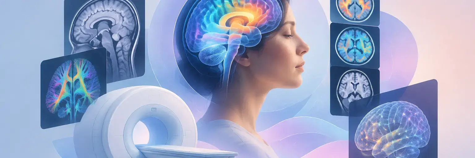
Explore how advanced brain imaging tools are transforming mental health research, offering unprecedented insights into the neural underpinnings of conditions like depression, anxiety, and PTSD. From functional MRI (fMRI) to PET scans, these technologies allow scientists to observe real-time brain activity, connectivity, and chemical changes—bridging the gap between subjective symptoms and objective biomarkers. Whether you’re a researcher, clinician, or curious mind, this section demystifies the techniques that drive precision psychiatry, from diffusion tensor imaging (DTI) mapping white matter pathways to magnetoencephalography (MEG) capturing millisecond neural dynamics. Discover how these tools not only deepen our understanding of mental illness but also pave the way for personalized treatments and early detection. Dive in to see how cutting-edge neuroimaging is reshaping the future of mental wellness, one scan at a time. 🧠✨
🔍 Ensuring brain imaging privacy research ethics is vital for safeguarding individual rights and fostering trust in mental health advancements. This article…
Read article
🧠 Discover how PTSD brain imaging techniques reveal the profound impact of trauma on neural circuits, offering vital insights for diagnosis and…
Read article
🌟 Discover how personalized mental health imaging trends are revolutionizing early diagnosis and tailored treatments, paving the way for a future where…
Read article
🧠 Discover how PET and SPECT brain scans revolutionize mental health diagnostics by revealing real-time brain chemistry, enabling more accurate diagnoses and…
Read article
🧠 Explore the complex landscape of brain imaging mental health ethics, balancing groundbreaking insights with moral responsibilities. This guide highlights the ethical…
Read article
🧠 Discover how autism brain imaging techniques reveal the unique structural and functional features of the autistic brain, advancing understanding and support…
Read article
🌟 Discover how brain scan recovery prediction is revolutionizing mental health treatment, enabling personalized approaches that improve outcomes and support recovery. This…
Read article
🔍 Discover how cutting-edge brain imaging mental health tech reveals the inner workings of the mind, advancing research and providing vital insights…
Read article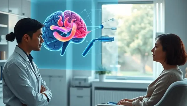
✨ Unlock the potential of brain imaging mental health insights to transform patient care. This article explores how cutting-edge research moves from…
Read article
🔍 Discover how the brain imaging mental health cost impacts diagnosis and treatment, highlighting the urgent need for accessible, affordable neuroimaging solutions…
Read article
🧠 Discover how imaging glial cells enhances our understanding of mental health, highlighting their crucial role beyond neurons. This innovative approach offers…
Read article
🧠 Discover the fascinating boundaries of brain imaging mental health limits, revealing why current scans can't fully decode mental health conditions—highlighting the…
Read articleWelcome to the Brain Imaging in Mental Health Research subcategory, part of our Neuroscience of Mental Health hub at Mind Care Guide. Here, we decode the advanced tools that let scientists "see" the living brain, revealing how thoughts, emotions, and mental health conditions emerge from neural activity. Whether you’re a student, a curious patient, or a professional, this page provides SEO-rich, accessible explanations of imaging methods like fMRI, EEG, PET, and specialized tractography—without dense jargon. Our goal is to empower you with knowledge, demystify research findings, and connect each technique to real-world mental health applications, from depression and anxiety to schizophrenia and PTSD.
fMRI is the workhorse of modern cognitive neuroscience, measuring blood flow changes tied to neuronal activity. It offers excellent spatial resolution (millimeters), allowing researchers to map brain regions activated during tasks like fear conditioning, memory recall, or emotional regulation. Clinically, fMRI helps identify altered connectivity in networks like the default mode network (DMN) in depression or the salience network in anxiety disorders. For a beginner, think of it as a live "heat map" of your brain’s oxygen usage during a given mental state. This technique is non-invasive and doesn’t require radiation, making it safe for repeated studies, though it requires the person to lie still inside a noisy tube.
But fMRI has trade-offs: its temporal resolution is poor (seconds), missing rapid neural spikes. Researchers often combine it with behavioral tasks or resting-state scans to infer network disruptions. For mental health, fMRI has revealed that chronic stress shrinks the hippocampus and hyperactivates the amygdala—findings that guide new therapies like real-time neurofeedback. When reading research, look for statistical corrections (e.g., FWE or cluster-level) to avoid false positives. This technique isn’t diagnostic in routine care yet, but it’s pivotal in experimental psychiatry, especially for predicting treatment response in major depressive disorder and guiding personalized interventions.
EEG records electrical activity via scalp electrodes, capturing brain waves in real time (millisecond precision). It shines in temporal resolution, making it ideal for tracking fast cognitive processes like attention, working memory, or emotional reactivity. In mental health, EEG patterns—such as theta/beta ratios in anxiety or reduced P300 ERP amplitude in schizophrenia—serve as promising biomarkers. Unlike fMRI, EEG is portable, affordable, and comfortable, allowing studies in children or during sleep. It’s also the foundation of brain-computer interfaces and neurofeedback training for ADHD and PTSD, where patients learn to modulate their own wave activity.
However, EEG’s spatial resolution is coarse, often blurring the source of signals deep in the brain. Modern techniques like source localization (e.g., LORETA) help estimate origins, but they’re approximations. For research on mental health, time-frequency analysis of EEG can reveal beta/gamma desynchronization linked to cognitive control deficits. Combining EEG with fMRI (simultaneous acquisition) provides both high temporal and spatial detail—though such setups are complex and expensive. For anyone exploring this, note that even simple metrics like frontal alpha asymmetry have shown moderate reliability as indicators of depression vulnerability, making EEG a grassroots tool in translational mental health science.
PET scans track radioactive tracers that bind to specific receptors, enzymes, or proteins—giving a molecular view of the brain. In mental health research, PET has quantified dopamine D2/3 receptor availability in schizophrenia, serotonin transporter density in depression, and neuroinflammation via TSPO ligands in bipolar disorder. This technique answers questions about neurotransmitter systems that fMRI cannot. For example, a PET study might show reduced dopamine release in the striatum during reward anticipation in anhedonia. It’s invasive (involves injection and radiation), but doses are kept low and safe for research participants, with protocols approved by ethics boards.
Despite its power, PET has challenges: low temporal resolution (minutes), high cost, and limited availability of radiotracers for every protein of interest. Additionally, interpretation is complex because receptor density changes may reflect disease or compensatory mechanisms. Researchers often combine PET with fMRI to correlate molecular availability with brain activity during tasks. For mental health, PET has been revolutionary in demonstrating that standard antidepressants alter serotonin binding sites within weeks—yet clinical improvement takes longer—pointing to downstream plasticity. Future work involves developing tau and amyloid PET for Alzheimer’s risk, but for primary psychiatric disorders, PET remains primarily a research tool, not a routine diagnostic, due to radiation exposure.
Diffusion MRI (DTI or DSI) measures the microscopic movement of water molecules in brain tissue, revealing white matter pathways—the brain’s "highways" connecting regions. Tractography algorithms reconstruct these fiber tracts, allowing researchers to examine the integrity of the uncinate fasciculus in depression or the cingulum bundle in obsessive-compulsive disorder. Reduced fractional anisotropy (FA) in these pathways indicates disrupted connectivity, often correlating with symptom severity and cognitive deficits. Unlike fMRI, this technique is purely structural, offering a stable anatomical readout that doesn’t require task performance, making it ideal for studies in non-compliant populations like severe psychosis.
However, diffusion MRI has significant limitations. Fiber crossing and kissing regions can confuse tractography, leading to false negatives or false reconstructions. Also, it only measures water movement, not actual neural firing—so "disrupted connectivity" must be interpreted cautiously with neurobiological plausibility. Newer models like NODDI and QBI attempt to disentangle intra- and extracellular compartments, improving specificity. In mental health, tractography has identified altered structural connectivity in chronic PTSD, possibly reflecting stress-induced myelin changes. It’s also used in pre-surgical planning for epilepsy and in monitoring treatment-related white matter changes (e.g., after psychotherapy or medication), offering a non-invasive window into brain plasticity over months.
Magnetoencephalography (MEG) detects magnetic fields from neural currents, offering both good spatial (~centimeters) and excellent temporal (milliseconds) resolution—superior to EEG for localizing sources. It’s used to study gamma-band synchronization in schizophrenia and neural oscillations in autism. Functional near-infrared spectroscopy (fNIRS) uses light to measure hemodynamics, being portable and tolerant of motion, making it suitable for infants or post-stroke patients. In mental health research, fNIRS has tracked prefrontal activation during social cognition tasks in borderline personality disorder. Meanwhile, optogenetics—though animal-only—enables precise control of specific neurons using light, translating to novel targets (e.g., DREADDs) for future human gene therapy.
Each emerging tool addresses gaps: MEG is expensive and requires magnetic shielding but yields richer oscillatory data; fNIRS is affordable but has shallow depth penetration (cortex only). For readers, understanding these techniques helps interpret press releases about "brain-computer interfaces" or "light-activated antidepressants." Future directions include fUS (functional ultrasound) for all-optical brain monitoring, and 7T MRI for microstructural cortical layers. These are not yet standard in mental health clinics but are accelerating experimental psychiatry, particularly in identifying subtypes of depression based on distinct neurophysiological profiles—paving the way for precision psychiatry.
Not yet as a routine diagnostic tool. Though fMRI, EEG, and PET reveal group-level differences, individual variability is high, and no single scan is specific or sensitive enough for a diagnosis. However, imaging helps rule out structural causes (e.g., tumors) and is used in research to guide treatment choices, but psychiatrists rely primarily on clinical interviews and standardized scales.
fMRI and EEG/fNIRS are completely non-invasive with no radiation, making them safe for repeated use, including children and pregnant women. PET involves low-dose radioactive tracers but still holds ethical constraints. MEG is also safe but less available. Always consult the study’s ethics approval for specifics.
Some research suggests that pre-treatment fMRI activation in the anterior cingulate cortex or subgenual cingulate may predict response to SSRIs or therapy. EEG-based theta activity has also shown promise. However, these biomarkers are not yet validated for clinic-wide use—they’re promising but require replication before becoming standard.
For fMRI, expect 45–60 minutes including preparation and tasks. EEG can take 30–90 minutes, depending on electrodes and tasks. PET scans require a tracer injection plus a 60–90-minute waiting period, then a 15–30 minute scan. DTI (diffusion) usually lasts 10–15 minutes as part of an MRI session.
MRI has no known biological risks if you’re screened for metallic implants or claustrophobia. EEG may cause mild skin irritation from gel do not persist. PET involves radiation exposure comparable to a few chest X-rays, considered low risk. Always disclose pregnancy or metal devices before scanning.